Vitamins
now browsing by category
Remarkable Progress in Regression of Liver Cancer
Reproduced from original OMNS article (OrthoMolecular News Service):
http://orthomolecular.org/
Subscribe to the free Orthomolecular Newsletter: http://orthomolecular.org/subscribe.html
Go to the OMNS Archive: http://orthomolecular.org/resources/omns/index.shtml
Orthomolecular Medicine News Service, September 30, 2022
Less than six months to decrease tumor mass by 85%
by Andrew W. Saul, Robert G. Smith, PhD and Ron Hunninghake, MD
OMNS (Sept. 30, 2022) We report a liver cancer patient’s success with intravenous vitamin C, 70,000 mg twice weekly, coupled with chemotherapy.
Following a 65 year old male’s diagnosis of stage four colon cancer and a partial bowel resection, scans revealed a tumor in the left lobe of the liver (Feb 23, 2022): 6.5 cm x 4.3 cm. The patient received 5FU chemotherapy, which although generally regarded as palliative for bowel cancer, is not considered curative. Indeed, both the patient’s oncologists told him that his condition was not curable and he might expect to live two years with treatment. 5FU is even less effective, if effective at all, against liver carcinoma. This prognosis motivated the patient to begin intravenous vitamin C treatments, starting at 20 grams (20,000 milligrams) and working up to 70g (70,000 mg) twice weekly.
Less than three months later, the liver tumor was reduced to 3.8 x 3.3 cm (May 16, 2022). The patient continued IV vitamin C, 70g twice weekly, and scans revealed a further reduction to 3.1 x 2.4 cm Aug 15, 2022. At this point he had completed a total of 48 IVC treatments, eight to work up from 20g to 60g, then 40 treatments at 70g.
Given an approximately ellipsoidal volume, in which the 3rd dimension was equal to the average of the other 2 dimensions, the measurements show:
Volume = 4/3 * PI * radius A * radius B * radius C
Patient’s initial tumor volume: 4/3 * PI * 3.25 * 2.15 * 2.7 => 79 cm3
Volume at second scan: 4/3 * PI * 1.9 * 1.65 * 1.775 => 23.3 cm3
Tumor volume at third scan: 4/3 * PI * 1.55 * 1.2 * 1.375 => 10.7 cm3
We see this result, from 79 cm3 down to 10.7 cm3, a reduction in volume by more than 85% as important, as it represents a major decrease in less than 6 months.
Additionally, we report this observation: there was an amazing stability of all his blood work, but especially his WBCs, his neutrophil/lymphocyte ratios, platelet counts, and the rapid improvement in the initially elevated alkaline phosphatase.
“There is no evidence that anecdotal information is any less accurate then clinical information. Where are the good old days, when honest physicians honestly reported what they saw in language that any doctor could understand?” (Abram Hoffer, MD, PhD)
For more information about the use of intravenous vitamin C to treat and reverse cancer, we recommend reading additional reports and scientific literature on this topic. [1-6]
References
1. Passwater M (2021) Summary of Recent Research with Three Nutrients in the War Against Cancer. Orthomolecular Medicine News Service. http://orthomolecular.org/resources/omns/v17n26.shtml
2. Yuen RCF (2021) High Dose Vitamin C for Cancer: The Struggle with “Non-Evidence-Based” Medical Practice. Orthomolecular Medicine News Service. http://orthomolecular.org/resources/omns/v17n20.shtml
3. Galindo Salom H, Carrillo C, Galvis G (2020) Impact of intravenous vitamin C megadose on the quality of life of terminal cancer patients. Orthomolecular Medicine News Service. http://orthomolecular.org/resources/omns/v16n45.shtml
4. Saul AW (2019) Why vitamin C fights cancer so well, and why more and more oncologists are now using it. Orthomolecular Medicine News Service. http://orthomolecular.org/resources/omns/v15n11.shtml
5. Cantley L, Yun J (2020) Intravenous High-Dose Vitamin C in Cancer Therapy. National Cancer Institute. https://www.cancer.gov/research/key-initiatives/ras/ras-central/blog/2020/yun-cantley-vitamin-c
6. Ngo B, Van Riper JM, Cantley LC, Yun J (2019) Targeting cancer vulnerabilities with high-dose vitamin C. Nat Rev Cancer. 2019 May; 19: 271-282. https://pubmed.ncbi.nlm.nih.gov/30967651
Nutritional Medicine is Orthomolecular Medicine
Orthomolecular medicine uses safe, effective nutritional therapy to fight illness. For more information: http://www.orthomolecular.org
Find a Doctor
To locate an orthomolecular physician near you: http://orthomolecular.org/resources/omns/v06n09.shtml
The peer-reviewed Orthomolecular Medicine News Service is a non-profit and non-commercial informational resource.
Editorial Review Board:
Albert G. B. Amoa, MB.Ch.B, Ph.D. (Ghana)
Seth Ayettey, M.B., Ch.B., Ph.D. (Ghana)
Ilyès Baghli, M.D. (Algeria)
Ian Brighthope, MBBS, FACNEM (Australia)
Gilbert Henri Crussol, D.M.D. (Spain)
Carolyn Dean, M.D., N.D. (USA)
Ian Dettman, Ph.D. (Australia)
Susan R. Downs, M.D., M.P.H. (USA)
Ron Ehrlich, B.D.S. (Australia)
Hugo Galindo, M.D. (Colombia)
Martin P. Gallagher, M.D., D.C. (USA)
Michael J. Gonzalez, N.M.D., D.Sc., Ph.D. (Puerto Rico)
William B. Grant, Ph.D. (USA)
Claus Hancke, MD, FACAM (Denmark)
Tonya S. Heyman, M.D. (USA)
Patrick Holford, BSc (United Kingdom)
Suzanne Humphries, M.D. (USA)
Ron Hunninghake, M.D. (USA)
Bo H. Jonsson, M.D., Ph.D. (Sweden)
Dwight Kalita, Ph.D. (USA)
Felix I. D. Konotey-Ahulu, MD, FRCP, DTMH (Ghana)
Jeffrey J. Kotulski, D.O. (USA)
Peter H. Lauda, M.D. (Austria)
Alan Lien, Ph.D. (Taiwan)
Homer Lim, M.D. (Philippines)
Stuart Lindsey, Pharm.D. (USA)
Pedro Gonzalez Lombana, MD, MsC, PhD (Colombia)
Victor A. Marcial-Vega, M.D. (Puerto Rico)
Juan Manuel Martinez, M.D. (Colombia)
Mignonne Mary, M.D. (USA)
Jun Matsuyama, M.D., Ph.D. (Japan)
Joseph Mercola, D.O. (USA)
Jorge R. Miranda-Massari, Pharm.D. (Puerto Rico)
Karin Munsterhjelm-Ahumada, M.D. (Finland)
Tahar Naili, M.D. (Algeria)
Zhiyong Peng, M.D. (China)
Isabella Akyinbah Quakyi, Ph.D. (Ghana)
Selvam Rengasamy, MBBS, FRCOG (Malaysia)
Jeffrey A. Ruterbusch, D.O. (USA)
Gert E. Schuitemaker, Ph.D. (Netherlands)
Han Ping Shi, M.D., Ph.D. (China)
T.E. Gabriel Stewart, M.B.B.CH. (Ireland)
Thomas L. Taxman, M.D. (USA)
Jagan Nathan Vamanan, M.D. (India)
Garry Vickar, M.D. (USA)
Anne Zauderer, D.C. (USA)
Andrew W. Saul, Ph.D. (USA), Editor-In-Chief
Associate Editor: Robert G. Smith, Ph.D. (USA)
Editor, Japanese Edition: Atsuo Yanagisawa, M.D., Ph.D. (Japan)
Editor, Chinese Edition: Richard Cheng, M.D., Ph.D. (USA)
Editor, Norwegian Edition: Dag Viljen Poleszynski, Ph.D. (Norway)
Editor, Arabic Edition: Moustafa Kamel, R.Ph, P.G.C.M (Egypt)
Editor, Korean Edition: Hyoungjoo Shin, M.D. (South Korea)
Editor, Spanish Edition: Sonia Rita Rial, PhD (Argentina)
Contributing Editor: Thomas E. Levy, M.D., J.D. (USA)
Contributing Editor: Damien Downing, M.B.B.S., M.R.S.B. (United Kingdom)
Contributing Editor: W. Todd Penberthy, Ph.D. (USA)
Contributing Editor: Ken Walker, M.D. (Canada)
Assistant Editor: Helen Saul Case, M.S. (USA)
Technology Editor: Michael S. Stewart, B.Sc.C.S. (USA)
Associate Technology Editor: Robert C. Kennedy, M.S. (USA)
Legal Consultant: Jason M. Saul, JD (USA)
Comments and media contact: drsaul@doctoryourself.com OMNS welcomes but is unable to respond to individual reader emails. Reader comments become the property of OMNS and may or may not be used for publication.
This article may be reprinted free of charge provided 1) that there is clear attribution to the Orthomolecular Medicine News Service, and 2) that both the OMNS free subscription link http://orthomolecular.org/subscribe.html and also the OMNS archive link http://orthomolecular.org/resources/omns/index.shtml are included.
Riordan Clinic | Orthomolecular.org
3100 N Hillside Ave
Wichita, Kansas 67219
United States
NIACIN for COVID
Reproduced from original OMNS article (OrthoMolecular News Service):
http://orthomolecular.org/resources/omns/v18n25.shtml
Subscribe to the free Orthomolecular Newsletter: http://orthomolecular.org/subscribe.html
Go to the OMNS Archive: http://orthomolecular.org/resources/omns/index.shtml
Orthomolecular Medicine News Service, September 20, 2022
How niacin, niacinamide, and NAD can help with Long COVID-19
by W. Todd Penberthy, PhD
OMNS (Sept. 20, 2022) Just 1,000 milligrams per day of niacinamide, which costs about 5 cents, has been shown to clearly reduce mortality in patients with COVID-19 related acute kidney injury (AKI) by 25% [1] Investigators observed that “niacinamide administered for the prevention of COVID-19-related AKI progression was safe and associated with reduced estimated risk of death or the need for renal replacement therapy compared with historical controls. The association was strongest in severe AKI.”
Given that niacinamide and/or niacin (vitamin B3) are routinely dosed at much higher levels, typically involving 1,000 mg taken 3x/d (T.I.D.) and also that these higher levels provide greater benefits, it is likely that even greater COVID-19 benefits may occur when using these higher niacin/niacinamide doses after regular administration for patients with COVID19. [2]
Higher needs addressed by higher doses
I personally take 2,000 mg T.I.D., totaling 6,000 mg/day niacin — and my lipids are always perfect and also my liver enzymes are fine. In fact niacin is now known to reduce fatty liver and it is under development to treat non-alcoholic fatty liver disease. [3,4] So do not be misled by the myth that niacin is toxic to the liver. This was only shown for timed release formulations and not for the most common form, the less expensive immediate release form. [5]
As Stephen McConnell and I detailed in a previous orthomolecular publication, 500 mg of niacin 3x daily (TID) can effectively reverse the stages of chronic kidney disease, especially when used together with sodium bicarbonate (baking soda). [6] This activity has been proven in multiple clinical trials and in case studies. Still, the use of these common pennies-a-day agents are unfortunately not common in nephrology practice as somehow myths persist that it must be more complicated.
Without equivocation, the benefits of high doses of vitamin B3 as niacinamide or niacin for treating both COVID-19 and CKD are now proven. Moreover, these are safe therapeutics that have been used for over 50 years with unparalleled safety. In fact, niacin is so important to basic human health that the U.S. government mandated fortification of processed flour and rice in the 1940s, saving countless thousands of lives. [7]
Network pharmacology points to niacin
In the emerging field of network pharmacology, the benefit of treating multi-morbidity with niacin has been identified as exceptionally promising. The network pharmacology and bioinformatics screen was performed with criteria set to identify small molecules that may have favorable anti-viral, anti-inflammatory, and immunomodulatory activities. Niacin treatment was prominently identified based on five core desirable targets and the expression of 14 genes that it favorably modified. [8] The investigators concluded that niacin may provide therapeutic benefit as a treatment for patients with COVID-19 based on favorable effects on important signaling pathways controlling outcome, thus paving the way for clinical trials.
COVID-19 basics & NAD
Viral infectious disease like coronavirus disease 2019 (COVID-19), caused by severe acute respiratory syndrome coronavirus 2 (SARS-CoV-2) stimulate an immune response that can have a disastrous effect when uncontrolled, leading to deadly cytokine storms. Ultimately this process actively depletes NAD (nicotinamide adenine dinucleotide) in infected cells. Accordingly, many NAD precursors have been tested to combat this virus-caused deficiency of the essential NAD molecule. [9]
Risk factors for increased susceptibility to COVID-19 are age, insulin resistance, and diabetes. Low NAD concentrations are commonly observed with aging and in diabetes. Depletion of NAD has been reported in severe COVID-19 patients. Moreover, the expression of genes controlling NAD biosynthesis from precursors (vitamin B3s) are known to be changed after infection with SARS-CoV-2. [10] Many cellular enzymes utilize NAD in their biochemical reactions. When these enzymes are modulated with drugs, NAD can control the immune responses, so investigators conclude that boosting NAD is “an actionable component.” Pennies-a-day niacin administered in divided doses may serve best to prevent catastrophic NAD deficiency.
Standard medical practice and the lack of trials examining the basics
Unfortunately, in standard of care medicine there is often little scientific consideration of the possibility that we simply need more of what keeps most of us from even succumbing to COVID-19 sickness in the first place. Adequate amounts of essential nutrients and sufficient high quality sleep can enable the body to recover from virus-induced deficiencies of essential molecules and prevent death due to sepsis.
The majority of COVID-19 clinical drug trials are primarily profit-driven and involve patentable xenobiotic small molecules or biologics, with relatively little systemic investigation of the basics that are responsible for keeping the non-COVID-19-succumbing population resistant to developing disease.
With only a few exceptions, patients are served best if we start by following the acronym K.I.S.S. (“keep it simple, stupid”), making certain that all biochemical pathways are replete with all requisite co-factors and essential molecules. By following a basic K.I.S.S. orthomolecular approach, we are assisting all the steps in any given biochemical pathway needed to optimally fight pathogens in a manner consistent with a healthy functioning immune system. Monotherapeutic approaches are needed for making advances in basic science, but they rarely benefit patients, and are of ethical concern in clinical trial settings. Again, it is best to make sure adequate levels of all essential nutrients are present so multi-step biochemical pathways can function through to completion.
Vitamin B3 to NAD
Niacin and niacinamide are precursors for the NAD molecule, which is required for over 400 different gene functions, many of which are essential for keeping the cell alive. [11] Genetic polymorphisms within the NAD-binding domain confer different vitamin B3 dependencies. Some individuals have genes that endow a lower than usual binding affinity for the enzyme binding to the NAD molecule. Accordingly, these individuals need higher amounts of vitamin B3 to achieve the higher levels of NAD required to achieve competent gene function. [12]
Mechanisms of depletion
Vitamin B3 is unique and our knowledge of it distinguished compared to the other vitamins in the amount of relevant scientific research. Perhaps more is known about the molecular details of protein degradations actively causing acute and chronic depletion of NAD levels from vitamin B3 than for any other vitamin. In contrast, the consequences of vitamin B1 (thiamine) deficiency and the mechanisms thereof are woefully understudied. Much more attention should be devoted to these questions especially given that deficiencies of vitamin B1 clearly cause a clinical presentation that resembles Alzheimer’s disease, a condition for which we have almost no effective treatment.
One function of NAD is to serve as a substrate in the poly ADP-ribose polymerase (PARP) reaction, which is involved in many essential cell functions including repair of DNA in response to DNA damage. However, hyper-activation of PARP1 rapidly depletes NAD leading to catastrophic cell death. It has long been known that PARP1 hyper-activated cell death due to ischemic shock, DNA damage, or other stimuli can be prevented by pre-incubation or supplementation with vitamin B3 as NAD or niacin or niacinamide. This effect is particularly dramatic in neurons which are energetically demanding. SARS-CoV-2 has been shown experimentally to upregulate PARP enzymes, which degrade NAD. [10] Again, the most rational intervention would be to increase NAD+ supply. This may be accomplished inexpensively by supplemental niacin or niacinamide.
Knowledge of the mechanisms causing active depletion of NAD and even genetically inherited greater dependence on NAD is increasingly garnered as research on NAD biology advances. COVID-19 is a disease condition in which known pathways are activated to cause a greater requirement for vitamin B3. However, many other pathways requiring high levels of vitamin B3 are also known.
Three major mechanisms causing NAD depletion are well understood. Firstly, depletion of NAD is likely most acutely caused by hyper-activation of poly-ADP ribose polymerase (PARP1) enzyme, which can rapidly cause depletion of NAD in the PAR polymerization reaction, ultimately leading to quick cell death. PARP1 activity is stimulated by DNA damage. Many other stresses and under defined conditions cell death can be prevented by either increasing NAD levels or by inhibiting the PARP1 enzyme.
Secondly, a slower decrease in NAD levels is likely caused by persistent activation of the enzyme indoleamine 2,3-dioxygenase (IDO), which depletes the source of de novo NAD biosynthesis. IDO enzyme activity is elevated in COVID-19, sepsis, and severe inflammatory response syndrome with higher values predicting mortality. [13,14] IDO is also excessively and persistently activated in autoimmune diseases and cancer — and for these many IDO-targeted therapeutics are under development. [15] This pathway likely may be involved in some of the symptoms of long COVID-19.
Thirdly, NAD is depleted via activation of CD38 enzyme expressed on many white blood cells. Therefore, CD38 is likely to be exceptionally significant in the context of infectious disease and so investigators have been targeting it as a potential approach to treat a variety of conditions.
Genetic aspect of greater dependency
Everyone has their own unique needs for vitamin B3 and, by extension, the NAD needed to enable approximately 400 gene functions. Some individuals need much greater doses of vitamin B3 to achieve the higher levels of NAD required to achieve competent gene function. [12] These individuals are said to have a greater “dependency on vitamin B3” in order to avoid symptoms of deficiency. This biochemical aspect of genetics is an advanced concept — at the peak of western biomedical scientific achievements — but it is not generally taught in medical schools with only a few exceptions, such as with glucose-6-phosphate dehydrogenase.
However, with advances in genetics and associated biochemistry, more examples of the variety of vitamin B3 dependencies are regularly discovered. One of the most impressive and important to consider is the mitochondrial aldehyde dehydrogenase (ALDH2) enzyme, which functions to remove toxic acetaldehyde after drinking alcohol. As many as 50% of the Asian population have the Glu487→Lys variant, which confers a roughly 150-fold reduced affinity for NAD. Accordingly, the ALDH2 Glu487→Lys variant enzyme requires much higher NAD concentrations to function at a healthy level and ultimately prevent hyper-sensitivity to drinking alcohol.
Abram Hoffer, MD. PhD treated thousands of patients with schizophrenia by using high doses of niacin in over 40 years of clinical practice (1950s-1990s). Hoffer finally settled on the observation that some patients required as much as 18,000 mg of niacin a day to avoid schizophrenic symptoms. [16] He described this genetic response as an inter-individual dependence. This dose of 18,000 mg for good health is more than one-thousand-fold higher than the RDA of 16mg for adult males. Accordingly, we can expect that these inter-individual variations may play prominent roles in controlling COVID19 susceptibility.
Not surprisingly, schizophrenia has been determined to be the second highest risk factor for dying of COVID-19, just after age. [17,18] Again, the evidence strongly supports that high doses of niacin should be administered after a COVID-19 or related diagnosis. It is safe and may be the difference that saves lives.
SARS-CoV-2 COVID19 basics, pathogenesis
One of the potential catastrophic activities initiated by SARS-CoV-2 infection is a cytokine storm. The levels of circulating proinflammatory factors IL-1, IL-6, and TNFα are strongly associated with mortality in COVID-19 patients. Niacin (pennies-a-day doses) is well known to reduce IL-6 and TNF alpha. Meanwhile, IL-6 antibodies costing thousands of dollars are being tested for use against COVID-19. [19]
A great amount of focus has been devoted to the NAD precursors nicotinamide mononucleotide (NMN) and/or nicotinamide riboside (NR) in many clinical trials owing to their potential for return on financial investments. However, the truth is that in the rare occasions that plain old niacin or niacinamide (both discovered in the 1930s) are tested alongside these precursors, these older largely non-patentable forms of NAD precursor often give superior results.
In my own experience directing research comparing all NAD precursors, I observed the most impressive ischemic stress-associated zebrafish embryo life-saving activities using preincubations with niacin as compared to NMN, NR, tryptophan, quinolinic acid, or NAD itself (unpublished experimental observations). These various molecules cross the intestinal mucosa. Their molecular sizes, cell-specific transporters, ability to cross the blood-brain barrier, and more are all factors controlling their ultimate activities in addressing disease that must be empirically compared and tested. Biochemical evolutionary studies of niacin suggest that it is among the oldest of all NAD precursors.
Between 1945 and 1961 several studies found vitamin B3 to be useful in the treatment of TB of the lung, but this potential role was superseded by modern antibiotics. More recently it has been investigated and found to be promising for the treatment of HIV. A recent paper stated, “… this small molecule could emerge at the beginning of the 21st century either as a therapeutic agent in itself or as the lead compound for a new class of agents with activity against both TB and HIV.” [20]
Metabolic analysis of SARS-CoV-2 of COVID-19 patients determined that there is a tryptophan deficiency caused by infection with a correlative increase in pathogenic interleukin-6 (IL-6) levels. [21] Decreased tryptophan causes decreased NAD biosynthesis with concomitant NAD deficiency. High dose niacin supplementation can correct this NAD+ deficiency and the multitude of associated clinical presentations. Chronic fatigue syndrome may be an IDO trap, the result of constant NAD deficiency causing symptoms similar to long COVID-19 that are likely to be preventable with regular high doses of niacin taken in divided doses. [22]
Niacin addressing specific aspects of COVID pathogenesis
Investigators based in South Africa reviewed changes of macrophages in the context of sepsis with consideration of COVID-19 and pointed out the important role of the immunosuppressive molecule IDO in depleting vitamin B3-derived NAD concluding “…we strongly suggest that vitamin B3 be investigated as a therapy for sepsis, including that caused by COVID-19, ideally as a single agent at high dose rather than within a multivitamin,” which is likely to be insufficient. [13]
Hallmark features of pathological SARS-CoV-2 infection are marked elevations in pro-inflammatory cytokines and chemokines-including, most notably, interleukin-6 (IL-6), in addition to interleukin-1β (IL-1β), tumor necrosis factor-α (TNF-α), and monocyte chemoattractant protein-1 (MCP-1). Thus, a commonly proposed effective treatment of COVID-19 is targeting the blockade of IL-6, through inhibition of its most prominent transcription factor, nuclear factor kappa B (NF-κB) ). [23]
Sufficient doses of niacin have consistently been shown to markedly reduce production of pro-inflammatory cytokines (IL-6, TNF-α, MCP-1) in human monocytes, and can substantially inhibit TNF-α-induced NF-κB activation along with MCP-1 secretion in cultured human aortic endothelial cells. Niacin also suppresses TNF-α and IL-6 expressions through down-regulation of the NF-κB pathway. [24] Since vitamin B3 is highly lung protective, it should be used as soon as coughing begins.
Physicians in India have recommended “the mass scale distribution and use of nicotinamide (NAM) supplementation to decrease COVID- 19 prevalence,” based on recent Phase II clinical studies observing that nicotinamide supplementation with the standard of care demonstrably reduced COVID-19 patient recovery time by nearly 30% compared to standard of care alone. [25]
In a mouse model COVID-19 study, injected NAD kept them alive. [26] Investigators examining the effect of treatment with NAD+ found that the pneumonia phenotypes, including excessive inflammatory cell infiltration and embolization in SARS-CoV-2 infected lungs were significantly rescued by boosting NAD+ levels. Most notably, cell death was suppressed by greater than 65% with NAD+ supplementation!
Niacin attenuates lung inflammation and improves survival during sepsis by down-regulating the nuclear factor-κB pathway. A pilot phase of the COVID-19 trial showed an effect of nicotinamide on the time to complete resolution of COVID-19 symptoms.
Landmark clinical trials completed recently in Finland proved that niacin cures systemic NAD deficiency, reduces fatty liver, and also improves muscle performance. [27] Why this study isn’t yet common knowledge in clinical practice may be due to the unfortunate excessive distraction of life in 2022. However, the results of this Finnish study were unparalleled in their positive outcome — the data supporting once again that more research needs to be focused on better determining the full range of clinical indications that respond favorably to high doses of niacin, e.g. 1,000 mg taken 2-3x/d.
Conclusion and doses
The ideal niacin dosage varies depending on an individual’s genetics and their infections and/or stresses. Ultimately, perhaps the best gauge of the most effective niacin dosage regimen may involve the flush response. The niacin-eliciting flush pathway is known to be independent of the NAD biosynthetic pathway in our understanding thus far, and the niacin dose that just causes flushing is currently known to be exceptionally therapeutic for several indications. Still, some will flush with as little as 50 mg while others still do not even experience a flush after taking 4,000 mg at one time! The latter situation is expected to involve an individual with other health problems.
A simple straightforward recommendation is to take whatever dosage of niacin that elicits a flush response, starting with low doses and increasing gradually over several days to weeks until a flush appears. Remember that 1,000 mg taken 3 times a day in the plain old immediate release form has been successfully used in medical practice for over 50 years. It reproducibly corrects the lipodystrophic profile favorably, to reduce cardiovascular events and is expected to provide benefits in the context of COVID-19 — and much more!
(OMNS Contributing Editor W. Todd Penberthy received his PhD in biochemistry from the University of Tennessee College of Medicine in 1997. He then conducted research and taught at the university level. He is presently a continuing medical education writer https://https://www.cmescribe.com/resume/ who prepares courses for physicians to maintain their certification.)
References
1. Raines NH, Ganatra S, Nissaisorakarn P, et al. (2021) Niacinamide May Be Associated with Improved Outcomes in COVID-19-Related Acute Kidney Injury: An Observational Study. Kidney360 2:33-41. https://pubmed.ncbi.nlm.nih.gov/35368823
2. Pieper JA (2002) Understanding niacin formulations. Am J Manag Care 8:S308-S314. https://pubmed.ncbi.nlm.nih.gov/12240702
3. Kashyap ML, Ganji S, Nakra NK, et al. (2019) Niacin for treatment of nonalcoholic fatty liver disease (NAFLD): novel use for an old drug? J Clin Lipidol. 13:873-879. https://pubmed.ncbi.nlm.nih.gov/31706905
4. Ganji S, Hoa N, Kamanna J, et al. (2022) Niacin regresses collagen content in human hepatic stellate cells from liver transplant donors with fibrotic non-alcoholic steatohepatitis (NASH). Am J Transl Res. 14:4006-4014. https://pubmed.ncbi.nlm.nih.gov/35836902
5. Guyton JR. (2007) Niacin in cardiovascular prevention: mechanisms, efficacy, and safety. Curr Opin Lipidol. 18:415-420. https://pubmed.ncbi.nlm.nih.gov/17620858
6. McConnell S, Penberthy WT (2021) Reversing Chronic Kidney Disease with Niacin and Sodium Bicarbonate. Orthomolecular Medical News Service. http://orthomolecular.org/resources/omns/v17n22.shtml
7. Park YK, Sempos CT, Barton CN, et al. (2000) Effectiveness of food fortification in the United States: the case of pellagra. Am J Public Health 90:727-738. https://pubmed.ncbi.nlm.nih.gov/10800421
8. Li R, Li Y, Liang X, et al. (2021) Network Pharmacology and bioinformatics analyses identify intersection genes of niacin and COVID-19 as potential therapeutic targets. Brief Bioinform. 22:1279-1290. https://pubmed.ncbi.nlm.nih.gov/33169132
9. Zheng M, Schultz MB, Sinclair DA. (2022) NAD+ in COVID-19 and viral infections. Trends Immunol. 43:283-295. https://pubmed.ncbi.nlm.nih.gov/35221228
10. Heer CD, Sanderson DJ, Voth LS, et al. (2020) Coronavirus infection and PARP expression dysregulate the NAD metabolome: An actionable component of innate immunity. J Biological Chem. 295:17986-17996. https://pubmed.ncbi.nlm.nih.gov/33051211
11. Penberthy WT, Axelsen KB. (2022) Table of NAD-Utilizing Enzymes. https://www.cmescribe.com/vitamin-dependent-gene-databases
12. Ames BN, Elson-Schwab I, Silver EA. (2002) High-dose vitamin therapy stimulates variant enzymes with decreased coenzyme binding affinity (increased K(m)): relevance to genetic disease and polymorphisms. Am J Clin Nutr. 75:616-658. https://pubmed.ncbi.nlm.nih.gov/11916749
13. Suchard MS, Savulescu DM. (2022) Nicotinamide pathways as the root cause of sepsis – an evolutionary perspective on macrophage energetic shifts. FEBS J. 289:955-964. https://pubmed.ncbi.nlm.nih.gov/33686748
14. de Assis Barros D’Elia Zanella LGF, de Lima Galvão L (2021) The COVID-19 Burden or Tryptophan Syndrome: Autoimmunity, Immunoparalysis and Tolerance in a Tumorigenic Environment. J Infect Dis Epidemiol. 7:195. https://doi.org/10.23937/2474-3658/1510195
15. Penberthy WT. (2007) Pharmacological targeting of IDO-mediated tolerance for treating autoimmune disease. Curr Drug Metab. 8:245-66. https://pubmed.ncbi.nlm.nih.gov/17430113
16. Hoffer A, Prousky J. (2008) Successful treatment of schizophrenia requires optimal daily doses of vitamin B3. Altern Med Rev. 13:287-291. https://www.researchgate.net/publication/24036385_The_proper_treatment_of_schizophrenia_requires_optimal_daily_doses_of_vitamin_B3
17. Nemani K, Li C, Olfson M, et al. (2021) Association of Psychiatric Disorders With Mortality Among Patients With COVID-19. JAMA Psychiatry 78:380-386. https://pubmed.ncbi.nlm.nih.gov/33502436
18. Dembosky A. (2022) Having schizophrenia is the second biggest risk factor for dying from COVID-19. NPR. https://www.npr.org/2022/03/20/1087766160/having-schizophrenia-is-the-second-biggest-risk-factor-for-dying-from-covid-19
19. NIH COVID-19 Treatment Guidelines. (2022) Interleukin-6 Inhibitors. https://www.covid19treatmentguidelines.nih.gov/therapies/immunomodulators/interleukin-6-inhibitors
20. Murray MF. (2003) Nicotinamide: an oral antimicrobial agent with activity against both Mycobacterium tuberculosis and human immunodeficiency virus. Clin Infect Dis. 36:453-460. https://pubmed.ncbi.nlm.nih.gov/12567303
21. Thomas T, Stefanoni D, Reisz JA, et al. (2020) COVID-19 infection alters kynurenine and fatty acid metabolism, correlating with IL-6 levels and renal status. JCI Insight 5:e140327. https://pubmed.ncbi.nlm.nih.gov/32559180
22. Kashi AA, Davis RW, Phair RD. (2019) The IDO Metabolic Trap Hypothesis for the Etiology of ME/CFS. Diagnostics (Basel) 9:82. https://pubmed.ncbi.nlm.nih.gov/31357483
23. Kats D. (2021) Sufficient Niacin Supply: The Missing Puzzle Piece to COVID-19, and beyond? Preprint. https://doi.org/10.31219/osf.io/uec3r
24. Kwon WY, Suh GJ, Kim KS, et al. (2011) Niacin attenuates lung inflammation and improves survival during sepsis by downregulating the nuclear factor-κB pathway. Crit Care Med. 39:328-334. https://pubmed.ncbi.nlm.nih.gov/20975550
25. Gharote M. (2021) POTENTIAL ROLE OF NICOTINAMIDE SUPPLEMENTATION IN PREVENTION OF COVID-19 TRANSMISSION-A PERSPECTIVE. https://www.researchgate.net/publication/350800103_POTENTIAL_ROLE_OF_NICOTINAMIDE_SUPPLEMENTATION_IN_PREVENTION_OF_COVID-19_TRANSMISSION-A_PERSPECTIVE
26. Jiang Y, Deng Y, Pang H, et al. (2022) Treatment of SARS-CoV-2-induced pneumonia with NAD+ and NMN in two mouse models. Cell Discov. 8:38. https://pubmed.ncbi.nlm.nih.gov/35487885
27. Pirinen E, Auranen M, Khan NA, et al. (2020) Niacin Cures Systemic NAD+ Deficiency and Improves Muscle Performance in Adult-Onset Mitochondrial Myopathy. Cell Metab. 31:1078-1090.e5. https://pubmed.ncbi.nlm.nih.gov/32386566
Nutritional Medicine is Orthomolecular Medicine
Orthomolecular medicine uses safe, effective nutritional therapy to fight illness. For more information: http://www.orthomolecular.org
Find a Doctor
To locate an orthomolecular physician near you: http://orthomolecular.org/resources/omns/v06n09.shtml
The peer-reviewed Orthomolecular Medicine News Service is a non-profit and non-commercial informational resource.
Editorial Review Board:
Albert G. B. Amoa, MB.Ch.B, Ph.D. (Ghana)
Seth Ayettey, M.B., Ch.B., Ph.D. (Ghana)
Ilyès Baghli, M.D. (Algeria)
Ian Brighthope, MBBS, FACNEM (Australia)
Gilbert Henri Crussol, D.M.D. (Spain)
Carolyn Dean, M.D., N.D. (USA)
Ian Dettman, Ph.D. (Australia)
Susan R. Downs, M.D., M.P.H. (USA)
Ron Ehrlich, B.D.S. (Australia)
Hugo Galindo, M.D. (Colombia)
Martin P. Gallagher, M.D., D.C. (USA)
Michael J. Gonzalez, N.M.D., D.Sc., Ph.D. (Puerto Rico)
William B. Grant, Ph.D. (USA)
Claus Hancke, MD, FACAM (Denmark)
Tonya S. Heyman, M.D. (USA)
Patrick Holford, BSc (United Kingdom)
Suzanne Humphries, M.D. (USA)
Ron Hunninghake, M.D. (USA)
Bo H. Jonsson, M.D., Ph.D. (Sweden)
Dwight Kalita, Ph.D. (USA)
Felix I. D. Konotey-Ahulu, MD, FRCP, DTMH (Ghana)
Jeffrey J. Kotulski, D.O. (USA)
Peter H. Lauda, M.D. (Austria)
Alan Lien, Ph.D. (Taiwan)
Homer Lim, M.D. (Philippines)
Stuart Lindsey, Pharm.D. (USA)
Pedro Gonzalez Lombana, MD, MsC, PhD (Colombia)
Victor A. Marcial-Vega, M.D. (Puerto Rico)
Juan Manuel Martinez, M.D. (Colombia)
Mignonne Mary, M.D. (USA)
Jun Matsuyama, M.D., Ph.D. (Japan)
Joseph Mercola, D.O. (USA)
Jorge R. Miranda-Massari, Pharm.D. (Puerto Rico)
Karin Munsterhjelm-Ahumada, M.D. (Finland)
Tahar Naili, M.D. (Algeria)
Zhiyong Peng, M.D. (China)
Isabella Akyinbah Quakyi, Ph.D. (Ghana)
Selvam Rengasamy, MBBS, FRCOG (Malaysia)
Jeffrey A. Ruterbusch, D.O. (USA)
Gert E. Schuitemaker, Ph.D. (Netherlands)
Han Ping Shi, M.D., Ph.D. (China)
T.E. Gabriel Stewart, M.B.B.CH. (Ireland)
Thomas L. Taxman, M.D. (USA)
Jagan Nathan Vamanan, M.D. (India)
Garry Vickar, M.D. (USA)
Anne Zauderer, D.C. (USA)
Andrew W. Saul, Ph.D. (USA), Editor-In-Chief
Associate Editor: Robert G. Smith, Ph.D. (USA)
Editor, Japanese Edition: Atsuo Yanagisawa, M.D., Ph.D. (Japan)
Editor, Chinese Edition: Richard Cheng, M.D., Ph.D. (USA)
Editor, Norwegian Edition: Dag Viljen Poleszynski, Ph.D. (Norway)
Editor, Arabic Edition: Moustafa Kamel, R.Ph, P.G.C.M (Egypt)
Editor, Korean Edition: Hyoungjoo Shin, M.D. (South Korea)
Editor, Spanish Edition: Sonia Rita Rial, PhD (Argentina)
Contributing Editor: Thomas E. Levy, M.D., J.D. (USA)
Contributing Editor: Damien Downing, M.B.B.S., M.R.S.B. (United Kingdom)
Contributing Editor: W. Todd Penberthy, Ph.D. (USA)
Contributing Editor: Ken Walker, M.D. (Canada)
Assistant Editor: Helen Saul Case, M.S. (USA)
Technology Editor: Michael S. Stewart, B.Sc.C.S. (USA)
Associate Technology Editor: Robert C. Kennedy, M.S. (USA)
Legal Consultant: Jason M. Saul, JD (USA)
Comments and media contact: drsaul@doctoryourself.com OMNS welcomes but is unable to respond to individual reader emails. Reader comments become the property of OMNS and may or may not be used for publication.
This article may be reprinted free of charge provided 1) that there is clear attribution to the Orthomolecular Medicine News Service, and 2) that both the OMNS free subscription link http://orthomolecular.org/subscribe.html and also the OMNS archive link http://orthomolecular.org/resources/omns/index.shtml are included.
Riordan Clinic | Orthomolecular.org
3100 N Hillside Ave
Wichita, Kansas 67219
United States
What Are the Health Benefits of Vitamin K2?
Dr Mercola’s web site has now been moved to https://mercola.substack.com.
Reproduced from original article:
https://articles.mercola.com/sites/articles/archive/2022/09/22/health-benefits-of-vitamin-k2.aspx
The original Mercola article may not remain on the original site, but I will endeavor to keep it on this site as long as I deem it to be appropriate, and will not be bullied into removing it.
Analysis by Dr. Joseph Mercola Fact Checked September 22, 2022
STORY AT-A-GLANCE
- Vitamin K2 is highly beneficial as it ensures calcium is not deposited in your arteries but transported to your bones and teeth where it belongs, reducing your risk of atherosclerosis and kidney disease, and supporting your hormonal health
- Vitamin K2 is important to balance testosterone production in men and women, reducing a man’s risk of low testosterone levels and a woman’s risk of polycystic ovarian syndrome
- Vitamin K2 also helps reduce inflammation and improve insulin sensitivity, and is a cofactor for vitamin D and calcium to support bone health
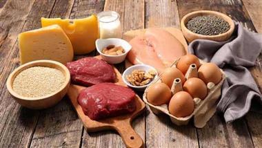
- Vitamin K2 MK-4 is found in animal foods such as organic egg yolk, dark chicken meat and goose liver. MK-7, the most effective form, is found in fermented foods such as natto, and in both soft and hard cheeses such as Brie and Gouda
Vitamin K is a fat-soluble vitamin essential for the functioning of several proteins involved in physiological processes. Naturally occurring forms are vitamin K1 (referred to as phylloquinone) and vitamin K2 (called menaquinones).1,2
Vitamin K1, which is derived from green plants, is best known for the role it plays in blood clotting, while vitamin K2, derived primarily from fermented foods and animal products such as eggs, meat and liver,3 is important to hormone production and utilization, as well as bone and heart health.
Vitamin K2 does not get the attention it deserves, and it’s highly likely you’re not getting enough in your diet. Although fat-soluble, the body stores very little and it’s rapidly depleted without regular dietary intake.
Before discussing the numerous areas of your health where vitamin K2 has a significant impact, it’s first important to understand the different forms of the vitamin.
The Different Forms of Vitamin K2
Vitamin K2 is needed to activate the protein osteocalcin, which is found in your bones. Without vitamin K2, this and other vitamin K2-dependent proteins remain inactivated, and cannot perform their biological functions.4
According to Leon Schurgers, a world-renowned researcher in vitamin K2, the difference between vitamins K1 and K2 was clearly established in The Rotterdam Study,5 published in 2004, of which Schurgers was a co-author.
While the study focused on the difference between how K1 and K2 affect the heart (K1 has no effect at all), the researchers also looked at what types of foods supply each of the vitamins. In an interview, he said:
“I measured a variety of food items for vitamin K content … Vitamin K1 is highly available in green, leafy vegetables — spinach, kale, broccoli and cabbage.
However, the absorption of vitamin K1 from food is extremely low. Only 10 percent of the vitamin K which is found in green leafy vegetables, is absorbed in your body … And there’s no variable or modification of the consumption that will significantly increase the absorption …”
Vitamin K2, on the other hand, was only present in fermented foods. It’s actually produced by specific bacteria during the fermentation process. Certain bacteria in your gut naturally produce vitamin K2 in your body as well. Interestingly, while the K1 in vegetables is poorly absorbed, virtually all of the K2 in fermented foods is readily available to your body. Vitamin K2 can be further broken down into:6,7,8
• MK-4 (menaquinone-4), a short-chain form of vitamin K2 found in animal-based foods such as meats dairy and eggs. MK-4 also has a very short biological half-life — about 2.5 hours — making it a poor candidate as a dietary supplement.
That said, natural MK-4 from food is important for good health,9 as MK-4 plays a role in gene expression, turning some genes off and others on, and is therefore important for cancer prevention.10
• MK-7 (menaquinone-7), longer-chain forms found in fermented foods. There’s a variety of these long-chain forms, but the most common one is MK-7. This is the one you’ll want to look for in supplements, as this form is extracted from real food, specifically natto,11 a fermented soy product.
The MK-7, which forms in the fermentation process, has two major advantages. It stays in your body longer and has a longer half-life, which means you can take it just once a day in very convenient dosing.12 Research13 has shown MK-7 helps prevent inflammation by inhibiting proinflammatory markers that can cause autoimmune diseases like rheumatoid arthritis.
While vitamin K1 has been found to “moderately reduce” the risk of bone fractures,14 MK-7 is more effective than vitamin K1 at reaching your bone.15,16 In your bones, vitamin K2 is used to produce osteocalcin, improving metabolic and hormonal health and exercise performance.

Cardiovascular Protection and Cofactor for Vitamin D, Calcium
Calcium deposits in the arteries are responsible for atherosclerosis, hardening and narrowing of the arteries, slowly blocking blood flow.
It’s a common cause of heart attacks, strokes and peripheral vascular disease. Vitamin K2 is important in the activation of important proteins, such as matrix GLA-protein (MGP), found in your vascular system,17 and studies have demonstrated K2 inhibits arterial calcification and improves arterial flexibility,18,19 thereby reducing your risk of atherosclerosis.
One recent study20 proposed therapeutic supplementation with vitamin K2 could prevent or halt progression of vascular calcification that may occur with excessive calcium intake, and other studies have demonstrated those eating the highest amount of vitamin K2 have the lowest risk of developing cardiovascular disease.21
Vitamin K2 also functions as an important cofactor for calcium and vitamin D. While calcium is important for strengthening bones and overall skeletal health, it only works when it gets to the right place. Vitamin K2 prevents calcium from being deposited along the walls of the blood vessels and directs it into the bone.22
So, it’s important to remember that vitamin K2 and vitamin D (along with calcium and magnesium) have a synergistic relationship when it comes to your bone and heart health. To maximize your benefits of oral vitamin D supplementation, it’s important to maintain optimal levels of vitamin K2.23 Studies indicate supplementing with both vitamins D3 and K2 offers greater cardiovascular health benefits than consuming either vitamin alone.24
K2 Helps Prevent Osteoporosis and Protects Teeth
As mentioned earlier, vitamin K2 activates osteocalcin, a protein hormone made by your bone that is dependent on K2 for activation via carboxylation. One of the primary roles osteocalcin plays is in binding calcium and transporting it to the bone.25
Osteocalcin is a small non-collagenous protein measured in the serum and synovial fluid samples of people with osteoarthritis,26 and researchers believe serum levels of osteocalcin may be useful as a screening tool to assess osteoporosis risk.27
Vitamin K2 is also important for your oral health. Preserving enamel helps reduce cavity formation. Your enamel is made of hollow tubes that extend into the dentin. The enamel is made of nonliving cells, while the dentin has live cells called odontoblasts.
These cells release proteins using vitamin K2 to help prevent tooth decay through an immune response.28 Research has demonstrated osteocalcin may be found in inflamed tissue, and researchers consider the protein to have a reparative role in dental pulp.29
Vitamin K2 Plays a Role in Reducing Inflammation
The inflammatory response is a crucial component in many chronic diseases and diseases of aging. Scientific evidence suggests both vitamin K1 and K2 have anti-inflammatory activity, in part by inhibiting nuclear factor kappa-B.30
For example, in one meta-analysis,31 researchers found evidence vitamin K has a protective role in chronic aging conditions, inflammation and cardiovascular disease. Another study32 confirmed the role the vitamin K2 MK-7 form specifically has in the modulation of inflammatory biomarkers.
The researchers evaluated natural vitamin K2 for its potential to inhibit gene expression and production of pro-inflammatory markers in vitro, finding the MK-7 form inhibited gene expression in a dose-dependent manner.
Other research33 has demonstrated a reduction in C-reactive protein, a common biomarker used to evaluate inflammation in patients with rheumatoid arthritis. C-reactive protein is a risk marker for a wide range of diseases and the researchers concluded higher intake of vitamin K2 may lower overall risk.
There is also accumulating evidence vitamin K2 may play a role in chronic kidney disease. A pre- and post-intervention clinical trial,34 which included patients with a diagnosis of chronic kidney disease who took a vitamin K2 MK-7 supplement for four weeks, found it significantly reduced calcification in the patients’ blood vessels. Patients with chronic kidney disease with a subclinical vitamin K deficiency may also benefit from a greater intake of the vitamin.35
Deficiency Affects Testosterone Production
Yet another role vitamin K2 plays in your body is the optimal production of sex hormones. Research shows vitamin K2 MK-4, found in animal foods such as meat and dairy products, can help balance testosterone production.36 Animal studies show inducing a vitamin K deficiency reduces testosterone production.37
Vitamin K2 has an effect in women as well, as the vitamin may be helpful in the management of polycystic ovarian syndrome (PCOS). This is now the most common hormonal disorders in young women, causing enlarged ovaries and potentially leading to fertility problems.38
In one randomized study, 60 vitamin D deficient women with PCOS took either a placebo or a combination of calcium, vitamin D and vitamin K for eight weeks. Those in the treatment group enjoyed greater decrease in testosterone and symptoms than the placebo group.39
How K2 May Influence Insulin Sensitivity
An unexpected discovery was made when researchers found bone was an endocrine organ, contributing to the regulation of a number of physiological processes, including glucose homeostasis.40
There’s even evidence to suggest vitamin K2 supplementation may improve insulin sensitivity via osteocalcin metabolism,41 and may help prevent the development of Type 2 diabetes. 42 The evidence is not conclusive, however, as a systematic review of controlled trials found K2 supplementation had no effect on insulin sensitivity.43
Part of the problem may have something to do with the form of vitamin given. Numerous studies have demonstrated synthetic vitamin K2 supplements are not as effective as natto-derived K2 supplements.
Statins Contribute to the Depletion of Vitamin K2
Nearly 25% of Americans over the age of 45 are taking cholesterol-lowering statin drugs, despite the high risks of the drugs. In addition to other side effects, statins inhibit the production of vitamin K2.
A study published in Expert Review of Clinical Pharmacology44 revealed statins may actually stimulate the development of atherosclerosis and heart failure, and one of the mechanisms by which this occurs is the inhibition of the synthesis of vitamin K2, which protects your arteries from calcification.
How to Boost Your Intake of Vitamin K2 Naturally
If you decide to take a statin or use a vitamin D3 supplement, it is highly recommended you include a vitamin K2 supplement in the MK-7 form. Seek out a supplement extracted from the Japanese fermented soy product called natto. Professor Cees Vermeer,45 one of the world’s top vitamin K2 researchers, recommends between 45 micrograms (mcg) and 185 mcg daily for adults.
Just remember that since vitamin K2 is fat-soluble, it’s best to take it with a small amount of fat to improve absorption. Consuming foods rich in vitamin K2 is your best bet. MK-4 is found in animal foods such as free-range, organic eggs (particularly the yolk), dark chicken meat and goose liver. MK-7 is found in fermented foods such as natto, a type of fermented soy, and in soft and hard cheeses such as Brie and Gouda.
- 1 Oregon State University, Vitamin K
- 2 Journal of Biological Chemistry, December 14, 2007
- 3, 12 Nutrition Journal, November 12, 2012
- 4 Integrative Medicine (Encinitas) February 2015
- 5 Journal of Nutrition November 1, 2004: 134(11); 3100-3105 (The Rotterdam Study)
- 6, 9 Journal of Agriculture and Food Chemistry, 2006
- 7 Biochemica et Biophysica Acta, 2002
- 8 Haemostasis, July 27, 2000
- 10 Journal of Hepatology, July 2007
- 11, 16 Nutraceutical Business Review, February 23, 2016
- 13 European Journal of Pharmacology, August 15, 2015
- 14 Medicine, 2017;96(17)
- 15 Osteoporosis International, March 2013
- 17 Thrombosis and Haemostasis, 2008; 100(4):393
- 18, 22 Integrative Medicine, 2015;14(1)
- 19 Oman Medical Journal, 2014;29(3)
- 20 Frontiers in C vardiovascular Medicine doi.org/10.3389/fcvm.2019.00006
- 21 Journal of Nutrition, 2004;134(11)
- 23 Int J Endocrinol. 2017; 2017: 7454376. September 12, 2017
- 24 International Journal of Endocrinology, 2017; 2017
- 25 Tissue Engineering, 2017; 23(3)
- 26 Science Direct, Osteocalcin
- 27 Journal of Clinical and Diagnostic Research, 2017;9(8)
- 28 Steven Lin, Use Vitamin K2 to Remineralize Your Teeth
- 29 Journal of Endodontics, 2013;39(7)
- 30 Anti-inflammatory Actions of Vitamin K March 22, 2017, DOI: 10.5771/63891
- 31 Current Nutrition Reports, 2016;5(2)
- 32 Journal of Medicinal Food, 2016;19(7)
- 33 Modern Rheumatology, 2012;23(5)
- 34 BMC Nephrology, 2017;18(191)
- 35 Nutrients, 2013;5(11)
- 36 Biochimica et Biophysica Acta, 2006;1760(10)
- 37 Lipids in Health and Disease, 2011; 10:158
- 38 BMC Medicine, 2010; 8:41
- 39 Hormone and Metabolic Research, 2016;48(7):446
- 40 Reviews of Endocrine and Metabolic Disorders, 2015;16(2)
- 41 Diabetes Care, 2011;43(9)
- 42 Diabetes Research and Clinical Practice, 2018; 136:39
- 43 Diabetes, Metabolic Syndrome and Obesity, 2017;10:169
- 44 Expert Review of Clinical Pharmacology, 2015;8(2)
- 45 Nutraceuticals World, June 11, 2013
Atherosclerosis is a Non-Healing Wound
Reproduced from original OMNS article (OrthoMolecular News Service):
http://orthomolecular.org/resources/omns/index.shtml
Subscribe to the free Orthomolecular Newsletter: http://orthomolecular.org/subscribe.html
Go to the OMNS Archive: http://orthomolecular.org/resources/omns/index.shtml
Orthomolecular Medicine News Service, September 8, 2022
Commentary by Thomas E. Levy MD, JD and Ron Hunninghake, MD
OMNS (Sept. 8, 2022) While chronic inflammation of the coronary artery wall is finally appreciated to be the direct cause of all coronary atherosclerosis by cardiologists and internists, there appears to be little curiosity as to what is causing this pandemic of inflammation that kills about 50% of the world population every year. [1,2] When a 50-year-old man has a heart attack but is completely free of all known risk factors for heart disease, the treating cardiologist just considers that individual to have had bad luck, although that sentiment is never directly expressed to that individual. Truth be known, luck is involved in whether someone becomes a heart patient. However, it pertains directly to the luck involved in whether that individual chose to see a well-informed biological dentist earlier in life rather than just a procedure/tooth structure-oriented mainstream dentist who has no awareness of, or concern with, the dynamic physiological interplay between the mouth and the rest of the body.
Coronary Atherosclerosis Pathophysiology
Over fifteen years ago, the main subspecialty journal for cardiologists, Circulation, reported on the presence of pathological bacterial species found in the atherosclerotic plaque specimens obtained from 38 coronary heart disease patients. The specimens were obtained via catheter-based atherectomy. In ALL 38 patients, polymerase chain reaction (PCR) testing revealed the DNA signatures of such bacteria, with over 50 different species being identified. [3] In 92% of those specimens the DNA of 2 to 9 different species of fungus was detected as well. Control specimens revealed no evidence of either bacteria or fungi. [4] Other studies have also shown a high incidence of pathogens, including viruses, in atherosclerotic plaques in the coronary arteries and elsewhere. [5-10]
Patients presenting with myocardial infarctions secondary to acute coronary occlusions precipitated by soft platelet clots depositing on atherosclerotic plaques were investigated as well. These clots were aspirated at angiography and analyzed by PCR testing. The overwhelming majority of these clots had DNA signatures from periodontal (oral) pathogens. Furthermore, the concentration of this DNA was 16-fold (1,600%) higher in those clots than in the surrounding blood. It would defy logic to consider these pathogens as anything other than the primary reason for that clot developing on the atherosclerotic plaque, although the authors would only go so far as to conclude that the clots “may be associated” with the oral bacteria that accumulated inside them. [11] In the medical literature it is almost impossible to find an article that definitively concludes a cause-and-effect relationship between any factor and any outcome. Rather, the boldest conclusion that is ever reached is that an association, correlation, or relationship of some sort is present, followed by the ever-present advice that more studies are needed.
Chronic Pathogen Colonization Causes All Atherosclerosis
Chronic pathogen colonization (CPC) in an arterial wall both initiates and sustains the evolution of atherosclerosis. Wherever a CPC gets established, pro-oxidant (toxic) molecules are continuously produced secondary to the metabolism of the pathogens. Furthermore, when some of those pathogens do finally die, they also release endotoxins, exotoxins, and an abundance of free iron, all of which are highly pro-oxidant in nature. As a direct result of this CPC insult, the arterial wall is quickly depleted of its antioxidant content, especially vitamin C. Once this occurs, the vessel wall can never again be restored to its normal antioxidant status until the focal infection sites that are continuously sustaining the CPC can be completely resolved or at least stabilized. Supplemental vitamin C will always help, but to a variable degree depending on how many focal infections are present and how “leaky” they are in releasing their pathogens and toxins into the blood. A small number of coronary patients can demonstrate a regression of atherosclerotic lesions by taking large doses of vitamin C while leaving the focal infections unaddressed. However, most coronary patients can only realistically hope for stopping or slowing the atherosclerotic process by taking vitamin C without resolving the seeding sites of infection.
Once the pathogen presence in the arterial wall has metabolized all of the vitamin C present and virtually no measurable amounts of vitamin C remain, a state of inflammation results. Inflammation cannot exist where vitamin C levels are normal or near-normal. Acute focal vitamin C deficiency and acute focal inflammation are actually synonyms for the same physiological state. And once the arterial wall becomes inflamed, an acute immune response ensues. Quite literally, CPC in the arterial wall results in a state of focal scurvy (severe vitamin C deficiency). Inflammation never occurs spontaneously, as it only develops where increased oxidative stress has consumed excess amounts of vitamin C.
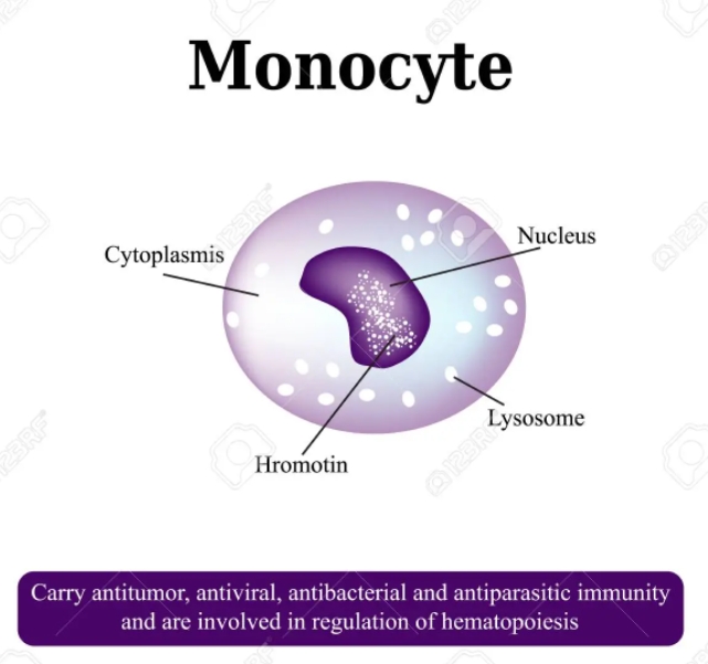
The acute immune response that seeks out sites of inflammation in the body has some noteworthy characteristics. The first cells that mobilize and reach the inflamed tissue are monocytes, which are heavily laden with vitamin C, more so than any other cells. The concentration of vitamin C in monocytes has been shown to be more than 80-fold (8,000%) higher than the surrounding plasma. Regular white blood cells, while still having relatively large concentrations of vitamin C, have only about one third of the concentration seen in monocytes. And unlike the white blood cells, the monocytes also have the ability to maintain their high vitamin C content in the face of lowered plasma concentrations. [12-14] In cell cultures, human monocytes were able to maintain a concentration gradient of 100-fold across the plasma membrane. [15]
The cells leading the acute immune response indicate that the prompt delivery of highly-concentrated vitamin C is a crucial part of the mechanism by which the immune response alleviates the inflammation that has developed. And as incredibly complex as the immune system is known to be, it is always inflammation that triggers the immune response, and an associated vitamin C deficiency is always present. Just as vitamin C deficiency results in inflammation, vitamin C repletion resolves it when enough can be delivered. In spite of its numerous metabolic pathways and components, the ultimate goal of the immune response appears to be the delivery of vitamin C to the site of inflammation and/or infection as quickly and substantially as possible. Quite simply, excess oxidation causes disease wherever it is found, and sufficient antioxidation resolves it.
If the initial pathogen presence is transitory, the acute immune response would be expected to promptly resolve the associated inflammation as the pathogens are killed and the increased oxidative stress is completely neutralized in the arterial walls. However, when remote focal infections, typically oral, remain unresolved and the seeding of the pathogens continues “24/7,” the acute immune response transitions into a chronic immune response trying to deal with a chronic inflammation. This brings other factors into play as the atherosclerosis evolves.
The earliest impact of the deficient vitamin C status in the arterial wall is seen in the extracellular matrix (ECM) surrounding the endothelial cells that coat the inner lining of the wall. The ECM is known as the basement membrane when associated with the endothelial cells. [16,17] This basement membrane normally has a high content of collagen and interconnected glycoproteins that give it a thickened, jelly-like consistency. Relatively large amounts of vitamin C are needed to maintain good levels of healthy collagen and glycoproteins in the basement membrane. When there is little to no vitamin C present, the jelly-like state degrades to a less intact, and even watery state, and the spaces between the endothelial cells expand, initiating what is known as the degenerative stage of atherosclerosis. This facilitates the uptake of calcium, cholesterol, fats, and even more pathogens/toxins from the bloodstream into these spaces. Monocytes then appear and transition into phagocytic macrophages which engulf these substances. These lipid-stuffed macrophages become the bubbly-appearing foam cells which collectively become visible to the naked eye as the fatty streaks which herald the clear beginning of atherosclerosis. [18,19] It is also important to know that “atherogenic” agents such as cholesterol can only promote the evolution of established atherosclerosis. It is unable to initiate atherosclerosis as long as there is no CPC present and the arterial wall has a normal content of vitamin C.
The degenerative stage of atherosclerosis then proceeds to the proliferative stage of atherosclerosis. This stage features the influx of fibroblasts, another cell type with a very high concentration of vitamin C. [20] Fibroblasts are crucial to producing new collagen and much of the protein content in the ECM and the basement membrane, and sufficient vitamin C is needed by the fibroblasts to accomplish this. [21] The recruitment of fibroblasts is a compensatory mechanism to fortify and strengthen the ECM that was weakened and rendered watery by the depletion of vitamin C. However, as long as the vitamin C remains depleted from the ongoing pathogen presence in the arterial wall, the fibroblast influx will continue unabated in a futile attempt to synthesize sufficient amounts of quality connective tissue to restore the wall to a normal state.
One hypothesis on the development of atherosclerosis is that since quality collagen and other forms of connective tissue will never be restored to normal levels in the absence of adequate vitamin C, the “next best” compensatory response for a wall that will never again have normal tensile strength is to make it thicker, as in the growth of atheromas (plaques). [22,23] Any compensatory mechanism in the body ultimately aims to keep the body alive, and a thickened arterial wall will be much less likely to develop arterial aneurysms that eventually rupture and cause sudden death. This actually works for an extended period of time in many people, at least until the atheromas grow so large that the coronary artery ends up becoming completely occluded. Basically, the atherosclerotic plaque grows where supportive connective tissue cannot.
Regardless of the mechanism, it is clear that as long as the vitamin C levels in the atherosclerotic arterial wall remain low to immeasurable, atheromas will continue to grow. And as long as the CPC in the arterial wall continues to consume all vitamin C present and remains unaddressed, atherosclerosis cannot resolve, or even slow down in its progression. Autopsy studies confirmed that vitamin C was always depleted, and often completely absent, in atherosclerotic arterial walls and plaques. [24] Furthermore, in an atherosclerotic animal model, a normal vitamin C level could be restored and atherosclerotic plaques would regress and even resolve. [25] A daily dose of only 1,500 mg of vitamin C in patients with peripheral atherosclerosis was able to stop progression and even induce regression in a majority of those treated, documented by angiography. [26]
Atherosclerosis, the Non-Healing Wound
The definition of a wound is damage to the integrity of biological tissue, including skin, mucous membranes, and organ tissues. When infection or pathogen colonization is present as well, versus an acutely-inflicted “clean” wound, healing is always dependent on resolving the pathogen-induced inflammation and focal vitamin C deficiency that always results. The resolution of atherosclerosis follows the principles of basic wound healing. Classical atherosclerosis is a non-healing wound due to the ongoing pathogen-induced vitamin C deficiency. As long as the inflammation-induced chronic immune response secondary to the focal vitamin C deficiency remain unaddressed, good healing will never occur.
Furthermore, the healing requirements for wounds inside and outside the body are much the same. [27] Fibroblasts, which are needed in order to achieve normal healing, cannot function normally in the absence of the vitamin C needed to regenerate a normal basement membrane or ECM. Chronic atherosclerosis and poorly-healing wounds all feature inflammation/infection-induced elevated oxidative stress, and vitamin C is always depleted at these sites.
In any vitamin C-depleted microenvironment, a normal healing will never be realized until the vitamin C status is restored. [28,29] However, healing of some wounds with a poor quality of resolution can occur when suboptimal amounts of vitamin C are available. Cancer, especially at metastatic sites, is a condition regarded as a non-healing or chronic wound in the literature, and very elevated levels of oxidative stress are always present. [30,31] Intravenous vitamin C has been reported to be an effective treatment for cancer patients, even when the cancer is advanced. [32]
A chronic bedsore is a classic example of an external open wound that often responds poorly to traditional wound treatment protocols. Patients with such lesions are typically invalids with an overall poor vitamin and nutrition status. However, such open wound healing is significantly enhanced by insulin, a hormone which greatly facilitates the uptake of vitamin C inside the cells of the infected tissue. [33,34] A sufficient presence of vitamin C is the most critical factor in healing anywhere in the body. [35-37]
Resolving Coronary Atherosclerosis
Stabilizing or reversing coronary atherosclerosis requires optimizing the healing response in the affected areas of the arteries. Like any other non-healing wound, atherosclerosis requires a normalized redox (reduction-oxidation) balance in order to allow healing to proceed. Any pathogen presence is a potent and continuous source of new pro-oxidants (toxins), which then provokes a chronic inflammatory process. As described above, this means that sufficient levels of vitamin C in the arterial wall can never be realized when the new pathogen-related toxins are not eliminated or at least severely curtailed. And until those vitamin C levels can be restored, a chronic inflammatory response with an influx of fibroblasts building up the plaque will continue unabated.
The goal of any effective treatment for the stabilization and/or resolution of coronary atherosclerosis is simple, although not necessarily easily achieved. New toxin presence (oxidative stress) must be prevented, and old oxidative damage must be repaired. As Dr. Hal Huggins, the father of biological dentistry, so elegantly expressed it: “You can’t dry off while you are still in the shower.”
While focal infections located anywhere in the body can potentially seed the coronary arteries and other sites remote from those infections, the simple fact is that oral cavity infections are responsible for the CPC in atherosclerotic coronary arteries well over 90% of the time. Focal infections outside of the mouth do not have the direct connection to venous and lymphatic drainage that infected gums, teeth, and tonsils have. Also, other sites of infection do not generate the high pressures that are seen when the teeth grind and chew. These pressures greatly increase the expression of pathogens into the blood and lymph.
As of the writing of this article, the routine diagnosis and elimination of infective foci in the mouth is rarely part of the routine treatment of a suspected or established coronary heart disease patient. Nevertheless, it remains the most important part of an atherosclerosis treatment protocol. Optimal supplementation while leaving such infections unaddressed will not protect the patient as much as removing all infective foci without an emphasis on optimal supplementation. But doing both will reliably stop atherosclerosis progression and frequently result in its regression.
Essential to achieving complete resolution of oral cavity infections is the administration of a cone beam computed tomography (CBCT) examination of the oral cavity. Also known as a 3D X-ray of the mouth, grossly abscessed but completely symptom-free teeth are OFTEN found, and just one of them can assure that coronary atherosclerosis begins and never ends until it is properly removed. Infected gums, tonsils, and sinuses are often found as well.
Whenever the infections of the oral cavity cannot be properly evaluated and treated, it is especially important to normalize critical hormone levels, including sex hormones, cortisol, and especially thyroid hormone. All hormones work to minimize body-wide oxidative stress. When such oxidative stress is sufficiently elevated, focal infections can effectively metastasize and set up shop anywhere in the body, but especially so in the coronary arteries. Quite literally, keeping completely normal levels of thyroid hormone in the cells of the body, along with normal sex hormone and cortisol levels, can keep focal infections focal. The work of Dr. Broda Barnes demonstrated the powerful ability of a normal thyroid hormone status in preventing heart attacks, which obviously meant that CPC of the coronary arteries in those patients was being kept in check as well. [38] It now appears that maintaining normal thyroid function in all of the cells in the body requires the maintenance of an optimal free T3/reverse T3 ratio. Generally, 18/1 or higher is the target value. Just measuring the “standard” thyroid blood tests will not detect the mild but significant compromise of thyroid function that allows focal infections to spread. Here is one example of how to calculate this ratio:
Free T3 (triiodothyronine): 3.1 pg/mL (reference range: 2.0-4.4)
Reverse T3: 18.4 ng/dL (reference range: 9.2-24.1)
To convert pg/mL to ng/dL: multiply by 100-3.1 x 100 = 310
Then divide the two numbers: 310/18.4 = 16.8
Generally, higher reverse T3 levels indicate a greater contribution of unchecked oxidative stress in the body and lower T3 levels indicate a greater contribution of decreased thyroid hormone. However, most individuals with a low ratio, as is seen here, have a combination of increased oxidative stress and decreased thyroid hormone. Long-term follow-up is essential to make sure as to the need and dosage of desiccated thyroid (not Synthroid or T4) after focal infections are resolved, since it is never clear how reversible a low thyroid hormone level in a given individual might be.
The protocols of dental revision, supplementation, and appropriate hormone application are covered in depth elsewhere, along with a clear-cut explanation of a lowered redox balance being the pathophysiology underlying all disease. [39,40]
Recap
Disease anywhere in the body is characterized by tissue with a continuing source of new pro-oxidants in excess of the arrival of new antioxidants. Focal areas of disease have the characteristics of a non-healing wound. A wound heals when new toxins (typically produced by pathogens) are curtailed and a large influx of antioxidants, especially vitamin C, can be assured.
Coronary heart disease accounts for about 50% of the deaths in the world today. Chronic pathogen colonization (CPC) is always found in the coronary arteries of such individuals, keeping the artery in a chronic state of scurvy with little or no vitamin C present. Until the vitamin C consumption is stopped or severely curtailed, atherosclerotic plaques grow and eventually cause heart attacks by completely blocking an affected artery.
When the oral focal infections seeding the coronary arteries can be identified and eliminated, and optimal supplementation and hormone levels can be maintained, atherosclerosis stabilization is almost a certainty to occur. Atherosclerosis reversal will often be seen as well.
A cone beam computed tomography (CBCT) examination of the oral cavity is as (or more) essential than any other testing involved in the proper evaluation and treatment of any suspected or documented heart disease patient. This 3D X-ray remains the only way to accurately know if deadly abscessed teeth are present.
(OMNS Contributing Editor Dr. Thomas E. Levy is board certified in internal medicine and cardiology. Dr. Ron Hunninghake is Chief Medical Officer at the Riordan Clinic, Wichita Kansas. Both authors are members of the Orthomolecular Medicine Hall of Fame. The views presented in this article are the authors and not necessarily those of all members of the Orthomolecular Medicine News Service Editorial Review Board.)
Corresponding author: Thomas Levy televymd@yahoo.com
References
1. Legein B, Temmerman L, Biessen E, Lutgens E (2013) Inflammation and immune system interactions in atherosclerosis. Cellular and Molecular Life Sciences 70:3847-3869. PMID: 23430000
2. Rosenfeld M (2013) Inflammation and atherosclerosis: direct versus indirect mechanisms. Current Opinion in Pharmacology 13:154-160. PMID: 23357128
3. Ott S, El Mokhtari N, Musfeldt M et al. (2006) Detection of diverse bacterial signatures in atherosclerotic lesions of patients of patients with coronary heart disease. Circulation 113:929-937. PMID: 16490835
4. Ott S, El Mokhtari N, Rehman A et al. (2007) Fungal rDNA signatures in coronary atherosclerotic plaques. Environmental Microbiology 9:3035-3045. PMID: 17991032
5. Haraszthy V, Zambon J, Trevisan M et al. (2000) Identification of periodontal pathogens in atheromatous plaques. Journal of Periodontology 71:1554-1560. PMID: 11063387
6. Mahendra J, Mahendra L, Kurian V et al. (2010) 16S rRNA-based detection of oral pathogens in coronary atherosclerotic plaque. Indian Journal of Dental Research 21:248-252. PMID: 20657096
7. Rosenfeld M, Campbell L (2011) Pathogens and atherosclerosis: update on the potential contribution of multiple infectious organisms to the pathogenesis of atherosclerosis. Thrombosis and Haemostasis 106:858-867. PMID: 22012133
8. Tufano A, Di Capua M, Coppola A et al. (2012) The infectious burden in atherothrombosis. Seminars in Thrombosis and Hemostasis 38:515-523. PMID: 22660918
9. Bale B, Doneen A, Vigerust D (2017) High-risk periodontal pathogens contribute to the pathogenesis of atherosclerosis. Postgraduate Medical Journal 93:215-220. PMID: 27899684
10. Priyanka S, Kaarthikeyan G, Nadathur J et al. (2017) Detection of cytomegalovirus, Epstein-Barr virus, and Torque Teno virus in subgingival and atheromatous plaques of cardiac patients with chronic periodontitis. Journal of Indian Society of Periodontology 21:456-460. PMID: 29551863
11. Pessi T, Karhunen V, Karjalainen P et al. (2013) Bacterial signatures in thrombus aspirates of patients with acute myocardial infarction. Circulation 127:1219-1228. PMID: 23418311
12. Evans R, Currie L, Campbell A (1982) The distribution of ascorbic acid between various cellular components of blood, in normal individuals, and its relation to the plasma concentration. The British Journal of Nutrition 47:473-482. PMID: 7082619
13. Moser U (1987) Uptake of ascorbic acid by leukocytes. Annals of the New York Academy of Sciences 498:200-215. PMID: 3475998
14. Schorah C (1992) The transport of vitamin C and effects of disease. The Proceedings of the Nutrition Society 51:189-198. PMID: 1438327
15. Bergsten P, Amitai G, Kehrl J et al. (1990) Millimolar concentrations of ascorbic acid in purified human mononuclear leukocytes. Depletion and reaccumulation. The Journal of Biological Chemistry 265:2584-2587. PMID: 2303417
16. Walma D, Yamada K (2020) The extracellular matrix in development. Development 147:dev175596. PMID: 32467294
17. Sherwood D (2021) Basement membrane remodeling guides cell migration and cell morphogenesis during development. Current Opinion in Cell Biology 72:19-27. PMID: 34015751
18. Chistiakov D, Melnichenko A, Myasoedova V et al. (2017) Mechanisms of foam cell formation in atherosclerosis. Journal of Molecular Medicine 95:1153-1165. PMID: 28785870
19. Li J, Meng Q, Fu Y et al. (2021) Novel insights: dynamic foam cells derived from the macrophage in atherosclerosis. Journal of Cellular Physiology 236:6154-6167. PMID: 33507545
20. Welch R, Bergsten P, Butler J, Levine M (1993) Ascorbic acid accumulation and transport in human fibroblasts. The Biochemical Journal 294:505-510. PMID: 8373364
21. Diller R, Tabor A (2022) The role of the extracellular matrix (ECM) in wound healing: a review. Biomimetics 7:87. PMID: 35892357
22. Rath M, Pauling L (1990a) Hypothesis: lipoprotein(a) is a surrogate for ascorbate. Proceedings of the National Academy of Sciences of the United States of America 87:6204-6207. PMID: 2143582
23. Rath M, Pauling L (1990b) Immunological evidence for the accumulation of lipoprotein(a) in the atherosclerotic lesion of the hypoascorbemic guinea pig. Proceedings of the National Academy of Sciences of the United States of America 87:9388-9390. PMID: 2147514
24. Willis G, Fishman S (1955) Ascorbic acid content of human arterial tissue. Canadian Medical Association Journal 72:500-503. PMID: 14364385
25. Willis G (1957) The reversibility of atherosclerosis. Canadian Medical Association Journal 77:106-108. PMID: 13446787
26. Willis G, Light A, Gow W (1954) Serial arteriography in atherosclerosis. Canadian Medical Association Journal 71:562-568. PMID: 13209447
27. Bainbridge P (2013) Wound healing and the role of fibroblasts. Journal of Wound Care 22:407-412. PMID: 23924840
28. Bechara N, Flood V, Gunton J (2022) A systematic review on the role of vitamin C in tissue healing. Antioxidants 11:1605. PMID: 36009324
29. Lepucki A, Orlinska K, Mielczarek-Palacz A et al. (2022) The role of extracellular matrix proteins in breast cancer. Journal of Clinical Medicine 11:1250. PMID:35268340
30. Menter D. Kopetz S, Hawk E et al. (2017) Platelet “first responders’ in wound response, cancer, and metastasis. Cancer Metastasis Reviews 36:199-213. PMID: 28730545
31. Deyell M, Garris C, Laughney A (2021) Cancer metastasis as a non-healing wound. British Journal of Cancer 124:1491-1502. PMID: 33731858
32. Padayatty S, Riordan H, Hewitt S et al. (2006) Intravenously administered vitamin C as cancer therapy: three cases. Canadian Medical Association Journal 174:937-942. PMID: 16567755
33. Qutob S, Dixon S, Wilson J (1998) Insulin stimulates vitamin C recycling and ascorbate accumulation in osteoblastic cells. Endocrinology 139:51-56. PMID: 9421397
34. Martinez-Jimenez M, Valadez-Castillo F, Aguilar-Garcia J et al. (2018) Effects of local use of insulin on wound healing in non-diabetic patients. Plastic Surgery 26:75-79. PMID: 29845043
35. Moores J (2013) Vitamin C: a wound healing perspective. British Journal of Community Nursing Suppl:S6, S8-11. PMID: 24796079
36. Li X, Teng L, Lin Y, Xie G (2018) Role of vitamin C in wound healing after dental implant surgery in patients treated with bone grafts and patients with chronic periodontitis. Clinical Implant Dentistry and Related Research 20:793-798. PMID: 30039526
37. Gunton J, Girgis C, Lau T et al. (2021) Vitamin C improves healing of foot ulcers: a randomized, double-blind, placebo-controlled trial. The British Journal of Nutrition 126:1451-1458. PMID: 32981536
38. Barnes B, Galton L (1976) Hypothyroidism: The Unsuspected Illness. New York, NY: Harper & Row
39. Levy T (2017) Hidden Epidemic: Silent oral infections cause most heart attacks and breast cancers. Henderson, NV: MedFox Publishing. Free download: www.hep21.medfoxpub.com
40. Levy T (2021) Rapid Virus Recovery: No need to live in fear! Henderson, NV: MedFox Publishing. Free download: www.rvr.medfoxpub.com
Nutritional Medicine is Orthomolecular Medicine
Orthomolecular medicine uses safe, effective nutritional therapy to fight illness. For more information: http://www.orthomolecular.org
Find a Doctor
To locate an orthomolecular physician near you: http://orthomolecular.org/resources/omns/v06n09.shtml
The peer-reviewed Orthomolecular Medicine News Service is a non-profit and non-commercial informational resource.
Editorial Review Board:
Albert G. B. Amoa, MB.Ch.B, Ph.D. (Ghana)
Seth Ayettey, M.B., Ch.B., Ph.D. (Ghana)
Ilyès Baghli, M.D. (Algeria)
Ian Brighthope, MBBS, FACNEM (Australia)
Gilbert Henri Crussol, D.M.D. (Spain)
Carolyn Dean, M.D., N.D. (USA)
Ian Dettman, Ph.D. (Australia)
Susan R. Downs, M.D., M.P.H. (USA)
Ron Ehrlich, B.D.S. (Australia)
Hugo Galindo, M.D. (Colombia)
Martin P. Gallagher, M.D., D.C. (USA)
Michael J. Gonzalez, N.M.D., D.Sc., Ph.D. (Puerto Rico)
William B. Grant, Ph.D. (USA)
Claus Hancke, MD, FACAM (Denmark)
Tonya S. Heyman, M.D. (USA)
Patrick Holford, BSc (United Kingdom)
Suzanne Humphries, M.D. (USA)
Ron Hunninghake, M.D. (USA)
Bo H. Jonsson, M.D., Ph.D. (Sweden)
Dwight Kalita, Ph.D. (USA)
Felix I. D. Konotey-Ahulu, MD, FRCP, DTMH (Ghana)
Jeffrey J. Kotulski, D.O. (USA)
Peter H. Lauda, M.D. (Austria)
Alan Lien, Ph.D. (Taiwan)
Homer Lim, M.D. (Philippines)
Stuart Lindsey, Pharm.D. (USA)
Pedro Gonzalez Lombana, MD, MsC, PhD (Colombia)
Victor A. Marcial-Vega, M.D. (Puerto Rico)
Juan Manuel Martinez, M.D. (Colombia)
Mignonne Mary, M.D. (USA)
Jun Matsuyama, M.D., Ph.D. (Japan)
Joseph Mercola, D.O. (USA)
Jorge R. Miranda-Massari, Pharm.D. (Puerto Rico)
Karin Munsterhjelm-Ahumada, M.D. (Finland)
Tahar Naili, M.D. (Algeria)
Zhiyong Peng, M.D. (China)
Isabella Akyinbah Quakyi, Ph.D. (Ghana)
Selvam Rengasamy, MBBS, FRCOG (Malaysia)
Jeffrey A. Ruterbusch, D.O. (USA)
Gert E. Schuitemaker, Ph.D. (Netherlands)
Han Ping Shi, M.D., Ph.D. (China)
T.E. Gabriel Stewart, M.B.B.CH. (Ireland)
Thomas L. Taxman, M.D. (USA)
Jagan Nathan Vamanan, M.D. (India)
Garry Vickar, M.D. (USA)
Anne Zauderer, D.C. (USA)
Andrew W. Saul, Ph.D. (USA), Editor-In-Chief
Associate Editor: Robert G. Smith, Ph.D. (USA)
Editor, Japanese Edition: Atsuo Yanagisawa, M.D., Ph.D. (Japan)
Editor, Chinese Edition: Richard Cheng, M.D., Ph.D. (USA)
Editor, Norwegian Edition: Dag Viljen Poleszynski, Ph.D. (Norway)
Editor, Arabic Edition: Moustafa Kamel, R.Ph, P.G.C.M (Egypt)
Editor, Korean Edition: Hyoungjoo Shin, M.D. (South Korea)
Editor, Spanish Edition: Sonia Rita Rial, PhD (Argentina)
Contributing Editor: Thomas E. Levy, M.D., J.D. (USA)
Contributing Editor: Damien Downing, M.B.B.S., M.R.S.B. (United Kingdom)
Contributing Editor: W. Todd Penberthy, Ph.D. (USA)
Contributing Editor: Ken Walker, M.D. (Canada)
Assistant Editor: Helen Saul Case, M.S. (USA)
Technology Editor: Michael S. Stewart, B.Sc.C.S. (USA)
Associate Technology Editor: Robert C. Kennedy, M.S. (USA)
Legal Consultant: Jason M. Saul, JD (USA)
Comments and media contact: drsaul@doctoryourself.com OMNS welcomes but is unable to respond to individual reader emails. Reader comments become the property of OMNS and may or may not be used for publication.
This article may be reprinted free of charge provided 1) that there is clear attribution to the Orthomolecular Medicine News Service, and 2) that both the OMNS free subscription link http://orthomolecular.org/subscribe.html and also the OMNS archive link http://orthomolecular.org/resources/omns/index.shtml are included.
Riordan Clinic | Orthomolecular.org
3100 N Hillside Ave
Wichita, Kansas 67219
United States
The Poorly-Understood Role of Copper in Anemia
Reproduced from original article.
Dr Mercola’s web site has been moved to https://mercola.substack.com.
Analysis by Dr. Joseph Mercola Fact Checked September 11, 2022
STORY AT-A-GLANCE
- Iron and copper are highly interdependent and need to be considered together. If you don’t have enough copper in your diet, hemoglobin production becomes impaired, along with many other aspects of iron metabolism
- Being anemic does not automatically mean that you’re iron deficient. Anemia typically relates to impaired iron recycling, not deficiency, and impaired iron recycling is caused by copper and retinol deficiency
- The best way to lower excess iron is to donate blood, one to four times a year. Most adult men and postmenopausal women have high iron and could benefit from regular blood donation, as high iron is extremely toxic and destroys health. An even better strategy is to remove smaller amounts of blood every month
- To raise your copper level, you could use a copper supplement, but foods like grass-fed beef liver, bee pollen and whole food vitamin C are better
- If you’re a farmer or grow your own food, the best way to put copper back into the soil, to get it into the food, is to add copper sulfate. Before you plant, simply spray the soil with copper sulfate, 10 to 15 pounds per acre, or use a copper sulfate foliage spray
Morley Robbins, MBA, CHC,1 a repeat guest, is the founder of the Magnesium Advocacy Group. He’s best known as the Magnesium Man, and is the author of “Cu-RE Your Fatigue: The Root Cause and How to Fix It on Your Own,” in which he explains the roles of magnesium, copper, iron, vitamins A and D and other essential nutrients.
His Root Cause Protocol2 is the implementation of that information. We’re currently planning to write a book together, which will focus on the little-understood importance of copper and its interaction with iron.
As explained by Robbins, if copper is lacking in your diet, iron will build up in your liver, which changes its physiology and immunoproperties. Liver metabolism is highly dependent on copper and retinol, and there’s not a lot of awareness of that.
“It’s a very sophisticated process of interaction between copper and iron, and if that interaction doesn’t go well, iron is going to start to accumulate in the tissues. It’s going to start in the liver, but it’s going to go elsewhere too,” he says.
“I think that is kind of the takeaway of these conversations — to make sure people know that iron does accumulate, and that iron can be reduced through blood donations. Especially as you get into your 50s, 60s, 70s, it needs to be a regular part of your health routine.”
Three Ways to Measure Iron Status
The clinical term for excess iron in the liver is hemosiderosis, and it’s so pervasive as to be near-universal. But where does the excess iron actually come from? And why is it that many with high iron stores have low serum ferritin?
As Robbins explains, oftentimes, low serum ferritin is not at all a sign of iron deficiency, but rather a deficiency in copper and retinol. The deficiency in copper basically locks iron in the liver and prevents it from being recycled as it should:
“It’s important for practitioners to not measure iron status with just one marker. I think a lot of practitioners are falling into that trap of just using serum ferritin. There are three key ways to measure iron status: Hemoglobin, serum iron and serum ferritin.
The biggest concentration of iron in the body is in our hemoglobin; 70% of the iron is in our red blood cells … Hemoglobin is essential to understand what is going on with the biggest bulk of iron.
The second marker that I really focus on is called serum iron. It’s less than 1% of the iron, but it’s a very important measure of iron because it’s really getting at the iron recycling program. Every second of every day, we have to turn over 2.5 million red blood cells. That’s a lot of activity. In the course of 24 hours it’s 200 billion red blood cells that need to be turned over.
But what’s a surprise is to learn that only 25 milligrams (mg) of iron are needed to support that 24-hour cycle, but 24 of those 25 milligrams, 95% of the iron, is coming from this recycling program. So, it’s a very significant understanding that the serum iron only represents a small percent, but it represents the efficiency of the iron recycling.”
Ideally, hemoglobin should be between 12.5 and 13.5 for women, 14.5 to 15.5 for men. Serum iron should ideally be about 100 for women and 120 for men. The closer serum iron is to these, the more efficient your recycling is.
What You Need to Know About Serum Ferritin
The third iron measure is serum ferritin. There are four different types of ferritin in your body, broadly categorized as heavy chain and light chain. Heavy chain ferritin refers to ferritin protein inside cells and mitochondria that require copper to work properly. Serum ferritin refers to ferritin in your plasma, is outside the cell — and also outside the red blood cells, the hemoglobin.
“What is not well-known is that this ferritin that shows up in the blood is very iron-poor. It doesn’t have iron in it. The iron has been discharged in the liver and then the protein gets secreted out … So, serum ferritin is not representative of iron per se. The iron was discharged in the liver …
I would never use ferritin only as an indication of iron status. You need to see hemoglobin, serum iron, and serum ferritin. You need to see them in relationship to each other …
[When] serum ferritin starts to get high, it’s highly correlated with inflammation or an infection. And again, it makes sense. The liver is taking it on the chin. Iron is not being metabolized properly. Pathogens might be involved. And so, the body starts to secrete the ferritin in a more significant way …
Serum ferritin should be between 20 and 50. That seems to be a nice sweet spot for people. When the serum ferritin begins to get up in the hundreds, there’s a significant likelihood that there’s pathology in the liver that’s causing that …
For women, the serum ferritin red flag goes up at 150. For men, the red flag goes up at 300. It can go up into the 5,000s and even higher, with severe chronic disease and inflammation …
Low ferritin is an indication of metabolic breakdown in the spleen … it’s some kind of parasitic dynamic that’s affecting protein production. The ferritin protein is not getting transcribed properly …
So, low ferritin … means low recycling. Something in the iron recycling system is out of balance and needs attention. I would argue that, almost without exception, it’s a lack of bioavailable copper. The spleen organ is intensely copper dependent. The liver intensely copper dependent. That’s not well known in clinical circles.”
So, to summarize, one of the most common errors doctors will make is to prescribe iron pills when serum ferritin is low. More than likely, what’s needed is copper, retinol and other factors to support iron recycling. Unfortunately, articles and textbooks on iron metabolism rarely if ever mention the copper side, even though copper plays a far more important role in the recycling of iron.
Why Blood Donation Is so Important
Most all men and non-menstruating women have excess iron. The reason for this is because many foods are fortified with iron, and your body has no excretory system for iron besides blood loss. So, it accumulates, and if the recycling mechanism doesn’t work properly, the iron gets lodged in tissues.
This is why regular blood donations are so important. Less iron means less oxidative stress, which is going to create less metabolic dysfunction, which results in fewer health problems and less tissue damage.
Every day you’re alive, your body accumulates about 1 mg of iron. Add up 1 mg of iron for every day of your life, and you’ll realize you won’t be able to eliminate all of it even if you tried. When you take out one unit of blood (500 cc), you remove about 250 mg of iron.
If donating a full pint (half a liter, 500 ml or about 8 ounces) of blood three to four times a year is problematic, you can remove blood in smaller amounts once a month on the schedule listed below.
| Men | 150 ml |
| Postmenopausal Women | 100 ml |
| Premenopausal Women | 50 ml |
If you have congestive heart failure or severe COPD, you should discuss this with your doctor, but otherwise this is a fairly appropriate recommendation for most. I personally remove 60 cc or 2 ounces of blood once a week, which is about 7 pints per year. This is a large amount but because it is done slowly it is far better tolerated. Robbins adds:
“I think what’s amazing is the sheer simplicity of doing a blood donation, and what it does to revitalize the body. When you have that blood loss, it trips a wire for erythropoietin, a very important hormone that triggers the production of new red blood cells.
And the beauty is it actually has two signals. The second signal is, let go of the iron in the tissue. It has a very powerful effect of releasing the iron to get it back down to the bone marrow, where it’s needed to make the new red blood cells.”
Hepcidin and Hemosiderin
To fully understand iron metabolism, you also need to understand the roles of hepcidin and hemosiderin. A ferritin protein can hold as many as 4,500 atoms of iron. Each iron atom has four unpaired electrons, which causes oxidative stress. Hemosiderin, an iron-storage complex composed of partially digested ferritin and lysosomes, can hold 10 times more iron than ferritin.
This also means it holds 10 times more unpaired electrons. When hemosiderin builds up in your tissue, that’s when you start having serious issues with iron regulation.
“What’s wildly confusing is hepcidin, [which is encoded by] the HAMP gene,” Robbins says. “Hepcidin is an antimicrobial peptide. So, it’s got some connection to pathogens. And what’s it trying to do? Hepcidin is trying to get iron out of the circulation, to get it away from the pathogens. But it’s a bit of a slippery fish because it reacts to iron status. It reacts to inflammation status. It reacts to hormonal status.
Estrogen and testosterone have significant influence over hepcidin. It’s reacting to copper status … Elevated levels of active hormone D [vitamin D] can suppress hepcidin. Retinol deficiency can increase hepcidin …
The key is understanding that this constant recycling system of the red blood cells, the iron doorway, is being opened by a copper doorman. And if copper isn’t doing its job, we’re going to have a problem. And what is hepcidin’s job?
Hepcidin shuts down the iron doorway. So, we have this significant dynamic between this very important iron egress that needs bioavailable copper, and if bioavailable copper’s not there, this hepcidin protein is going to shut it down.
That’s where a lot of the confusion is because the true anemia that exists on the planet isn’t one of iron deficiency. It’s one of copper deficiency not allowing for proper iron recycling. That’s a very important nuance.
And the misunderstanding is that iron may look low in the blood, ferritin looks low or hemoglobin looks low, serum iron looks low, but it’s high in the tissue. There’s no blood test that measures iron status in the tissue.”
Excess Iron Can Cause Dysfunction in Many Organs
A colleague of Robbins in Miami, Florida, has developed a way to measure iron in the liver and brain using a Tesla 2 MRI and a novel scoring technique. This technique was catalyzed by my previous interview with Robbins, because the doctor in question was initially skeptical, but after measuring his own liver iron, he was shocked at the amount stored in there.
But iron is also stored in other organs, including your heart, which can have serious consequences. Robbins explains:
“Jerry Sullivan, a pathologist — his real focus was on cardiology — developed what became known as the iron heart hypothesis. It’s not a very popular thesis with cardiologists, but he was able to prove that it was accumulation of iron in the heart muscle cells that were causing the wide spectrum of all the issues, whether it’s atrial fibrillation, enlarged heart, any kind of myocardial infarct.
He was able to link it back to the accumulation of iron, and what that was doing to kill energy production in that incredibly important organ in our body. So, the accumulation of iron in our organs is very significant, because these organs are supposed to be producing energy to do their function …
Too much iron syncs up with the symptoms that are laid out in the Merck Manual. You can trace just almost every one of them back to this iron-copper dysregulation, because copper’s supposed to be regulating the iron. And when it doesn’t do that, it starts to accumulate … and then cause dysfunction and dysregulation in the body.”
Food Supply Nearly Devoid of Copper
As mentioned, iron recycling depends on copper and retinol, both of which are lacking in our food supply, thanks to depleted soils. Copper is further eliminated through processing and refining. Making matters worse, our food supply is chockful of iron, sugar and seed oils (which are loaded with linoleic acid) and this triad suppresses copper and retinol function.
For these reasons, copper supplementation can be a good idea, especially if you’re anemic, in addition, of course, to reducing or eliminating iron, sugar and seed oils.
“My new phrase, and I don’t know whether it’s going to get traction or not, but I’m coming to the opinion that sugar is white iron,” Robbins says. “People don’t realize how glucose metabolism influences iron metabolism, especially accumulation of iron, and it’s absolutely staggering when you get into it.
So, I think it’s important for people to just be aware that sugar isn’t just bad, it’s really bad. And I think the coupling with the linoleic acid, it’s out of control.”
Facts About Retinol
Retinol is vitamin A, which is not the same as beta carotene. They are two different distinct molecules, not to be confused, although nutrition labels get away with conflating beta carotene with vitamin A. Vitamin A also should not be confused with retinyl palmitate.
What people believe is “vitamin A toxicity” is actually a sign of iron toxicity in the liver, caused by copper deficiency. What happens is, when you get vitamin A from your diet, be it cod liver oil, beef liver or free range eggs, the retinol is turned into retinyl palmitate and gets stored in the stellate cells in your liver.
To function properly, the retinyl palmitate then needs to be turned back into retinol, so it can be transported on the transthyretin (TTR) protein, composed of T4 and retinol. Without the retinol, TTR becomes destructive. While Robbins cannot prove it yet, he’s convinced that copper is required for the conversion of retinyl palmitate to retinol.
“And while we’re talking about retinol, it’s probably important for people to know [that] retinol as a key component of the movement of electrons from complex 3 to complex 4. The electron actually rides the back of the retinol structure. That alone is mind blowing to think about that.
If retinol is not in our diet, then it’s not in our electron transport chain, then it’s not able to support the optimal generation of energy. So think of retinol as an energy-focused nutrient. It’s very unusual, because most of its [known benefits are] around immune system or vision.”
Retinol is best obtained through whole foods, such as cod liver oil, beef liver, free range egg yolks (the deeper orange the yolk, the more retinol it contains), cacao and hot cocoa, and organic grass fed butter, ghee and heavy cream.
More on Copper
Copper also plays an interesting role in energy production. Your mitochondria produce melatonin in response to natural sunlight, and melatonin in turn catalyzes and upregulates glutathione, which is important for energy production, antioxidant protection against free radical damage and more.
As explained by Robbins, glutathione is also the “greeter” of copper in the cell’s cytoplasm. There are two metabolic steps to make glutathione, and one of the steps involve cysteine, which has a very tight relationship with copper.
Furthermore, a copper-dependent enzyme is required to convert serotonin into melatonin. Copper is also instrumental in regulating immune function. All of these functions are yet more reasons to make sure your copper level is optimal.
How to Optimize Your Copper Level
As for raising your copper intake, it’s best to get your copper from food and not a supplement. Good sources include bee pollen, grass fed beef liver and other organ meats. You also want plenty of saturated fats in your diet, as copper is a fat-soluble mineral. If you don’t have fat in your diet, your ability to absorb copper plummets.
If you do opt for an oral supplement, Robbins suggests 3 to 4 mg of copper bisglycinate per day, taken with a fatty food (as it’s a fat-soluble mineral). The upper tolerable limit is 10 mg. This is so good because it has no charge on the molecule and readily penetrates cell membranes.
If you’re a farmer or grow your own food, you can put copper back into the soil, to get it into the food, by adding copper sulfate. Before you plant, simply spray the soil with copper sulfate, 10 to 15 pounds per acre. Alternatively, use a copper sulfate foliar spray, which is what I do.
Most farmers merely use NPK (nitrogen, phosphorous and potassium) fertilizer and NPK actually blocks copper uptake in the plants, which was highlighted by Andre Voisin, Ph.D., in his 1957 classic (which, sadly, is now out of print): “Soil, Grass & Cancer.”
Vitamin C Can Help Augment Copper Level
Whole food vitamin C can also boost your copper level, as vitamin C contains an enzyme called tyrosinase, which has 2 atoms of copper in it. Acerola cherry is one excellent source. A single acerola cherry contains about 80 mg of whole food vitamin C. Ascorbic acid is prooxidant, while vitamin C complex is actually an antioxidant. Anything that has copper is going to be antioxidant.
Do not make the mistake of taking ascorbic acid, however, as it is NOT the same as whole food vitamin C. If you were to compare the two to a car, vitamin C would be the whole car, fully functional, and the engine is an enzyme called tyrosinase, while ascorbic acid is the car frame, with no moving parts.
Importantly, ascorbic acid chelates copper out of tyrosinase, which is exactly what proton pump inhibitors do. It’s my view that ascorbic acid is a “pharmacomimetic.” While it’s a natural molecule, it has drug-like effects. It acts differently from vitamin C because it’s been taken out of the vitamin C complex. For example, ascorbic acid does not prevent or treat scurvy. Only whole food vitamin C does.
Ascorbic acid was identified by two scientists who also discovered ceruloplasmin, the major copper-carrying protein in your blood, and ascorbic acid can affect the structure and the copper composition of this crucial copper protein too.
The ideal ratio of copper to ceruloplasmin is copper around 100, and ceruloplasmin at 30, giving us a ratio of 3.33. If that ratio starts to rise or fall, then you likely have some kind of pathology going on.
A ratio in the fours and fives is often indicative of inflammation or an infection of some sort. When it starts to drop precipitously, it’s a clear sign that there isn’t adequate copper to fuel the function of ceruloplasma protein. So, to summarize the key take-home, if you’re going to take vitamin C, use whole food vitamin C, not ascorbic acid.
More Information
If you missed our last interview, where we went deep into iron metabolism and recycling, you can find it here. You can also learn more on Robbins’ website, RCP123.org, which stands for Root Cause Protocol.
“We have an RCP community that you can join, where every other week we have Q&As. People get to ask questions and we do our best to answer them. And then we offer training through the RCP Institute. We’re about halfway through the class now. Historically, we’ve had 20 or 30 students in each class.
It started to creep up, and this class is 220 students. So, word is getting out. And it’s a very switched on group of people. I’m absolutely blown away by the caliber. But we have intakes and the classes are in the beginning of the year and then the second half of the year, for 16 weeks.”
Vitamin C Lowers Mortality in Severe Sepsis
Reproduced from original article:
https://articles.mercola.com/sites/articles/archive/2022/09/06/vitamin-c-thiamine-and-hydrocortisone-for-sepsis.aspx
The original Mercola article may not remain on the original site, but I will endeavor to keep it on this site as long as I deem it to be appropriate, and will not be bullied into removing it.
Analysis by Dr. Joseph Mercola Fact Checked September 06, 2022
STORY AT-A-GLANCE
- Sepsis is a last-ditch effort by your immune system to fight an infection in your body; it can lead to multiple organ failure and death unless promptly treated
- While viruses, fungi and parasites all have the ability to trigger sepsis, bacterial infections are the most common cause. The most common types of infection triggering sepsis are respiratory and urinary tract infections
- Each year, an estimated 1.7 million Americans get sepsis and nearly 270,000 die as a result. The reason for its high mortality rate is because sepsis is often overlooked; many are unfamiliar with its signs and symptoms. It’s also notoriously difficult to treat
- In 2017, a critical care physician announced the discovery of a simple and inexpensive way to treat sepsis using an intravenous cocktail of vitamin C and thiamine in combination with hydrocortisone
- A 2019 study using vitamin C only found it lowered mortality risk in patients with severe sepsis, and reduced the number of days spent in intensive care and overall hospital stay
One of the leading causes of death in American hospitals is something many are still unfamiliar with: septicemia (sepsis or septic shock). Also known as blood poisoning among lay people, sepsis1 is a last-ditch effort by your immune system to fight an infection in your body, which can lead to multiple organ failure and death unless promptly treated. As explained by the National Institute of General Medical Sciences:2
“The body releases immune chemicals into the blood to combat the infection. Those chemicals trigger widespread inflammation, which leads to blood clots and leaky blood vessels. As a result, blood flow is impaired, and that deprives organs of nutrients and oxygen and leads to organ damage.
In severe cases, one or more organs fail. In the worst cases, blood pressure drops, the heart weakens, and the patient spirals toward septic shock. Once this happens, multiple organs — lungs, kidneys, liver — may quickly fail, and the patient can die.”
While viruses, fungi and parasites all have the ability to trigger sepsis, bacterial infections are currently the most common cause. The most common types of infection triggering sepsis are respiratory and urinary tract infections.3 That said, research4 has demonstrated the number of fungal-induced sepsis infections is on the rise.
The problem is that sepsis is often overlooked as many are unfamiliar with its signs and symptoms. It’s also notoriously difficult to treat. A successful outcome relies on early detection and rapid treatment.
Sepsis Is the Costliest Condition Treated in the US
Each year, an estimated 1 million Americans get sepsis5 — a dramatic increase from 1.1 million just 14 years ago in 20086,7 — and nearly 270,000 of them die as a result, a substantial dip from the nearly half a million that were dying every year in 2014.8,9,10 According to data11 from two hospital cohorts, 34.7% to 55.9% of American patients who died in hospitals between 2010 and 2012 had sepsis at the time of their death (depending on which inpatient population they were in).
Experts are now calling for recognition12 of sepsis as a distinct cause of death, hoping this will result in better clinical practice guidelines. They also stress the importance of awareness in the community and the emergency room. To this end, September 13 has been designated as the annual “World Sepsis Day” to raise awareness.13
Conventional treatment, which is typically focused on high doses of antibiotics that further contribute to antibiotic resistant bacteria, is also a tremendous financial burden. A U.S. government report14,15 published in 2016 found sepsis was the most expensive condition treated in the U.S., racking up $23.7 billion in health care costs each year; by 2022 that number had risen to $62 billion just for treatment and care.16
The good news is there’s an inexpensive treatment that has been shown to be very effective against sepsis. The bad news is the number of hospitals that have adopted it as standard of care is still limited.
Vitamin C Concoction — An Inexpensive Cure for Sepsis
In 2017, news emerged about a critical care physician who claimed to have discovered a simple and inexpensive way to treat sepsis using an intravenous (IV) cocktail of vitamin C and thiamine (vitamin B1) in combination with the steroid hydrocortisone.17,18
The precise protocol used was 200 mg of thiamine every 12 hours, 1,500 mg of ascorbic acid every six hours, and 50 mg of hydrocortisone every six hours.19
The doctor in question, Dr. Paul Marik, former chief of pulmonary and critical care medicine at Sentara Norfolk General Hospital in East Virginia, published a small retrospective before-after clinical study20,21,22 showing that giving septic patients this simple IV cocktail for two days reduced mortality from 40% to 8.5%.
Sentara Norfolk General Hospital, where Marik worked, has since made the protocol its standard of care for sepsis, and others are starting to join in. Unfortunately, many hospitals are still dragging their heels, waiting for more clinical trials to be completed.
This despite the fact that the treatment is harmless in and of itself, meaning it won’t make the patient any worse than he or she already is. A 2018 review23 of the available research presents a hypothetical model for why and how the Marik protocol actually works, discussing how each of the three components are known to impact the biological processes involved in sepsis.
As noted in that review,24 reception of the treatment has been mixed, with some critical care leaders embracing it while others aren’t using it at all. What this means is that your ability to receive this potentially life-saving treatment is dependent on the hospital where you end up.
On the upside, “Enthusiasm for this drug combination in sepsis has grown rapidly” since the release of Marik’s initial study results, and much larger studies are now underway.
One of them was the VICTAS study25 (Vitamin C, Thiamine and Steroids in Sepsis), sponsored by Emory University, which expected to have about 2,000 participants, but ended up with only 501.26 The projected completion date for this study was October 2021, but it ended early for unexplained “administrative” reasons, and concluded that the treatment didn’t “significantly increase ventilator- and vasopressor-free days within 30 days.”
However, the authors noted that the study may not have lasted long enough to detect a “clinically important difference,” so the lack of “significant” effects could be attributed to that. While I wouldn’t ordinarily include a study like this in one of my articles, I think it’s important to note that they didn’t complete it, so the presumed lack of positive results shouldn’t be used to discredit Marik’s work.
What to Do if Your Doctor Refuses to Administer This
If your doctor refuses to consider Marik’s protocol offhand, convince him or her to review the recent studies cited here that show this works.27,28,29,30,31,32,33,34,35,36 Simply look up the references in the endnotes to the previous sentence (references 24 through 33) and make copies to take to your doctor.
Alternatively, you can go to PubMed37 directly and type in “vitamin C” and “sepsis” in the search engine and you will get a list of the available research.
These articles are completely free to download. I hope you never need to access them, but if you do, you can print them and use the information to convince your medical team to use these simple life-saving strategies. If they refuse, I would strongly suggest you take control of the situation and find another doctor and/or hospital that will.
Vitamin C Alone May Lower Mortality Risk
More recently, a study38,39,40,41 led by Dr. Alpha “Berry” Fowler was published in the October 2019 issue of JAMA. The study is not reflective of the Marik protocol per se, as it only used IV vitamin C, but its results are still tantalizing.
Fowler and his team sought to investigate the effectiveness of vitamin C infusion on organ failure scores and biomarkers of inflammation and vascular injury in patients with severe sepsis and acute respiratory failure.
Curiously, while the vitamin C infusion had no detectable influence on these end points, those who received the treatment did have a higher chance of survival, and spent less time in the hospital. As reported by NPR:42
“If you read the study summary, vitamin C didn’t help the patients. But if you dig deep into the paper, you will find that the people who got the treatment were much more likely to survive … The rub comes from the way the study … was designed.”
While vitamin C alone had no impact on organ failure scores and biomarkers of inflammation, when the researchers looked at 46 secondary endpoints, they discovered the mortality rate for the treatment group actually dropped from 46% to 30%. As noted by NPR:43
“If death had been the primary endpoint of the study, this result would have been highly significant. The conclusion would strongly support the hypothesis that vitamin C is an effective treatment of sepsis.
But there’s a catch. Since Fowler and his colleagues looked at 46 secondary endpoints, it’s likely that something would randomly pop up as statistically significant. It’s as though they had 46 bites at the apple to find something meaningful …
What patients really care about, of course, is … whether they live or die. Fowler tells NPR that he now rues his decision to select an endpoint that seemed more likely to show a benefit …
Though he’s now bound by the rules of experimental design to downplay the mortality results, he personally feels a sense of success. ‘We’re all whooping and hollering because of what we found,’ he says.”
Fowler’s team also found that, on average, those who received vitamin C had by day 28 spent three fewer days in the intensive care unit than the placebo group (seven days compared to 10). By day 60, the treatment group had also spent seven fewer days in the hospital overall —15 days compared to 22.44
Vitamin C, Thiamine and Steroids Have Synergistic Effects
When asked for comment on Fowler’s study, Marik pointed out vitamin C and corticosteroids have a synergistic effect. In other words, Fowler’s study cannot really be used to judge the effectiveness of vitamin C, thiamine and steroids in combination, as it only used one of the three ingredients.
Vitamin C is well-known for its ability to prevent and treat infectious diseases on its own. Influenza,45 encephalitis and measles46 have all been successfully treated with high-dose vitamin C, and previous research has shown it effectively lowers proinflammatory cytokines and C-reactive protein.47,48,49
To investigate the mechanism of action for vitamin C in sepsis with and without steroids, Marik, in collaboration with John Catravas, Ph.D., a pharmacology researcher at Old Dominion University, and others performed a study50 in which endothelial cells from lung tissue were exposed to lipopolysaccharide — a type of endotoxin found in patients with sepsis — in the absence or presence of ascorbic acid and hydrocortisone.
Interestingly, when either vitamin C or the steroid were administered in isolation, very little improvement in endothelial barrier function occurred. When administered together, however, the infection was successfully eradicated and the cells were restored to normal.
The addition of thiamine is also important. Not only is thiamine required for metabolism of some of the metabolites of vitamin C, thiamine deficiency syndrome (beriberi) has many similarities to sepsis, and thiamine deficiency is relatively common in critically ill patients.51
Studies have also shown thiamine can be helpful for a long list of diseases and disorders, including mitochondrial disorders,52 heart failure,53 delirium,54 thyroid fatigue and Hashimoto’s (a thyroid autoimmune disorder).55 These and other health effects may help explain why thiamine works so well in conjunction with vitamin C and hydrocortisone for sepsis.
Marik told NPR that Fowler’s study does highlight two important things, though. First, that there are no side effects of vitamin C infusion in critically ill patients and, second, a lowered mortality risk. “You can argue about all the statistical nuances, but that’s what the study showed,” Marik told NPR.56
Potential Contraindication
While vitamin C and thiamine administration is incredibly safe, it may be contraindicated if you happen to be glucose-6-phosphate dehydrogenase (G6PD) deficient, which is a genetic disorder.57 G6PD is an enzyme your red blood cells need to maintain membrane integrity.
High-dose IV vitamin C is a strong prooxidant, and giving a prooxidant to a G6PD-deficient individual can cause their red blood cells to rupture, which could have disastrous consequences.
Fortunately, G6PC deficiency is relatively uncommon, and can be tested for. People of Mediterranean and African descent are at greater risk of being G6PC deficient. Worldwide, G6PD deficiency is thought to affect 400 million individuals, and in the U.S. an estimated 1 in 10 African-American males have it.58
Know the Signs and Symptoms of Sepsis
One of the most important steps you can take to protect your health is to recognize the symptoms of sepsis and seek immediate medical attention if you suspect it.
It is important not to make a diagnosis at home. Instead communicate your concerns with a medical professional so that proper testing and treatment can be implemented. Common signs and symptoms of sepsis include the following.59,60,61 Many of these symptoms may be confused with a bad cold or the flu. However, they tend to develop much more rapidly than you would normally expect.
| A high fever with chills and shivering | Rapid heartbeat (tachycardia) |
| Rapid breathing (tachypnea) | Unusual level of sweating (diaphoresis) |
| Dizziness | Confusion or disorientation |
| Slurred speech | Diarrhea |
| Difficulty breathing, shortness of breath | Severe muscle pain |
| Low urine output | Cold and clammy skin |
| Skin rash | Nausea and/or vomiting |
The Sepsis Alliance recommends using the acronym TIME to remember some of the more common symptoms:62
- T — Temperature higher or lower than normal?
- I — Have you now or recently had any signs of an infection?
- M — Are there any changes in mental status, such as confusion or excessive sleepiness?
- E — Are you experiencing any extreme pain or illness; do you have a “feeling you may die?”
Post-Sepsis Syndrome
While some will recover fully from sepsis, for many the problems do not end at discharge from the hospital. Survivors may suffer physical, psychological and/or neurological consequences for the rest of their lives. For some survivors, their immune function can remain depressed for as long as a year after their recovery, resulting in frequently recurring infections.
The combination of symptoms is called post-sepsis syndrome and usually last between six and 18 months. Symptoms of post sepsis syndrome may include:63,64
| Lethargy (excessive tiredness) | Changes in peripheral sensation | Repeated infections at the original site or a new infection |
| Poor mobility | Muscle weakness | Shortness of breath |
| Chest pains | Swollen limbs | Joint and muscle pains |
| Depression, mood swings, anxiety or sadness | Hair loss | Dry flaking skin and nails |
| Taste changes | Poor appetite | Changes in vision |
| Difficulty swallowing | Reduced kidney function | Feeling cold |
| Excessive sweating | Post-traumatic stress disorder | Flashbacks and nightmares |
| Poor concentration and clouded thinking | Insomnia | Short-term memory loss |
There is no specific treatment for post-sepsis syndrome, but most get better over time. The U.K. Sepsis Trust65 recommends managing individual symptoms and supporting optimal health as you’re recovering.
Not all medical professionals are aware of post-sepsis syndrome, so it may be helpful to talk about your symptoms and ask for a referral to someone who may help manage your mental, physical and emotional challenges.
How to Reduce Your Risk of Sepsis
Again, part of what makes sepsis so deadly is people typically do not suspect it, and the longer you wait to treat it, the deadlier it gets.66 If you develop an infection, stay alert to symptoms of sepsis and seek immediate medical attention if they appear. Even health care workers can miss the signs and delay treatment.
While health care workers have a responsibility to prevent infections that could potentially turn septic and to educate patients about warning signs of sepsis, you can lower your own risk by:
• Promptly treating urinary tract infections (UTIs) — UTIs are the second most common type of infection,67 and one-quarter of sepsis cases are related to UTIs.68
Conventional treatment typically involves antibiotics, but research69,70 shows that UTIs caused by E. coli — which comprise71 90% of all UTIs — can be successfully treated with D-Mannose, a naturally occurring sugar that’s closely related to glucose.
• Properly cleaning skin wounds — About 1 in 10 sepsis cases are due to skin infections, so always take the time to properly clean and care for wounds and scrapes. Wash the wound with mild soap and water to clean out dirt and debris, then cover with a sterile bandage. Diabetics should follow good foot care to avoid dangerous foot infections.
• Caring for any chronic illness affecting your risk of sepsis — Research has found illnesses that increase your risk may include chronic lung disease, chronic kidney disease, diabetes, stroke and cardiovascular disease.72
• Avoiding nail biting — One study found 46.9% of the participants were nail biters.73 Exposure of the delicate skin underneath the nail, transferred from your mouth or acquired from the environment, increases your risk of infection.
• Avoiding infections in hospitals — When visiting a health care facility, be sure to wash your own hands, and remind doctors and nurses to wash theirs (and/or change gloves) before touching you or any equipment being used on you.
If you have to undergo a colonoscopy or other testing using a flexible medical scope, remember to call and ask how they clean their scopes and what kind of cleaning solution they use.
If the answer is glutaraldehyde (brand name Cidex), find another hospital or clinic — one that uses peracetic acid. This preliminary legwork will significantly decrease your risk of contracting an infection from a contaminated scope.
- 1 Centers for Disease Control and Prevention, What is Sepsis?
- 2, 59 National Institute of General Medical Sciences, Sepsis
- 3, 64 World Sepsis Day, Sepsis
- 4 Expert Review of Anti-Infective Therapy 2012; 10(6):701
- 5 NIH. Sepsis
- 6 CDC Inpatient Care for Septicemia or Sepsis
- 7 Mayo Clinic, Sepsis
- 8 PharmacoEconomics. 2004;22(14):895-906
- 9 NIH.gov Sepsis Fact Sheet
- 10 Advisory Board, May 21, 2014
- 11 JAMA, 2014; 312(1):90
- 12, 60 World Sepsis Day, Sepsis FAQ
- 13 World Sepsis Day
- 14 HCUP Statistical Brief #204, May 2016, Table 1
- 15 Sepsis Alliance News, July 30, 2016
- 16 Persown. The Economic Burden of Sepsis
- 17 NPR March 23, 2017
- 18 NBC4i.com March 23, 2017
- 19, 20 Chest June 2017; 151(6): 1229-1238
- 21 Dr. Malcolm Kendrick, January 28, 2017
- 22 Science Daily June 26, 2017
- 23 Critical Care 2018; 22: 283
- 24 Critical Care 2018; 22: 283, Ascorbic acid, corticosteroids and thiamine for the treatment of sepsis in current practice
- 25 Clinical Trials VICTAS, Clinical trial identifier: NCT03509350
- 26 JAMA. February 23, 2021
- 27 J Clin Med. 2019 Apr 9;8(4). pii: E478
- 28 Ann Intensive Care. 2019 May 20;9(1):58
- 29 J Thorac Dis. 2019 Apr;11(4):1562-1570
- 30 Trials. 2019 Jul 11;20(1):420
- 31 Crit Care. 2019 Jul 30;23(1):265
- 32 Nutrients. 2019 Aug 23;11(9). pii: E1994
- 33 Journal of Clinical Medicine January 16, 2019’ 8(1): E102
- 34 Indian Journal of Anaesthesia December 2018; 62(12): 934-939
- 35 Nutrients November 14, 2018; 10(11): E1762
- 36 Critical Care October 29, 2018; 22(1): 283
- 37 Pubmed
- 38 JAMA October 1, 2019;322(13):1261-1270
- 39, 44 Health Canal October 1, 2019
- 40 Science Daily October 1, 2019
- 41 Medical Xpress October 1, 2019
- 42, 43, 56 NPR October 1, 2019
- 45 Journal of Manipulative and Physiological Therapeutics 1999 Oct;22(8):530-3
- 46 Clinical Guide to the Use of Vitamin C by Fredrick R. Klenner, MD
- 47 Journal of Translational Medicine 2012; 10: 189
- 48 Riordan Clinic Press Release October 2012
- 49 Naturalhealth365.com November 22, 2016
- 50 Chest 2017 Nov; 152(5): 954–962
- 51 Critical Care 2018; 22: 283, Thiamine
- 52 Mayo Clinic, Thiamine
- 53 Heart Failure July 2013, DOI: 10.1111/chf.12037
- 54 Innovations in Clinical Neuroscience 2013 Apr; 10(4): 26–32
- 55 Thyroidpharmacist.com February 3, 2015
- 57, 58 NIH.gov Glucose-6-Phosphate Dehydrogenase Deficiency
- 61 Medical News Today, October 23, 2018
- 62 Sepsis Alliance, December 14, 2018
- 63, 65 Sepsis Trust, Post Sepsis Syndrome
- 66 Consumer Reports, August 23, 2016
- 67 Medline Plus UTI’s
- 68 Journal of Global Infectious Diseases 2009 Jan-Jun; 1(1): 57–63
- 69 Urology Research 1983;11(2):97-102
- 70 Kresser Institute September 22, 2016
- 71 Transl Androl Urol 2017 Jul; 6(Suppl 2): S142–S152.
- 72 Plos|One, 2012;7(10)
- 73 Acta Derm Venereol 2014; 94: 67–71
Top 9 Nutrients for Better Brain Health
Reproduced from original article:
https://articles.mercola.com/sites/articles/archive/2022/07/26/nutrients-for-brain-health.aspx
The original Mercola article may not remain on the original site, but I will endeavor to keep it on this site as long as I deem it to be appropriate, and will not be bullied into removing it.
Analysis by Dr. Joseph Mercola Fact Checked July 26, 2022

STORY AT-A-GLANCE
- Nine nutrients that are vital for brain health and cognition are the marine-based omega-3 fats DHA and EPA, choline, phosphatidylserine, acetyl-L-carnitine, vitamins D and B12, MCT oil and probiotics
- By using a lysophospholipid form of EPA (LPC-EPA), researchers were able to increase the EPA level in the brain of mice 100-fold. LPC-EPA also doubled the DHA level in the brain, whereas free EPA had no effect on DHA levels
- Choline helps protect against Alzheimer’s by reducing your homocysteine level, an amino acid associated with neurodegeneration and formation of amyloid plaques, and inhibiting microglia activation, which inhibits brain inflammation
- Supplementing with 400 mg of phosphatidylserine increased the speed of calculations done in short-term memory by 20% in one study. In another, 300 mg/day improved cognitive function of geriatric patients
- Low vitamin D is associated with poorer brain function, and increasing levels may help keep older adults mentally fit. Vitamin D has also been shown to improve a number of brain disorders, including dementia and Alzheimer’s disease
This article was previously published January 24, 2019, and has been updated with new information.
Like autism among children, Alzheimer’s among seniors has reached epidemic proportions, with no slowdown in sight. On the contrary, evidence suggests the trend is worsening. In 2022, Alzheimer’s affects about 6.5 million Americans age 65 and older; of this number, 73% are over age 75. Alzheimer’s is the fifth leading cause of death in the U.S.1 The World Health Organization predicts that by 2050, 139 million adults worldwide will be living with dementia.2
It’s important to realize that dementia is a lifestyle-predicated disease, and there are many strategies that will help prevent this kind of neurodegeneration. In essence, anything that will help optimize your mitochondria will also help prevent Alzheimer’s.
Here, the focus is on specific nutrients shown to play an important role in brain health and cognition, starting with marine-based omega-3, which contains two long-chained fatty acids that are vital for brain health: docosahexaenoic acid (DHA) and eicosapentaenoic acid (EPA).
1. Marine-Based Omega-3 Fat: DHA
The influence of marine-based omega-3 fats on physical and mental health has been the subject of intense research for decades, and there’s compelling evidence they can help ameliorate a variety of psychiatric illnesses and degenerative brain disorders, including Alzheimer’s.
In my book, “Superfuel,” cowritten with James DiNicolantonio, Pharm. D., we explain how DHA is an essential structural component of your brain, and is found in high levels in your neurons, the cells of your central nervous system. When your omega-3 intake is inadequate, your nerve cells become stiff and more prone to inflammation as the missing omega-3 fats are substituted with omega-6 instead.
Once your nerve cells become rigid and inflamed, proper neurotransmission from cell to cell and within cells become compromised. Low DHA levels have been linked to memory loss and Alzheimer’s disease, and some studies suggest degenerative brain diseases may potentially be reversible with sufficient DHA.
For example, in one study,3 elderly volunteers suffering from memory deficits saw significant improvement after taking 900 milligrams (mg) of DHA per day for 24 weeks, compared to controls. Another study4 found significant improvement in verbal fluency scores after taking 800 mg of DHA per day for four months, compared with placebo. Furthermore, memory and rate of learning were significantly improved when DHA was combined with 12 mg of lutein per day.
I never previously realized this but DHA also stimulates one of the most important transcription factors that regulates cellular oxidation and reduction, and aids in detoxification, the Nrf2 pathway.5 Additionally, it also increases heme oxygenase 16 (a protein produced in response to oxidative stress) and upregulates antioxidant enzymes — all of which are important for brain health.
In addition, DHA is a precursor for the signaling molecule protectin, which is synthesized in response to oxidative stress. (When found in your central nervous system it’s referred to as neuroprotectin, or NPD1.) As explained in a 2011 paper in the journal Nutrients:7
“NPD1 induces nerve regeneration, reduce leukocyte infiltration and maintains homeostasis through ageing by reducing pro-apoptotic and pro-inflammatory signaling. NPDI is induced by oxidative stress and protects retinal and neuronal cells from oxidative stress-induced apoptosis.
Many mechanisms have been implicated, including suppression of the IL-1β induced stimulation of COX. The discovery of NPD1 offers new therapeutic opportunities for a range of neurodegenerative conditions, such as Alzheimer’s disease. It also provides an exciting potential for DHA in helping to delay or minimize the ‘normal’ cognitive decline during ageing.”
2. Marine-Based Omega-3 Fat: EPA
EPA, meanwhile, appears to be particularly beneficial in the treatment of depression,8 as it helps lower levels of tumor necrosis factor alpha, interleukin 1 beta and prostaglandin E2 — three immune chemicals that tend to be elevated in those with depression.
A stumbling block has been to get sufficiently large amounts into your brain. To raise your brain level of EPA, you’d need to consume impractically large amounts of fish oil, for example. One theory is that degradation by pancreatic enzymes renders the EPA incapable of passing the blood-brain-barrier.9
However, recent research10 has discovered something really interesting. By using a lysophospholipid form of EPA (LPC-EPA), they were able to increase the EPA level in the brain of mice 100-fold. Even more remarkable, LPC-EPA also doubled the DHA level in the brain, whereas free EPA had no effect on DHA levels. The authors further noted that:
“Only LPC-EPA increased EPA and DHA in retina, and expression of BDNF, CREB and 5-HT1A receptor in the brain. These novel results show that brain EPA can be increased through diet. Because LPC-EPA increased both EPA and DHA in the brain, it may help treat depression as well as neuroinflammatory diseases, such as Alzheimer’s disease.”
Papasani Subbaiah, professor of medicine and biochemistry and molecular genetics in the UIC College of Medicine and corresponding author, told MedicalXpress:11
“It seems that there is a transporter at the blood-brain barrier that EPA must pass through in order to get into the brain, but EPA in fish oil can’t get through, whereas LPC-EPA can. You don’t have to consume all that much LPC- EPA to have significant increases of EPA show up in the brain, so this could be a way to do rigorous studies on the effects of EPA in humans.”
The Importance of Phospholipid-Bound Omega-3
There are a number of drawbacks to fish oil, and the lack of phospholipids is one of them. DHA and EPA are water insoluble and therefore cannot be transported in their free form in your blood. They must be packaged into lipoprotein vehicles such as phospholipids. This is primarily why the bioavailability of krill oil is so much higher than fish oil, because in fish oil, the DHA and EPA are bound to triglycerides.
When you consume fish oil, your liver has to attach it to phosphatidylcholine in order for it to be efficiently utilized by your body and brain. Phospholipids are also one of the principal compounds in high-density lipoproteins (HDL), which you want more of, and by allowing your cells to maintain structural integrity, phospholipids help your cells function optimally.
Importantly, your brain cannot readily absorb DHA unless it’s bound to phosphatidylcholine, and while krill oil contains phosphatidylcholine naturally, fish oil does not. As the name implies, phosphatidylcholine is composed partly of choline, the precursor for the vital neurotransmitter acetylcholine, which sends nerve signals to your brain, and choline itself is crucial for brain development, learning and memory, as you’ll see in the next section.
Recent research12 by Rhonda Patrick, Ph.D., also highlights the value of DHA bound to phospholipids, showing this form may actually reduce the risk of Alzheimer’s disease in those with the apolipoprotein E4 (APOE4) gene, which lowers the typical age of onset of this degenerative brain disorder.
Two hallmarks of Alzheimer’s are amyloid beta plaques and tau tangles, both of which impair normal brain functioning. Alzheimer’s patients also have reduced glucose transport into their brains, and this is one of the reasons why plaque and tangles form and accumulate in the first place. According to Patrick,13 DHA encourages your brain’s uptake of glucose by regulating the structure and function of glucose transporters, proteins located at your blood-brain barrier.
While eating DHA-rich fish has been shown to slow the progression of Alzheimer’s in APOE4 carriers, taking fish oil has not demonstrated the same efficacy. According to Patrick, this variation in response appears to be related to the different ways in which the two forms of DHA are metabolized and ultimately transported into your brain.
When the triglyceride form of DHA is metabolized, most of it turns into nonesterified DHA, while the phospholipid form is metabolized primarily into DHA-lysophosphatidylcholine (DHA-lysoPC). While both of these forms can cross the blood-brain barrier to reach your brain, the phospholipid form does so far more efficiently.
According to Patrick, people with APOE4 have a faulty nonesterified DHA transport system, and this may be why they’re at increased risk for Alzheimer’s. The good news is that DHA-lysoPC can bypass the tight junctions, thereby improving DHA transport, and for those with one or two APOE4 variants, taking the phospholipid form of DHA may therefore lower their risk of Alzheimer’s more effectively.
3. Choline Helps Fight Alzheimer’s Disease
Other recent research14 demonstrates the importance of choline for brain health and prevention of neurodegenerative diseases such as Alzheimer’s. Choline is a precursor to acetylcholine, a neurotransmitter required for the proper function of your brain and nervous system, playing a role in fetal brain development, memory, muscle control, mood and gene expression. As reported by Science Daily:15
“The study focuses on mice bred to display AD-like symptoms. Results showed that when these mice are given high choline in their diet, their offspring show improvements in spatial memory, compared with those receiving a normal choline regimen in the womb.
Remarkably, the beneficial effects of choline supplementation appear to be transgenerational, not only protecting mice receiving choline supplementation during gestation and lactation, but also the subsequent offspring of these mice. While this second generation received no direct choline supplementation, they nevertheless reaped the benefits of treatment, likely due to inherited modifications in their genes.”
Choline helps protect against Alzheimer’s by:
- Reducing your homocysteine level, an amino acid that has been shown to cause neurodegeneration and is involved in the formation of amyloid plaques, two hallmarks of Alzheimer’s. Choline converts homocysteine into methionine, which has a number of beneficial effects.
- Inhibiting microglia activation. Microglia cells clear debris from your brain, and while this is a crucial function, in Alzheimer’s the microglia have a tendency to become overactivated, causing inflammation in the brain that can result in the death of neurons. By reducing activation of microglia, choline can help protect Alzheimer’s patients from further brain damage.
4. Phosphatidylserine Boosts Cognitive Function
Phosphatidylserine is another supplement that can help improve cognitive function16 and protect against Alzheimer’s disease.17 Phosphatidylserine is an amino acid derivative that is highly prevalent in neural tissue and plays an important role in the cellular function in your brain.
While your body can synthesize it on its own, you can also get it through food (such as mackerel, cod, egg yolks and organ meats) and/or a phosphatidylserine complex supplement.18 Phosphatidylserine has a unique structure that makes it both hydrophilic, meaning it’s attracted to water and hydrophobic, or repelled by water.
With this structure, phospholipids are able to arrange themselves into a phospholipid bilayer — two parallel layers that form a major component of the human cell membranes. In brain cell membranes, the phospholipid bilayer acts as a “gatekeeper,” regulating the entry beneficial substances, basically nutrients, water and oxygen, while eliminating metabolic waste.19
In one study,20 supplementing with 400 mg of phosphatidylserine increased the speed of calculations done in short-term memory by 20% in a group of healthy adults. In another, it improved cognitive function of geriatric patients at a dosage of 300 mg per day for six months.21
Taken with DHA, it’s also been shown to improve behavior and short-term auditory memory in children with ADHD.22 In patients with Alzheimer’s, phosphatidylserine helps inhibit amyloid beta and interferon-gamma-induced microglial activation.23
5. Acetyl-L-Carnitine Protects Against Neurotoxic Insults
Acetyl-L-carnitine (ALCAR) has many beneficial effects on brain metabolism, protects against neurotoxic insults, and has been shown to benefit certain forms of depression. In one study,24 healthy mice given ALCAR for 25 days at a dose of about half a gram per kilo, were found to have increased levels of the neurotransmitters noradrenaline and serotonin. According to the authors, this is “consistent with ALCAR’s potential efficacy for depressive symptoms.”
In another study,25 ALCAR was found to improve the clinical condition of patients with degenerative cerebellar ataxia (a condition resulting in the loss of control of bodily movements). According to the authors, “statistically significant improvement of some symptoms and a slow progression of the disease in both groups of patients” were observed.
6. Vitamin D Protects Against Memory Loss
Activated vitamin D receptors increase nerve growth in your brain, and researchers have also located metabolic pathways for vitamin D in the hippocampus and cerebellum, areas involved in planning, information processing and memory formation.
According to research26 published in the Journal of Neurology, Neurosurgery and Psychiatry, low vitamin Dis associated with poorer brain function, and increasing levels may help keep older adults mentally fit. Vitamin D has also been shown to improve a number of brain disorders, including dementia and Alzheimer’s disease.27
In one six-year study,28 those with severe vitamin D deficiency were more than twice as likely to develop dementia and Alzheimer’s than those with higher levels. Even those with moderate deficiency had a 53% increased risk for all forms of dementia, and 69% increased risk for Alzheimer’s specifically. According to the authors, “Our results confirm that vitamin D deficiency is associated with a substantially increased risk of all-cause dementia and Alzheimer disease.”
The findings also suggest there’s a threshold level of circulating vitamin D, below which your risk for dementia increases. This threshold was found to be right around 20 ng/mL (50 nmol/L). Higher levels were associated with good brain health.
I recommend getting your vitamin D level tested twice a year, and taking whatever dosage required to achieve and maintain a vitamin D level between 60 and 80 ng/mL (150 and 200 nmol/L). Ideally, you’d want to raise your level by getting sensible sun exposure, but if you cannot, consider an oral vitamin D3 supplement balanced with magnesium and vitamin K2.
7. Vitamin B12 Deficiency Wreaks Havoc on Cognitive Function
Vitamin B12 is another important player. Research29 shows people with high levels of markers for vitamin B12 deficiency are more likely to score lower on cognitive tests and have a smaller total brain volume, which suggests a lack of B12 may contribute to brain shrinkage.
Mental fogginess and problems with memory are two of the top warning signs that you have vitamin B12 deficiency, and this is indicative of its importance for your brain health.
Finnish researchers have also found that people who consume foods rich in B12 may reduce their risk of Alzheimer’s in their later years.30 For each unit increase in the marker for vitamin B12 (holotranscobalamin) the risk of developing Alzheimer’s was reduced by 2%. Another study31 found that supplementing with B vitamins, including B12, helps to slow brain atrophy in elderly people with mild cognitive impairment.
Vitamin B12 deficiency is widespread and many have trouble absorbing this nutrient properly from food sources. Blood tests for vitamin B12 are not always a reliable indicator of B12 status, so watching for symptoms of deficiency and increasing your dietary and supplemental intake is a practical alternative to blood testing.
B12 is available in its natural form only in animal food sources such as seafood, beef, chicken, pork, milk and eggs. If you rarely or never consume these animal products, you’re at particularly high risk of B12 deficiency.
The good news is vitamin B12 supplementation is completely nontoxic and inexpensive, especially when compared to the cost of laboratory testing. I recommend an under-the-tongue fine mist spray, as this technology helps you absorb the vitamin into the fine capillaries under your tongue.
8. MCT Oil Boosts Brain Performance
One of the primary fuels your brain needs is glucose, which is converted into energy. Your brain actually manufactures its own insulin to convert glucose in your bloodstream into the food it needs to survive.
If your brain’s production of insulin decreases, your brain literally begins to starve, as it’s deprived of the glucose-converted energy it needs to function normally. This is what happens to Alzheimer’s patients — portions of their brain start to atrophy, or starve, leading to impaired functioning and eventual loss of memory, speech, movement and personality.
In effect, your brain can begin to atrophy from starvation if it becomes insulin resistant and loses its ability to convert glucose into energy. Fortunately, your brain is able to run on more than one type of energy supply. It can run on both glucose and ketones, and ketones are actually better.
Ketones are what your body produces (when it converts fat as opposed to glucose) into energy, and a primary source of ketone bodies are medium chain triglycerides (MCT). While coconut oil is one healthy option, MCT oil is a more concentrated source of ketones, so it tends to be more appropriate for clinical uses.
Most commercial brands of MCT oil contain a 50/50 combination of C8 and C10 fats (the “C number” stands for the carbon length of the MCT). My personal preference is straight C8 (caprylic acid), as it converts to ketones far more rapidly than do C10 fats, and will give you higher levels of ketones.
Ketones appear to be the preferred source of energy for the brain in people affected by diabetes, Alzheimer’s, Parkinson’s and maybe even ALS, because in these diseases, certain neurons have become insulin resistant or have lost the ability to efficiently utilize glucose. As a result, neurons slowly die off.
The introduction of ketones may rescue these neurons and they may still be able to survive and thrive. In multiple studies, ketones have been shown to be both neurotherapeutic and neuroprotective. They also appear to lower markers of systemic inflammation. As noted by Mental Health Daily:32
“In small scale human trials,33 MCT supplementation boosted cognition in individuals with cognitive impairment and mild forms of Alzheimer’s disease after just a single dose. While not everyone improved from the MCT treatment, those with certain genetics experienced notable improvement.”
9. Probiotics Feed Your Second Brain
As your “second brain,” the state of your gut also plays an important role in your neurological and psychological health. Probiotics have been shown to reduce symptoms of depression and decrease pathological hallmarks of Alzheimer’s, including amyloid plaques and tangles. In one study,34 the probiotic strain L. plantarum MTCC1325 was identified as having these “anti-Alzheimer’s properties.”
In another study,35 published in Frontiers in Aging Neuroscience, 60 elderly patients diagnosed with Alzheimer’s received either a placebo or a probiotic milk products containing Lactobacillus acidophilus, Lactobacillus casei, Bifidobacterium bifidum and Lactobacillus fermentum for 12 weeks.
At the beginning and end of the study, participants underwent a standardized cognitive assessment and a highly sensitive c-reactive protein test, which is a powerful marker of inflammation. As reported by neurologist Dr. David Perlmutter:36
“The results of the study were stunning. The placebo group showed an increase in hs-CRP, the inflammation marker, by an impressive 45%. In the group taking the probiotic, on the other hand, hs-CRP didn’t just stay the same, but actually declined by 18%, indicating a dramatic reduction in inflammation.
But here’s the truly exciting news. Over the 12 weeks, the patients in the placebo continued to decline mentally, as you might expect. Their MMSE score dropped from 8.47 to 8.00, a substantial reduction.
But the group on the inflammation reducing probiotics actually demonstrated, not a decline in brain function, but an actual improvement, with their MMSE scores going from 8.67 up to 10.57, and that’s a huge improvement. Again, not only was their mental decline stopped in its tracks, these individuals regained brain function!
The message here is that inflammation is directly determined by the health and diversity of our gut bacteria, and this has major implications in terms of brain health, function and disease resistance.”
- 1 Neurology March 25, 2014; 82(12)
- 2 World Health Organization. Dementia. Key Facts. September 2, 2021
- 3 Alzheimer’s and Dementia 2010 Nov;6(6):456-64
- 4 Nutritional Neuroscience 2008 Apr;11(2):75-83
- 5 Journal of Diabetes Investigation September 14, 2018 [Epub ahead of print]
- 6 BMC Cancer. 2018; 18: 1042
- 7 Nutrients 2011 May; 3(5): 529–554
- 8 British Journal of Psychiatry 2016; 2009: 192-201
- 9, 11 Medical Xpress January 8, 2019
- 10 J Lipid Res. 2019 Mar; 60(3): 566–578
- 12 FASEB Journal October 5, 2018
- 13 PR Newswire October 25, 2018
- 14 Molecular Psychiatry January 8, 2019
- 15 Science Daily January 8, 2019
- 16 Selfhacked.com November 20, 2018
- 17, 23 Free Radic Biol Med. 2007 Apr 1;42(7):945-54
- 18 Examine.com, Phosphatidylserine
- 19 Be Brain Fit, 6 Ways Phosphatidylserine Benefits Memory, Stress and Cognitive Decline
- 20 J Int Soc Sports Nutr. 2011; 8: 16
- 21 Aging 1993 Apr;5(2):123-33
- 22 J Hum Nutr Diet. 2014 Apr;27 Suppl 2:284-91
- 24 Neurochemistry International 2012 Jul;61(1):100-7
- 25 Clinical Neuropharmacology 2000 Mar-Apr;23(2):114-8
- 26 J Neurol Neurosurg Psychiatry. 2009 Jul;80(7):722-9
- 27 Neurology October 21, 2014 [Epub ahead of print]
- 28 Neurology August 6, 2014
- 29 Neurology. 2011 Sep 27;77(13):1276-82
- 30 Neurology. 2010 Oct 19;75(16):1402-3
- 31 PLOS ONE September 8, 2010
- 32 Mental Health Daily April 13, 2015
- 33 Nutritional Metabolism August 2009;10(6):31
- 34 Journal of Clinical and Diagnostic Research 2017 Aug; 11(8): KC01–KC05
- 35 Front. Aging Neurosci., 10 November 2016
- 36 Drperlmutter.com Reversing Alzheimer’s With Probiotics?
Low-Dose Niacinamide Has Striking Antiobesity Effects
Reproduced from original article:
https://articles.mercola.com/sites/articles/archive/2022/07/07/low-dose-niacinamide-antiobesity-effects.aspx
The original Mercola article may not remain on the original site, but I will endeavor to keep it on this site as long as I deem it to be appropriate, and will not be bullied into removing it.
Analysis by Dr. Joseph Mercola Fact Checked July 07, 2022
STORY AT-A-GLANCE
- Niacinamide is a precursor to nicotinamide adenine dinucleotide (NAD+), a vital signaling molecule that’s believed to play an important role in longevity
- Niacinamide, also known as nicotinamide (NAM), supplementation increased cellular NAD+ level by 30%
- Niacinamide decreased fat accumulation in mice with diet-induced obesity by 47% while increasing lean mass by 1.4-fold
- It is likely because of financial interests that the best NAD+ precursor — niacinamide — is not recommended more frequently; it costs less than a penny a day
- Ideal dosing of niacinamide is from 25 to 50 mg three times a day
- Time-restricted eating, physical exercise and sauna bathing are examples of methods that can increase your NAD+ levels naturally
Niacinamide is a precursor to NAD+ (nicotinamide adenine dinucleotide), (a vital signaling molecule that’s believed to play an important role in longevity. In fact, the best single NAD+ precursor is niacinamide, and this is reason alone to educate yourself about its benefits, but niacinamide, also known as nicotinamide (NAM), is also valuable in its own right.
Supplementation with NAM has been found to improve insulin sensitivity in models of human diabetes, for instance, and it also decreases oxidative stress and prevents fatty liver.1 It also shows impressive promise for fighting obesity and associated diseases, including stroke, cardiovascular diseases, diabetes and certain cancers, which have reached epidemic levels worldwide.2
But the key with NAM is that you need the right “Goldilocks” dose. The most common mistake people make is taking too much and thereby actually sabotaging any metabolic benefits.
Niacinamide Reprograms Fat Metabolism, Ameliorates Obesity
NAD+ modulates energy production and many enzymes and in so doing controls hundreds of processes in your body, including the survival of cells and energy metabolism. NAD+ is influenced on a daily basis by what you eat, exercise levels and more, and also declines with age, leading to changes in metabolism and an increased risk of disease.3
Boosting NAD+ levels may be akin to a fountain of youth, which is where niacinamide — as an NAD+ precursor — comes in. More than 2 billion people worldwide are overweight, 35% of whom are severely obese.4 NAD+ has notable roles in obesity, with higher levels linked to decreases in fat tissue in obese rodents and humans.5
Writing in The Journal of Nutritional Biochemistry, researchers from Tsinghua University, Beijing, revealed that NAM reprograms adipose cellular metabolism and increases mitochondrial biogenesis to ameliorate obesity.6 In a study on obese mice, NAM supplementation led to significant reductions in fat mass and improved glucose tolerance, while increasing mitochondrial biogenesis in fat tissue.7
Niacinamide Activates NAMPT
In the study, NAM also activated the rate limiting enzyme for NAD+ NAMPT (nicotinamide phosphoribosyltransferase). Nichola Conlon, Ph.D., a molecular biologist, antiaging specialist and founder of a nutraceutical company that produces an NAD+ boosting supplement, explained in our past interview:8
“The reason the salvage pathway declines with age is because of this one key enzyme. NAMPT actually recycles niacinamide and converts it into NMN [nicotinamide mononucleotide], which then gets converted back into NAD. The rate limiting step, the bottleneck in that process, is NAMPT. And lo and behold, that is the key enzyme that declines as we get older.
Studies have demonstrated that you get a 50% decrease in this enzyme between the ages of 45 and 60. That’s a significant decline considering how important this is for new NAD production. The decline in the levels of this enzyme again correlate with the decline in NAD that we experience.
Many diseases and issues that are associated with NAD decline are found to be because of a reduction in this enzyme. So, it’s absolutely critical to try and improve the activation and expression of this enzyme in the body to enhance NAD. It worked brilliantly to give us high NAD levels when we were younger, so why not restore it back to that?”
Interestingly, this recent study on obese mice confirmed the dose that I recommended when I first posted it three months ago. They found the NAM supplementation increased cellular NAD+ level by 30% — and this was without the additional interventions I advised, such as exercise and time-restricted eating. The researchers noted:9
“Unexpectedly, we found that NAM also increased glucose derived amino acids to enhance glutathione synthesis for maintaining cellular redox homeostasis. Taken together, our results demonstrated that NAM reprogramed cellular metabolism, enhanced adipose mitochondrial functions to ameliorate symptoms associated with obesity.”
In summary, the study revealed impressive benefits yielded by NAM supplementation at a low dose of 2.5 milligrams (mg) per kilogram of body weight daily for three weeks10 which is about 170 mg for a 150-pound person. I had recommended 50 mg three times a day. In addition to increases in NAD+ they noted:11
| Decreased fat accumulation in mice with diet-induced obesity — by 47% | Increased lean mass by 1.4-fold |
| Enhanced mitochondrial biogenesis and functions | Increased glucose derived glutathione to maintain redox homeostasis |
| Increased fatty acid catabolism and amelioration of obesity12 | Decreased body weight of mice fed a normal diet |
Niacinamide Is the Best NAD+ Precursor
In my research for my landmark book, “EMF’D,” I read dozens, if not hundreds, of papers on NAD+ and now realize it is likely because of financial interests that the best precursor — niacinamide — is not recommended more frequently.
The immediate breakdown product of NAD+ is niacinamide and the enzyme NAMPT is the rate limiting enzyme in the salvage pathway to restore niacinamide back to NAD+. As you can see by the pathway below, niacinamide is first converted to NMN before NAD+. This is likely why researchers like David Sinclair with Harvard Medical School and others promote NMN.
However, the enzyme NMNAT1-3 that converts NMN to NAD+ is not the rate limiting enzyme. Recall that NAMPT is what controls how much NAD+ you make. So, flooding your body with NMN is not going to be as useful as using small amounts of niacinamide and activating NAMPT. Ideal dosing of niacinamide is from 25 to 50 mg three times a day. It is the rare person who will not respond favorably to this simple intervention for increasing NAD+.
Why You Haven’t Heard of Niacinamide
When it comes to NAD+ precursors, there’s a good chance you haven’t heard about niacinamide, despite its powerful benefits, and this is because it costs much less than other NAD+ precursors, such as nicotinamide riboside (NR).
No one is promoting niacinamide because it costs less than one cent a day and as a result there is simply no money to be made in promoting it. Ideally you buy niacinamide powder and use 1 to one-half of one-sixty-fourth of a teaspoon three times a day (25 to 50 mg).
If you would like a more precise dose you could use the dose in the study of 2.5 mg/kg or 1.13 mg/pound of body weight in three divided daily doses. If you are a 150-pound person this would be about 50 to 60 mg three times a day.
You will need special measuring spoons to measure this small a dose. This is important, as more is most definitely not better. If you use too much you will actually inhibit sirtuins, which are important longevity proteins.
Spending $11 on 250 grams of niacinamide powder will give you a nearly four-year supply. That pencils out to 23 cents a month — or less than one penny a day. It is basically free. NMN in therapeutic doses of 1 to 2 grams per day can be $100 to $200 a month, or 400 to 800 times more expensive than niacinamide powder.
Why Boosting NAD+ Levels Is Important
As mentioned, the reason why the research into niacinamide is so exciting is because it appears to efficiently and safely boost NAD+, which otherwise declines with age. In animal studies, restoring NAD+ levels in older or diseased individuals promoted better health and extended lifespan, which suggests NAD-boosting molecules have the potential to increase resilience, reduce disease and extend healthy lifespan in humans as well.13
Research into NAD+ is still in its infancy, but James Clement, author of “The Switch: Ignite Your Metabolism With Intermittent Fasting, Protein Cycling, and Keto,” conducted his own research, starting with a clinical trial testing intravenous NAD+ in elderly people, in collaboration with Dr. John Sturges.
Clement underwent the treatment himself, which involved an infusion of 1,000 mg of NAD+ per day for six straight days, and found it remarkably effective for tremors he’d had since he was 20 years old. While he believes 1,000 mg may be too much for those who don’t have a severe NAD+ depletion — and is also cost prohibitive, at $1,000 per IV — he noted that many lifestyle factors can reduce your levels, including alcohol consumption.
Aging is another factor and, according to Clement, NAD+ may be depleted by 50% by the time you’re 60 (compared to in your 20s or 30s), and when you’re 70 you may only have 10% of the amount you did when you were younger.
“And then at 80, there’s almost none,” he said, adding that this will seriously impair your body’s ability to repair broken DNA. “You can see how this huge build-up of damaged DNA in every cell of your body is potentially one of the driving forces of these morbidities that you see with aging, heart disease, cancer [and] Alzheimer’s …”
Natural Ways to Raise Your NAD+
Niacinamide supplementation can be beneficial, but for best results it should be done in conjunction with a healthy lifestyle. There are many natural strategies you can take to boost and maintain youthful NAD+ levels, including:14
- Physical exercise — Naturally increases NAMPT by activating AMPK, which in turn increases NAD+. Conlon cites research showing about three weeks of resistance training will boost NAMPT by about 127%, which again is far greater than what you can get from an NR supplement alone.
- Fasting or time-restricted eating — Naturally increases NAMPT by activating AMPK, which in turn increases NAD+.
- Circadian rhythm optimization, by going to bed at sunset and getting up at sunrise and avoiding blue light after sunset, will have profound impacts on increasing NAMPT.
- Avoid radiation exposure — To protect your NAD+ by preventing its consumption by PARPs (poly ADP ribose polymerases), consider avoiding EMFs in the form of your cellphone, Wi-Fi and other forms of DNA damaging radiation, such as unnecessary medical X-rays and CT scans.
- Sauna bathing — Heat stress helps boost NAD+.
It’s difficult and complicated to test NAD+ levels in a lab, and Conlon is skeptical that most companies that advertise NAD+ testing are getting accurate results. So how can you know if the strategies you’re using are actually increasing your levels? Pay attention to the following three items, which nearly always improve as NAD+ levels increase, according to Conlon:15
- Energy levels, more “get up and go”
- Mental clarity and focus
- Sleep
- 1, 2, 4, 5, 6, 7 The Journal of Nutritional Biochemistry May 21, 2022
- 3, 13 Cell Metabolism, 28 Feb 2018, 27(3):529-547
- 8, 14, 15 Substack March 28, 2022
- 9 The Journal of Nutritional Biochemistry May 21, 2022, Abstract
- 10 Haidut May 31, 2022
- 11 The Journal of Nutritional Biochemistry May 21, 2022, Result
- 12 The Journal of Nutritional Biochemistry May 21, 2022, Discussion
Is a Colonoscopy Worth the Risk?
Reproduced from original article:
https://articles.mercola.com/sites/articles/archive/2022/07/07/is-colonoscopy-worth-the-risk.aspx
The original Mercola article may not remain on the original site, but I will endeavor to keep it on this site as long as I deem it to be appropriate, and will not be bullied into removing it.
Analysis by Dr. Joseph Mercola Fact Checked July 07, 2022

STORY AT-A-GLANCE
- Based on the evidence, an international team of experts is recommending routine colonoscopies only if you have a 3% potential of developing a growth in the next 15 years. If it is less, then the pitfalls associated with the procedure are not worth the risks
- One study found a 17% risk of a missed cancer diagnosis with a colonoscopy. Other risks include perforation of the colon, which is raised when you have anesthesia with the procedure, gut microbial imbalance from preparation for the procedure, equipment contamination from improper sterilization between patients and death from complications
- David Lewis, Ph.D., states that up to 80% of hospitals sterilize their endoscopes with glutaraldehyde (Cidex), which exacerbates the issue by preserving human tissue in the endoscope picked up during the procedure. The best method of sterilizing the equipment to date is peracetic acid, which will dissolve protein tissue and clear the endoscope
- You may significantly reduce your risk of colorectal cancer by making lifestyle changes, including reducing or eliminating alcohol and smoking, maintaining a normal weight, eating a high fiber diet and exercising consistently
This article was previously published October 16, 2019, and has been updated with new information.
Knowing your potential risk for developing cancer can help you weigh the risks against the benefits of different tests for your situation. According to the American Cancer Society,1 more than 16.9 million people in the U.S. have a history of cancer. At least 1.9 million new cases will be diagnosed in 2022, which does not include a diagnosis of carcinoma in situ (noninvasive cancer).
The society estimates 609,360 people will die from cancer in 2022, which is about 1,670 deaths per day. The four most common types include lung, breast, prostate and colorectal cancer. One of the screening tests commonly prescribed to rule out colorectal cancer is a colonoscopy.
Colorectal cancer can start in the colon or in the rectum but the two types are grouped together since they have many of the same characteristics.2 The society estimates that in 2022 there will be 106,180 new cases of colon cancer diagnosed and 44,850 new cases of rectal cancer. Although it remains the third leading cause of cancer-related deaths in men and women, the rate has been dropping for several decades.3
According to the Colorectal Cancer Alliance,4 the five-year survival rate has been rising and there are more than 1 million colorectal cancer survivors alive in the U.S. Knowing your risk factors and making lifestyle changes to prevent the development of colorectal cancer are the basis of the most recently published Rapid Recommendation of The BMJ initiative.5
In Many Cases Routine Colonoscopies Are Unnecessary
The 2019 practice guidelines published in The BMJ6 recommend physicians use a tool to estimate an individual’s potential risk for developing colorectal cancer in the next 15 years. The team recommends that only those who have a risk of 3% or greater should undergo screening tests.
Current guidelines recommend screening for everyone over the age of 50, without regard to their individual risk. At the age of 50, this is typically less than 3%.7 The international panel reviewed scientific evidence and research data to evaluate the risks versus the benefits of colonoscopies.
They found their recommendations could accurately be applied to healthy people from 50 to 79 years who expected to live another 15 years. The Centers for Disease Control and Prevention recommends colorectal cancer screening for those over 50.8
Using data from across the U.S., they found the number who were up to date with their screening went up 1.4% from 2016 to 2018, representing an additional 3.5 million people.
In addition to asking if screening made a difference in health outcomes, The BMJ initiative team also attempted to differentiate the type of testing best used to screen for colorectal cancer.9 They recommended that those with a 3% risk or greater over the following 15 years could choose from one of four screening options.
The first was a fecal immunochemical test (FIT) done every year, or every two years depending upon their risk factors. Patients may also choose a single sigmoidoscopy or, the weakest recommendation from the team, a single colonoscopy.
Possibility of Cancer Compared to Screening Hazards
From their examination of the evidence, the team believed a yearly FIT, sigmoidoscopy or colonoscopy could reduce the incidence of cancer while a FIT every two years may not have an effect on incidence over 15 years. They wrote:
“Based on benefits, harms, and burdens of screening, the panel inferred that most informed individuals with a 15-year risk of colorectal cancer of 3% or higher are likely to choose screening, and most individuals with a risk of below 3% are likely to decline screening. Given varying values and preferences, optimal care will require shared decision making.”
The team determined that the risks associated with colorectal cancer screening outweighed the benefits in many cases. For instance, the risk of death from a colonoscopy from one source was 1 in 16,318 procedures evaluated.10 In the same analysis, the researchers also found 82 suffered serious complications.
Colonoscopies Are Not Risk Free
Although a colonoscopy is supposed to help find early tumor growth, one study reported a 17% rate of missed diagnosis.11 Other risks associated with a colonoscopy include worsening stool patterns and contributing to the growth of colorectal polyps or tumors.
Perforation of the colon during the exam has an incidence of 0.2% to 5% and is widely recognized as a serious complication associated with a high morbidity and mortality rate.12 The risk of perforation rises with the age of the patient and the presence of two or more other health conditions. One study showed 51.9 people per 1,000 whose colons were perforated died within the first 14 days.13
Dysbiosis may occur after using harsh laxatives to prepare for a colonoscopy. A study published in Cell14 suggested even a short-term course of laxative use could trigger an immune response. Research on an animal model15 found treatment eliminated one family of beneficial gut bacteria and allowed another to flourish. Even two weeks after completing the laxatives the bacteria showed reduced diversity.
Many experts agree you should opt for the lightest level of sedation possible, or none at all, as full anesthesia increases risks. Those who have sleep apnea, are obese, have high blood pressure or diabetes are at increased risk from the anesthesia.16 Across the U.S. 34.4% of those undergoing a colonoscopy used anesthesia.17
The use of anesthesia was associated with a 13% increased risk of experiencing complications within 30 days of the procedure and specifically associated with an increased risk of perforations of the colon and/or a stroke. The risk of complications varied by area, with those in the Northeast reporting an increase of any complication of 12%, but among those performed in the West this increased to 60%.
Researchers who conducted one study found an increased risk of aspiration pneumonia.18 Another19 sought to determine if the procedure could be successfully completed without any sedation. Patients were given the option of undergoing a colonoscopy without premedication and then evaluated immediately following the procedure, two and five days later.
The researchers asked about the severity of pain and willingness to consider the procedure again without sedation. When questioned, only 5% experienced no pain; 41% had mild pain; 34% reported moderate pain and 20% said they experienced severe pain. However, despite the level of pain experienced, 73% were willing to repeat it without sedation and only 18% said they would request sedation the next time.
Equipment Contamination Another Risk of Colonoscopies
A real risk of undergoing an endoscopy of any nature is chance of improper sterilization of the flexible scope. David Lewis, Ph.D., and I discuss this in the short video above. One issue is the inability to thoroughly clean the inside of the scope.
Lewis describes a problem that he states is commonly experienced by physicians. During the examination the physician may be unable to see through the scope and is unsuccessful in the attempt to flush it using the air/water channel as it is clogged with human tissue from a past exam.20 The scope must be retracted and another one used.
Since endoscopes have sensitive equipment attached, they cannot be heat sterilized. Unfortunately, manufacturers have not been made to produce a scope with the ability to be heat sterilized. As Lewis points out:21 “We can put a Rover on Mars, surely we can build a flexible endoscope that we can put in an autoclave.”
These expensive tools are not disposable but require sterilization between each patient. Lewis reports that up to 80% of hospitals are sterilizing the flexible endoscopes with glutaraldehyde (Cidex). On testing, he finds this has complicated the process as it does not dissolve tissue in the endoscope but rather preserves it.
When sharp biopsy tools are run through the tube, patient material from past testing is scraped off and potentially carried into your body. This is why it’s important to find a clinic or hospital that uses peracetic acid to thoroughly sterilize the equipment by dissolving proteins found in the flexible endoscopes. Before scheduling any endoscopic examination call to ask how the equipment is sterilized between patients.
Tailor Lifestyle Choices to Reduce the Risk of Colon Cancer
Like many other types of cancer, colorectal cancer is often preventable. Research suggests only 5% to 10% of all cancer cases are due to genetic defects, while the rest are linked to environmental and lifestyle factors.22 The Mayo Clinic writes that one-third of the most diagnosed cancers in the U.S. could be prevented through diet and nutrition alone, i.e., with plenty of fresh fruits and vegetables.23
The extent that diet contributes to cancer death varies by the type of cancer, which researchers find is associated by as much as 70% in the case of colorectal cancer.24 For example, long-term exposure to chlorinated drinking water can increase the risk of leukemia, colorectal cancer and bladder cancer.
There are several strategies you can use to lower your risk of developing this potentially deadly disease as it is impacted by your diet, vitamin D levels, exercise and alcohol intake.
- 1 American Cancer Society Cancer Facts and Figures 2022
- 2 American Cancer Society, What Is Colorectal Cancer
- 3 American Cancer Society, Key Statistics For Colorectal Cancer
- 4 Colorectal Cancer Alliance, Determine Your Risk and Practice Prevention
- 5, 6 The BMJ, 2019;367: l5515
- 7 MedicalXpress, October 2, 2019
- 8 Centers for Disease Control and Prevention
- 9 The BMJ, 2019;367: l5515 Abstract
- 10 Annals of Internal Medicine 2006 Dec 19;145(12):880
- 11 Annals of Gastroenterology, 2015;28(1):94
- 12 World of Gastroenterology, 2010;16(4):425 Introduction
- 13 Journal of the National Cancer Institute, 2003;95(3):230
- 14 Cell, 2018;173(7):p1742
- 15 Nature, June 19, 2018
- 16 Medical News Today, January 5, 2018
- 17 Gastroenterology, 2015;doi.org/10.1053/j.gastro.2015.12.018
- 18 JAMA, 2013;173(7):551
- 19 Journal of Clinical Gastroenterology, 1998;26(4):279
- 20, 21 Bitchute
- 22, 24 Pharmaceutical Research, 2008;25(9):2097
- 23 Mayo Clinic
More Evidence Vitamin D Is Effective in Preventing COVID-19
Reproduced from original article:
https://articles.mercola.com/sites/articles/archive/2022/07/07/vitamin-d-prevents-covid-19.aspx
The original Mercola article may not remain on the original site, but I will endeavor to keep it on this site as long as I deem it to be appropriate, and will not be bullied into removing it.
Analysis by Dr. Joseph Mercola Fact Checked July 07, 2022
STORY AT-A-GLANCE
- Recent research assessing the effectiveness of vitamin D supplementation for the prevention of COVID-19 in frontline health care workers found only 6.4% of those taking vitamin D tested positive for COVID, compared to 24.5% of controls
- The treatment group received 4,000 international units (IUs) of vitamin D per day for 30 days. The use of fixed dosing is a shortcoming of this study, as the most accurate way to assess vitamin D’s benefits is by comparing the effects of different serum vitamin D levels
- Data from GrassrootsHealth’s D*Action studies suggest the optimal level for disease prevention and good health is a vitamin D level between 60 ng/mL and 80 ng/mL. The cutoff for sufficiency appears to be around 40 ng/mL
- Sun exposure is exponentially superior to oral supplementation, as it not only triggers vitamin D production but also melatonin production inside your mitochondria
- Melatonin has been shown to be an important part of COVID treatment, reducing incidence of thrombosis and sepsis and lowering mortality. Evidence suggests sun exposure may help combat any number of respiratory infections, including COVID, and the production of melatonin in your mitochondria — not merely vitamin D production — appears to be a key part of how and why that works
Early on in the COVID-19 pandemic, it became apparent that vitamin D played an important role and could help reduce incidence of positive tests, infection severity and the risk of hospitalization and death.
The evidence of benefit was so clear, I published a scientific review1 in the journal Nutrients at the end of October 2020, co-written with William Grant, Ph.D., and Dr. Carol Wagner, both of whom are part of the GrassrootsHealth expert vitamin D panel.
This paper, “Evidence Regarding Vitamin D and Risk of COVID-19 and Its Severity” — which you can download and read for free — ended up being the second-most downloaded Nutrients paper in the 12 months following its publication.
The study with the most downloads that year and the all-time highest number of views (178,562) for any Nutrients paper was another vitamin D study2 by Bhattoa et.al., which found vitamin D supplementation reduced the risk of both influenza and COVID-19 infections and deaths. One of the coauthors of my paper, Grant, was a coauthor on this paper as well.
Several other vitamin D papers topped the lists of views, downloads and citations as well, which was a testament to the fact that many were in fact paying attention to the data rather than blindly believing media claims that there was “no scientific basis” for the recommendation of vitamin D for COVID.
More Evidence for Vitamin D
Since then, mounting data have continued to support and strengthen the recommendation of vitamin D against COVID. One of the most recent pieces of evidence is a paper3,4 that assessed the effectiveness of vitamin D supplementation for the prevention of COVID-19 in frontline health care workers, published in the June 2022 issue of Archives of Medical Research.
A total of 321 health care workers in four Mexican hospitals were enrolled, all of whom tested negative for COVID at the start of the study. They were then randomly assigned to receive 4,000 international units (IUs) of vitamin D per day for 30 days, or a placebo. Along with Real-Time PCR testing, participants’ vitamin D levels and antibody levels were also measured at baseline and again on day 45.
Follow-up was completed by 192 participants: 94 in the treatment group and 98 controls, and the difference in infection rates was pretty impressive. Only 6.4% of those taking vitamin D tested positive for COVID during the trial, compared to 24.5% of controls. (The p-value was less than 0.001, and anything below 0.05 is considered statistically significant.)
The researchers concluded that lower infection risk was associated with an incremental increase in the blood level of vitamin D, independently of vitamin D deficiency. This means, even if an individual started out with deficiency and still had a deficient or insufficient blood level of vitamin D at the end of the study, as long as it went up a little bit, it had a protective effect.
Shortcomings of This Study
As I’ve mentioned in most all previous vitamin D articles, for optimal benefit, it’s really your blood level that counts, not the dose of vitamin D used. Data from GrassrootsHealth’s D*Action studies suggest the optimal level for disease prevention and good health is between 60 ng/mL and 80 ng/mL, while the cutoff for sufficiency appears to be around 40 ng/mL.
In Europe, the measurements you’re looking for are 150 to 200 nmol/L and 100 nmol/L respectively. To convert ng/mL into the European measurement (nmol/L), simply multiply the ng/mL measurement by 2.5. It’s also worth noting that 4,000 IUs is a fairly low dose. If you’re deficient, you may need a whole lot more, perhaps 8,000 to 10,000 IUs a day, to reach an ideal vitamin D blood level.
The fact that they used fixed dosing and didn’t titrate doses based on vitamin D levels in the blood is a shortcoming of this study. To their credit, they did include vitamin D testing, even if it wasn’t the primary consideration. Ideally, a study of this kind would focus on getting the treatment group into a protective vitamin D level of 60 to 80 ng/ml, and then compare the effects with controls.
All of that said, this double-blind, randomized, controlled trial (RCT) was not available when I wrote my paper, and it’s good to know there’s now additional support for supplementation, even at relatively low, fixed doses.
Of course, sun exposure is exponentially superior to oral supplementation, and the only way to ensure you’re getting an ideal amount of sun exposure is by getting your vitamin D level tested on a regular basis (ideally twice a year, in winter and summer).
Why Sun Exposure Is the Best Way to Optimize Vitamin D
Why use sun exposure rather than oral vitamin D? The short answer is that sun exposure gives you far more bang for your buck. Not only is it free, but it also provides a plethora of biological health benefits over and beyond vitamin D production.
If the science of sun exposure interests you, check out Dr. Roger Seheult’s MedCram lecture above. In it, he explains the ins and outs of how sunlight impacts your health. One really important health benefit you can only get from sunlight and not oral supplementation is mitochondrial melatonin production.
The best review of the sun’s effect on melatonin is the February 2020 paper,5 “Melatonin in Mitochondria: Mitigating Clear and Present Dangers,” published in the Physiology journal. It’s written by the best researcher in melatonin, Russel Reiter, Ph.D., whom I first heard lecture on melatonin over 25 years ago.
Reiter’s key finding is that 95% of the melatonin your body produces is made inside your mitochondria in response to near-infrared radiation from the sun or other near IR sources.
Only 5% of melatonin is produced in your pineal gland. Melatonin is a master hormone,6 a potent antioxidant7 and antioxidant recycler,8 and a master regulator of inflammation and cell death.9 These functions are part of what makes melatonin such an important anticancer molecule.10
So, to produce melatonin inside your mitochondria actually makes perfect sense, as your mitochondria desperately need protection from the damage caused by oxidative stress produced in the electron transport chain.
In summary, your mitochondria produce ATP, the energy currency of your cells. A byproduct of this ATP production is reactive oxidative species (ROS), which are responsible for oxidative stress. Excessive amounts of ROS will damage your mitochondria, contributing to suboptimal health, inflammation and chronic health conditions such as diabetes, obesity and thrombosis (blood clots).
The good news is your body has a built-in way to counteract these ROS. Inside your mitochondria, you also have an antioxidant system, and the main antioxidant is melatonin. Melatonin also upregulates your glutathione pathway, which is another potent antioxidant pathway. So, your body is literally designed to address the destructive impact of energy production, but you need sun exposure in order for that mechanism to work.
Sun Exposure and the Prevention of Viral Infections
Interestingly enough, melatonin has also been shown to be an important part of COVID treatment, reducing incidence of thrombosis and sepsis11 and lowering mortality.12,13
Evidence suggests sun exposure may help combat any number of respiratory infections, including COVID, and the production of melatonin in your mitochondria appears to be a key part of how and why that works.
Evidence suggests sun exposure may help combat any number of respiratory infections, including COVID, and the production of melatonin in your mitochondria appears to be a key part of how and why that works.
There are a number of observations showing COVID rates across the world correlate to the solar index or the amount of sun striking the area. Positive case rates also correlate with vitamin D levels in the blood. Higher blood levels correlate with lower incidence of COVID and higher rates of survival for inpatients.
The same correlation does not always appear when using oral supplementation, however. Here, studies have produced mixed results. Some studies looking at the effect of giving vitamin D to patients with severe COVID, for example, found no benefit, even at very high doses. So, what’s going on?
In short, your vitamin D level may simply be a marker for sun exposure. Many of the benefits of sun exposure may actually be due to factors that are unrelated to vitamin D. For example, research14 looking at UVA levels and COVID mortality rates found areas of the U.S., the U.K. and Italy with higher UVA also had lower COVID mortality rates.
Vitamin D does not rise in response to UVA, only UVB, so, something in the sunlight other than vitamin D must be responsible for the beneficial impact. In that particular study, they speculated that nitric oxide, which is produced in response to UVA, could be the key, as nitric oxide has been shown to limit SARS-CoV-2 replication in vitro.
But while it’s true that nitric oxide rises in response to sunlight (specifically UVA and near-infrared), the primary mechanism at work may in fact be melatonin, because it’s produced in response to the infrared spectrum — which makes up a much greater portion of the solar spectrum than ultraviolet — and works regardless of the angle at which it hits the earth.
Hence the southern part of England can have lower COVID deaths than the northern part, even though the entire country is too far north for vitamin D production.
Melatonin and Sunlight Are Intimately Connected
Melatonin and sunlight are intimately linked and their relationship is unique in the fact that there are two forms of melatonin, circulatory and subcellular, or that produced by your pineal gland and secreted into the blood, and that produced by your mitochondria and used there locally.
Both appear to be controlled by either the absence of sunlight or the presence of sunlight. While circulatory melatonin may be the “hormone of darkness,” subcellular melatonin is the “hormone of daylight.”
Since the beginning of human history, people have lived and worked outdoors during the light of day, absorbing light energy from the sky. An average of 10 hours outdoors each day, 70 hours weekly, was common. Today, we spend an average of fewer than 30 minutes a day or a mere three hours per week in daylight, according to a study by Dr. Daniel Kripke, professor of psychiatry at UC San Diego.15
It is likely that near-infrared photons stimulate subcellular melatonin synthesis in your mitochondria through cyclic adenosine monophosphate (cAMP) or NF-kB activation, or alternatively by stimulating bone marrow stem cells.16
However, if you fail to expose your skin to sufficient near-infrared light from the sun, then your mitochondria will have seriously depleted melatonin levels that can’t be corrected through supplementation. This is why it is essential to get in the sun every day you can and have as much clothing removed as possible. This is the time of year where nearly everyone in the U.S. can do it. So, no excuses now, shoot for an hour a day in the sun.
Vitamin D Level Is Directly Correlated to COVID-19 Outcome
As mentioned, over the past two years, many published studies confirm the wisdom of using vitamin D against COVID and other respiratory infections, and while sun exposure is best, oral supplementation can still be beneficial.
According to a Spanish study17,18,19 published online October 27, 2020, in The Journal of Clinical Endocrinology & Metabolism, 82.2% of COVID-19 patients tested were found to be deficient in vitamin D, the medical term for which is 25-hydroxycholecalciferol (25OHD).
While this particular study failed to find a correlation between vitamin D levels and disease severity, other studies have shown patients with higher levels do tend to have milder disease. In fact, one such study20,21 found your risk of developing a severe case of, and dying from, COVID-19 is significantly lower once your vitamin D level gets above 30 ng/mL (75 nmol/L).
In the video above, Ivor Cummins, biochemist and chief program officer for Irish Heart Disease Awareness, reviews a number of studies showing how your vitamin D level can impact the outcomes from COVID-19.
One of the reasons why vitamin D is so important against COVID-19 has to do with its influence on T cell responses. Animal research22 published in 2014 explained how vitamin D receptor signals regulate T cell responses and therefore play an important role in your body’s defense against viral and bacterial infections.
As noted in that study, when vitamin D signaling is impaired, it significantly impacts the quantity, quality, breadth and location of CD8 T cell immunity, resulting in more severe viral and bacterial infections.
What’s more, according to a December 11, 2020, paper23 in the journal Vaccine: X, high-quality T cell response actually appears to be far more important than antibodies when it comes to providing protective immunity against SARS-CoV-2 specifically.
Epitopes associated with SARS-CoV-2 have been identified on CD4 and CD8 T-cells in the blood from patients who have successfully recovered from COVID-19, and according to the authors, these epitopes “are much less dominated by spike protein than in previous coronavirus infections.”24
Epitopes25 are sites on the virus that allow antibodies or cell receptors in your immune system to recognize it. This is why epitopes are also referred to as “antigenic determinants,” as they are the part that is recognized by an antibody, B-cell receptor or T-cell receptor.
Most antigens — substances that bind specifically to an antibody or a T-cell receptor — have several different epitopes, which allow it to be recognized by several different antibodies.
We May Be Seeing the Results of Pathogenic Priming
Importantly, some epitopes can cause autoimmunological pathogenic priming if you’ve been previously infected with SARS-CoV-2 or exposed via a COVID-19 vaccine.26
In other words, if you’ve had the infection once, and get reinfected (either by SARS-CoV-2 or a sufficiently similar coronavirus), the second bout has the potential to be more severe than the first. Similarly, if you get the COVID shot and are then infected with SARS-CoV-2, your infection could potentially be more severe than had you not gotten the shot.
We’re now also finding the COVID shot can make you more susceptible to repeated COVID reinfections, compared to people with natural immunity, and the more shots you get, the greater that risk.
COVID-19 is now actually more than twice as prevalent among the boosted, compared to those who quit after the initial series.27,28 Pfizer’s pediatric trial and Moderna’s adult trial29,30 also reveal the shots raise, rather than lower, the risk of reinfection31 (meaning catching COVID more than once). I review these and other evidences for this in “Why Three COVID Jabs Are Worse Than Two.”
How to Optimize Your Vitamin D
In closing, remember that the most crucial factor when it comes to vitamin D is your blood level, not the dose, as the dose you need is dependent on several individual factors, including your baseline blood level. Again, the level you’re shooting for is between 60 ng/mL and 80 ng/mL (150 nmol/L to 200 nmol/L).
I’ve published a comprehensive vitamin D report in which I detail vitamin D’s mechanisms of action and how to ensure optimal levels. I recommend downloading and sharing that report with everyone you know. A quick summary of the key steps is as follows:
1.First, measure your vitamin D level — One of the easiest and most cost-effective ways of measuring your vitamin D level is to participate in the GrassrootsHealth’s personalized nutrition project, which includes a vitamin D testing kit.
2.Once you know what your blood level is, you can assess the dose needed to maintain or improve your level. Your first and primary goal is to get in the sun for one hour a day close to solar noon with minimal clothes on as this will not only increase your vitamin D levels but your melatonin.
If you cannot get enough vitamin D from the sun (you can use the DMinder app32 to see how much vitamin D your body can make depending on your location and other individual factors), then you’ll need an oral supplement.
Ideally, take oral vitamin D3 together with magnesium and K2, as you need 244% more oral vitamin D if you’re not also taking magnesium and vitamin K2.33 What this means in practical terms is that if you take all three supplements in combination, you need far less oral vitamin D in order to achieve a healthy vitamin D level.
3.Assess your individualized vitamin D dosage — To do that, you can either use the chart below, or use GrassrootsHealth’s Vitamin D*calculator. (To convert ng/mL into the European measurement (nmol/L), simply multiply the ng/mL measurement by 2.5.) To calculate how much vitamin D you may be getting from regular sun exposure in addition to your supplemental intake, use the DMinder app.34
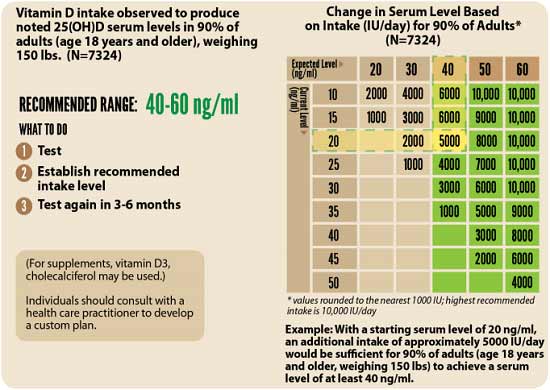
4.Retest in three to six months — Lastly, you’ll need to remeasure your vitamin D level in three to six months, to evaluate how your sun exposure and/or supplement dose is working for you.
- 1 Nutrients October 31, 2020; 12: 3361; DOI: 10.3390/nu12113361
- 2 Nutrients 2020; 12(11):3377
- 3 Archives of Medical Research June 2022; 53(4): 423-430
- 4 The Epoch Times April 26, 2022 (Archived)
- 5 Physiology February 5, 2020 DOI: 10.1152/physiol.00034.2019
- 6 Indian J. Exp Biol. May 1996; 34(5): 391-402
- 7 Frontiers in Pharmacology August 21, 2020 DOI: 10.3389/fphar.2020.01220
- 8 Allergy Research Group, Melatonin, the Antioxidant Recycler
- 9 Cell Death & Disease 2019; 10 article number 317
- 10 Oncotarget June 13, 2017; 8(24): 39896–39921
- 11, 12 Diseases, 2021; DOI: 10.1016/j.ijid.2021.10.012
- 13 Frontiers in Medicine, 2020; DOI: 10.3389/fmed.2020.00226
- 14 British Journal of Dermatology May 31, 2021 DOI: 10.1111/bjd.20093
- 15 Sleep Medicine Reviews August 2016; 28: 69–85
- 16 Melatonin Research February 2019; 2(1): 138-160
- 17 The Journal of Clinical Endocrinology & Metabolism, Volume 106, Issue 3, March 2021, Pages e1343–e1353
- 18 Endocrine.org October 27, 2020
- 19 Science Daily October 27, 2020
- 20 Infectious Diseases April 8, 2020 DOI: 10.21203/rs.3.rs-21211/v1
- 21 Orthomolecular Medicine News Service June 22, 2020
- 22 The Journal of Nutrition October 15, 2014; 144(12): 2073-2082
- 23, 24 Vaccine: X December 11, 2020; 6: 1000076
- 25 Creative Diagnostics Immunology
- 26 Journal of Translational Autoimmunity 2020; 3: 1000051
- 27 CDC COVID Dashboard
- 28 CBS June 6, 2022
- 29 medRxiv April 19, 2022 DOI: 10.1101/2022.04.18.22271936
- 30 The Defender May 4, 2022
- 31 Igor Chudov Substack June 19, 2022
- 32, 34 DMinder app
- 33 GrassrootsHealth Magnesium and Vitamin K2 Combined Important for Vitamin D Levels