Inflammation
now browsing by category
Astaxanthin — A Therapeutic Agent in Cardiovascular Disease
Reproduced from original article:
https://articles.mercola.com/sites/articles/archive/2026/09/12/astaxanthin-cardiovascular-benefits.aspx
Analysis by Dr. Joseph Mercola September 12, 2026

Story at-a-glance
- Heart disease develops quietly over years as blood vessels lose resilience under oxidative and inflammatory stress, long before symptoms appear
- Astaxanthin supports heart health by protecting cell membranes and blood vessels from damage at the cellular level, rather than targeting surface-level markers alone
- Research shows astaxanthin helps maintain flexible blood vessels, steady blood flow, and healthier heart tissue during periods of high stress or reduced oxygen
- The benefits of astaxanthin depend on using natural sources and having it present before and during cardiovascular strain, not after damage has already occurred
- Reducing oxidative stress through diet, improving sleep timing, and supporting natural vitamin D production all reinforce the same vascular repair pathways astaxanthin supports
Cardiovascular disease accounts for roughly 30% of all deaths worldwide.1 But before the dramatic event at the end of the timeline, there’s often a quiet biological drift that occurs. You rarely feel this shift as it unfolds. It typically shows up subtly in routine patterns, then compounds year after year until the damage becomes visible and difficult to reverse.
Much of that long arc traces back to how blood vessels respond to ongoing metabolic and environmental pressure. When the inner lining of your arteries loses resilience, circulation suffers, tissues receive less oxygen, and stress accumulates in places designed for constant flow.
Researchers have spent decades trying to interrupt this process, often focusing on single markers while missing the underlying drivers that keep the cycle active. That gap explains the renewed interest in compounds that work at the cellular level. Some nutrients influence how cells handle stress, repair themselves, and maintain structure under pressure.
When that foundation improves, downstream markers tend to follow. Astaxanthin, a naturally occurring red pigment found in certain marine foods and microalgae, stands out in this context because it supports the basic structures that protect cells under stress while reinforcing the systems that keep blood vessels and your heart functioning over time.
Astaxanthin Targets the Core Drivers of Heart Disease
A review published in the Journal of King Saud University examined astaxanthin as a nutraceutical for cardiovascular health, with a primary focus on atherosclerosis, blood pressure, lipid levels, and metabolic stress.2 The researchers evaluated how astaxanthin affects the biological processes that actually damage arteries over time, including oxidative stress and chronic inflammation, which directly affect your long-term heart risk.
The paper draws from animal research and human clinical trials involving adults with imbalanced blood fats, diabetes, high blood pressure, or elevated cardiovascular risk factors. These are the same categories many people fall into long before a diagnosis like heart attack or stroke appears on their chart.
• Astaxanthin consistently improved lipid patterns tied to plaque buildup — Across human trials summarized in the paper, astaxanthin supplementation lowered triglycerides and raised HDL cholesterol, often referred to as “good cholesterol,” because it helps remove excess cholesterol from artery walls.
• Improvements occurred without the tradeoffs seen with other antioxidants — Astaxanthin differs from beta-carotene and vitamin E, which failed to improve cardiovascular outcomes in large trials and, in some cases, worsened oxidative stress at higher doses. This comparison helps explain why astaxanthin shows benefits where other antioxidants fell short.
Why did vitamin E and beta-carotene fail where astaxanthin succeeds? Location matters. Most antioxidants float in the watery parts of cells or accumulate in fat stores, leaving membranes vulnerable. Astaxanthin is uniquely shaped to span the entire cell membrane, anchoring at both the inner and outer surfaces.
This positioning lets it intercept damage right where it starts — at the membrane itself. Additionally, high-dose vitamin E actually becomes a pro-oxidant under certain conditions, generating the very damage it’s supposed to prevent. Astaxanthin doesn’t share this liability.
• Blood vessel function improved alongside lipid changes — Animal studies reviewed in the paper showed reductions in systolic blood pressure and improved endothelial function. The endothelium is the thin inner lining of all your blood vessels — think of it as the “skin” inside your arteries. When it’s healthy, vessels relax easily, blood flows smoothly, and plaque has trouble gaining a foothold.
When it’s damaged, arteries stiffen, blood pressure rises, and the conditions for heart disease take hold. With astaxanthin, arteries stayed more flexible instead of stiffening under pressure. Most lipid and oxidative stress improvements appeared after four to 12 weeks of consistent supplementation.
• Astaxanthin works by neutralizing damaging molecules before they injure arteries — Oxidative stress involves unstable molecules that attack fats, proteins, and DNA inside your blood vessels. Think of oxidative stress like a fire spreading through dry brush. Unstable molecules called free radicals steal electrons from healthy cells, damaging them and triggering a chain reaction.
Astaxanthin acts like a fire break — it donates electrons freely, satisfying these unstable molecules before they damage your artery walls.
Unlike many antioxidants that float outside cells, astaxanthin spans the entire cell membrane, anchoring itself across the fatty layer that lines blood vessels. This positioning lets it shield vulnerable fats from oxidation, which slows plaque formation. Astaxanthin also activates a cellular switch that turns on your own antioxidant enzymes, including glutathione-related systems.
• Inflammation reduction plays a central role — Astaxanthin lowers inflammatory activity inside artery walls, slowing one of the earliest structural changes that leads to plaque formation. When immune cells absorb too much damaged cholesterol, they swell up and become trapped in artery walls — researchers call these bloated cells “foam cells” because of their bubbly appearance under a microscope.
These foam cells are the foundation of arterial plaque, the buildup that narrows arteries over time. Taken together, the paper shows that astaxanthin supports healthier cholesterol handling, steadier blood pressure, and stronger vessel walls through mechanisms that match how cardiovascular disease actually develops.
Why Form and Timing Matter for Real Cardiovascular Protection
A review published in the journal Marine Drugs set out to evaluate astaxanthin specifically through the lens of cardiovascular disease, with an emphasis on oxidative stress, inflammation, blood flow, and clot risk.3
Researchers studied what happens when blood flow to the heart stops and then suddenly returns — a situation doctors call ischemia-reperfusion injury. This mimics what occurs during a heart attack or heart surgery. Surprisingly, much of the damage happens not during the blockage itself, but when blood flow returns and floods oxygen-starved tissue. Astaxanthin showed remarkable protective effects during this key window.
• Astaxanthin protected the heart muscle during oxygen deprivation — In multiple animal studies summarized in the review, astaxanthin administration before an induced cardiac event limited the amount of heart tissue damaged during the period of extreme stress, helping preserve more healthy heart muscle.
• Protection increased with dose in controlled experimental settings — The review reports a clear dose-response relationship in several studies, where higher astaxanthin exposure led to greater reductions in tissue damage and oxidative markers.
Astaxanthin provided the strongest protection when present in tissues before vascular injury occurred. Human safety data summarized in the paper report no significant adverse effects across a wide dosing range, including effects on blood pressure, clotting, or liver markers.
• Blood flow dynamics improved independently of cholesterol changes — One unique finding emphasized in this paper is improved arterial blood flow and delayed clot formation in animal models, even when lipid levels were not the primary variable.
This finding separates astaxanthin from the cholesterol-focused approach that dominates conventional cardiology. Astaxanthin improves circulation directly, not just by changing lipid numbers on a lab report. In blood clot models, astaxanthin delayed vessel blockage without interfering with normal clotting needed for wound repair.
Why Astaxanthin Works Upstream of Cardiovascular Damage
Astaxanthin is a compound that strengthens heart cells, stabilizes blood vessels, and improves flow before damage escalates into clinical disease.4 Astaxanthin stands out because it targets oxidative stress and inflammation at their source, not downstream damage.
• Cell membranes remained intact under extreme oxidative pressure — Astaxanthin embeds across the lipid membrane, anchoring both ends and preventing breakdown when reactive molecules attack. This means your cells hold their shape instead of leaking and failing under stress.
Astaxanthin increased nitric oxide, the molecule that signals blood vessels to relax and widen. At the same time, it reduced peroxynitrite — a harmful compound formed when nitric oxide reacts with free radicals. Peroxynitrite stiffens arteries and damages tissue, so this dual action keeps vessels flexible while preventing collateral damage.
• Astaxanthin interrupts the inflammatory chain reaction in arteries — As explained in a review published in The American Journal of Cardiology, cardiovascular disease worsens when reactive oxygen and nitrogen species overwhelm blood vessels, activating inflammatory switches that drive endothelial dysfunction, plaque growth, and rhythm disturbances such as atrial fibrillation.5
Unlike vitamin E and beta-carotene, which failed in human trials, astaxanthin belongs to a class of oxygenated carotenoids that directly neutralize these reactive molecules and break destructive chain reactions before they damage vessel walls, making it a strong candidate for addressing a long-standing gap in cardiovascular treatment.
• Astaxanthin shows broad cardiovascular actions — A 2017 review published in Food & Function explains that astaxanthin influences multiple cardiovascular pathways at once, including oxidative stress control, inflammation reduction, blood pressure regulation, lipid handling, glucose balance, kidney protection, and plaque development.6
How to Address the Root Causes of Cardiovascular Damage
Many heart problems build over time through oxidative stress, chronic inflammation, damaged blood vessel lining, and poor cellular energy handling. Fixing those drivers first changes the trajectory, because lipid patterns, blood pressure, and circulation often improve as downstream effects. If you’re already seeing warning signs on labs, or you simply want stronger long-term protection, these steps keep the focus on causes, not symptoms.
1. Lower oxidative stress at the cellular level first — Oxidative damage injures artery walls and sets plaque processes in motion, and one of the biggest drivers is excess linoleic acid (LA) from seed oils. Many Americans consume far more LA than their tissues can safely handle, which fuels inflammation and mitochondrial dysfunction that pushes heart disease forward.
You lower that burden by cutting ultraprocessed foods and seed oils — keeping your LA intake below 5 grams per day. If you can get it under 2 grams, the benefit is even stronger.
To help measure your intake, I recommend you sign up for the Pax health platform, which contains the Seed Oil Sleuth. This feature helps calculate the LA in your food to a tenth of a gram. As oxidative stress drops, blood vessels regain flexibility instead of staying irritated and stiff.
Here’s where astaxanthin and seed oil reduction work together: Excess LA from seed oils makes your cell membranes more vulnerable to oxidation. Astaxanthin embeds in those same membranes and protects them. But if you’re constantly flooding your body with unstable fats, you’re fighting an uphill battle. Reducing seed oil intake lowers the oxidative burden, while astaxanthin reinforces the membranes you’re trying to protect.
2. Build blood vessel resilience — Reducing LA from seed oils and using more stable fats like grass fed butter, ghee, and tallow lowers oxidative stress in vessel walls, which helps preserve normal structure and function over time. Also support vessel flexibility with habits that reinforce circulation and relaxation.
Daily walking keeps blood moving across vessel walls, which signals them to stay elastic instead of stiff. Adequate magnesium intake also helps vessels relax appropriately rather than over-tightening under stress. When membranes are stable and vessels stay responsive, circulation improves naturally and the strain on artery walls drops.
3. If you use astaxanthin, choose the source that matches human nutrition — Astaxanthin occurs naturally in certain marine foods, including wild salmon, sardines, trout, shrimp, and krill, where it plays a protective role in those organisms. Including these foods regularly helps you obtain astaxanthin in a form your body already recognizes.
Krill oil offers a convenient way to obtain astaxanthin alongside omega-3 fats, since the astaxanthin in krill helps protect those delicate omega-3s from oxidation. This combination delivers cardiovascular benefits from multiple angles simultaneously.
If you decide to use an astaxanthin supplement, I strongly recommend products made from the Haematococcus pluvialis microalgae, not versions produced from petrochemicals or genetically engineered yeast. This keeps your intake aligned with the forms studied in cardiovascular research and avoids unnecessary exposures tied to synthetic production, while preserving the benefit profile linked to natural astaxanthin.
Most human studies showing cardiovascular benefits used doses between 4 and 12 milligrams (mg) daily of natural astaxanthin from Haematococcus pluvialis. Higher doses (up to 24 mg) have been studied safely, but the sweet spot for most people appears to be 8 to 12 mg daily, taken with a fat-containing meal to enhance absorption.
4. Use astaxanthin strategically during periods of higher cardiovascular stress — Astaxanthin provides the most value when oxidative and inflammatory stress rise. Intense exercise, emotional stress, frequent travel, and disrupted sleep all place extra strain on blood vessels and heart tissue. These are the moments when oxidative damage accumulates fastest — and when astaxanthin’s protective effects matter most.
Using astaxanthin consistently during these higher-demand phases helps reinforce cellular defenses when damage is most likely to occur. This shifts astaxanthin from a passive supplement to an active support tool matched to real-world stress patterns.
5. Restore circadian alignment with sunlight, vitamin D, and proper sleep — Your blood vessels follow a circadian rhythm. For instance, untreated sleep apnea and disrupted sleep cause a sharp nighttime drop in blood vessel function, increasing vulnerability to heart attacks and other cardiac events.7 Supporting healthy sleep timing and morning light exposure helps stabilize that rhythm.
As you begin to eliminate seed oils, avoiding harsh midday sun for at least six months gives your skin time to clear stored LA, which improves sun tolerance and lowers burn risk during peak midday exposure. As that process completes, gradual midday sun exposure supports natural vitamin D production, reinforcing vascular function, energy balance, and overnight repair.
FAQs About Astaxanthin and Heart Disease
Q: Why does cardiovascular disease develop so gradually?
A: Cardiovascular disease usually develops over many years through ongoing stress on blood vessels. Factors such as oxidative stress, chronic inflammation, and impaired cellular energy slowly weaken the artery lining, reducing circulation efficiency long before noticeable symptoms appear.
Q: What makes astaxanthin different from other antioxidants studied for heart health?
A: Astaxanthin acts at the cellular level, protecting cell membranes and interrupting damaging oxidative and inflammatory processes before they escalate. Unlike antioxidants that failed in large trials, astaxanthin works upstream by stabilizing cells and supporting normal vessel function under stress.
Q: How does astaxanthin support blood vessel and heart function?
A: Research shows astaxanthin helps preserve flexible blood vessels, supports healthy blood flow, and protects heart tissue during periods of reduced oxygen or increased stress. These effects align with how cardiovascular damage actually develops, rather than focusing on isolated markers alone.
Q: Why does the form and timing of astaxanthin matter?
A: The benefits of astaxanthin depend on using biologically appropriate forms and having it present during periods of higher oxidative or inflammatory stress. Natural sources and consistent use during demanding phases, such as intense exercise or disrupted sleep, align best with the research findings.
Q: How do lifestyle factors like diet, sleep, and sunlight fit into cardiovascular protection?
A: Lowering oxidative stress through diet, supporting daily circulation with movement, and maintaining healthy circadian rhythms all reinforce blood vessel repair. Proper sleep timing and regular sunlight exposure support vascular function and vitamin D production, helping protect your heart.
Q: How much astaxanthin should I take, and when?
A: Most research showing cardiovascular benefits used 4 to 12 mg daily of natural astaxanthin. Because astaxanthin is fat-soluble, taking it with a meal containing healthy fats (like grass fed butter or pastured eggs) improves absorption. Consistency matters more than timing — daily use builds tissue levels over weeks, providing ongoing protection rather than acute effects.
- 1, 2 Journal of King Saud University November 6, 2025
- 3, 4 Marine Drugs 2011, 9(3), 447-465
- 5 The American Journal of Cardiology May 22 2008, Volume 101, Issue 10, Supplement, S58-S68
- 6 Food & Function January 1, 2017, 8(1):39-63
- 7 Journal of the American Heart Association, November 6, 2025, Volume 14, Number 22
Microplastics Found to Trigger Cancer-Linked Changes in Lung Cells
Reproduced from original article:
https://articles.mercola.com/sites/articles/archive/2026/09/11/microplastics-lung-cancer-risk.aspx
Analysis by Dr. Joseph Mercola September 11, 2026
Story at-a-glance
- Microplastics were found to trigger cancer-like changes in healthy lung cells, including increased mobility, DNA damage, and activated survival pathways
- Inhaled particles bypass your body’s defenses and embed deep in your lungs, where they silently disrupt cellular function without causing obvious inflammation
- A review of 31 studies confirmed that microplastics harm your lungs, gut, and reproductive organs even at exposure levels that mimic everyday life
- Smaller nanoplastics are especially dangerous because they cross into your bloodstream, reaching your liver, brain, and other organs where they cause long-term damage
- Natural strategies like psyllium, chitosan, probiotics, and autophagy-enhancing nutrients help trap and remove plastic particles before they’re absorbed
Microplastics are no longer just a problem in the ocean — they’re showing up deep in your lungs, changing how your cells function, and raising red flags about cancer risk.
A study published in the Journal of Hazardous Materials revealed that when healthy lung cells absorb polystyrene micro- and nanoplastics — the same kind found in food containers and packaging — they adapt in dangerous ways.1
Instead of dying off, these cells become more mobile and activate pro-survival signals linked to tumor formation. In other words, plastic doesn’t kill your lung cells; it rewires them to behave more like cancer. You inhale thousands of these particles every day from indoor dust, car tires, synthetic fabrics, and degraded packaging.
They’re small enough to bypass your airways’ built-in defenses and embed themselves in the tissue, right where gas exchange happens. Once there, they generate oxidative stress — an internal firestorm of reactive molecules that attack your DNA, disrupt repair systems, and throw off normal cell function. The transformation into a more aggressive, unstable state starts earlier than anyone thought — not in tumors, but in the tissues you rely on to breathe.
These changes don’t cause symptoms right away. But left unchecked, they lay the groundwork for chronic inflammation, lung disease, or cancer later in life. The evidence is clear: plastic is interfering with the core biology of your lungs. Now let’s look at how these findings came to light — and what exactly plastic does once it enters your body.
Healthy Lung Cells Absorb More Plastic Than Cancer Cells — and Change in Dangerous Ways
For the Journal of Hazardous Materials study, researchers examined the effects of microplastics and nanoplastics on both healthy lung cells and three types of lung cancer cells.2 They wanted to see if these plastic particles — widely found in food packaging, household dust, and industrial waste — interfere with normal cell function or trigger biological changes tied to disease.
• Healthy cells were more affected than cancerous ones — The researchers exposed the cells to various sizes of microplastics and nanoplastics at low doses meant to reflect real-world conditions. Surprisingly, it was the healthy lung epithelial cells that absorbed more plastic than the cancer cell lines. These normal cells also showed a greater shift in behavior, including changes in shape, structure, and migration — all red flags for malignant transformation.
• Plastic exposure didn’t kill cells — it pushed them into survival mode — Unlike many toxins that kill off cells through apoptosis, or programmed cell death, microplastics didn’t trigger widespread cell death. Instead, they activated internal damage response systems, including DNA repair signals and antioxidant defense. This is concerning because it means the cells adapted to survive in a toxic environment — the first step in the chain reaction that leads to cancer.
• DNA damage and oxidative stress were key findings — The lung cells showed elevated markers of oxidative stress and significant DNA strand breaks after exposure to microplastics and nanoplastics. This kind of internal damage, if not properly repaired, leads to genetic instability — a known precursor to cancer development. The study also confirmed that oxidative stress was size-dependent, with smaller nanoparticles causing more harm than larger ones.
Plastic Exposure Made Healthy Cells More Mobile — a Cancer-Like Behavior
One of the most troubling findings was that lung cells increased their rate of migration after plastic exposure. In cancer biology, increased mobility is a marker for aggressive tumor cells, which invade surrounding tissues and spread throughout the body. The fact that noncancerous cells began behaving this way highlights the hidden risk of daily microplastic exposure.3
• Plastic particles disrupted the cell membrane and cytoskeleton — Researchers used imaging tools to show that both nano- and microplastics entered the cells and altered the internal structure. The actin cytoskeleton — a network that helps cells maintain their shape and movement — was significantly reorganized in exposed cells. This internal restructuring made the cells more fragile and unstable.
• Several survival pathways were activated in the lung cells — Exposure to microplastics and nanoplastics triggered signaling pathways that promote cell survival and resistance to stress. These same pathways are commonly overactive in tumor cells, and their activation in healthy lung cells suggests that plastic is not just a passive contaminant but an active disruptor of cell biology.
• Plastic-induced changes occurred without visible inflammation — One of the more insidious findings was that all of these harmful changes occurred without classic signs of inflammation or immune response. That means you wouldn’t feel anything or see any symptoms — but the long-term cellular effects could be serious. This stealth effect underscores why daily exposure to microplastics should not be dismissed.
• Cells exposed to plastic lost their ability to function normally — Overall, the study showed that plastic particles interfere with nearly every aspect of healthy lung cell behavior: from DNA integrity to cell shape, mobility, and stress response. While the research didn’t follow these changes to full tumor formation, the authors emphasized that these are precisely the kinds of shifts that lead to long-term disease.
Microplastics Damage Your Lungs, Gut, and Reproductive System — Even at Everyday Exposure Levels
A 2024 review in Environmental Science & Technology looked at 28 animal studies and three human studies to understand what happens when microplastics get inside you — whether you breathe them in or swallow them.4 The damage wasn’t limited to one area. It showed up in the lungs, digestive tract, and even reproductive organs.
• Plastic triggered inflammation, DNA damage, and hormonal disruption — Inhaling plastic particles caused inflammation in the lungs, scarring of airways, and changes in how immune cells responded.
Ingested plastics damaged the gut lining, disrupted the gut microbiome, and kicked off chronic inflammation. Some studies showed sperm damage, lower testosterone, changes in ovary structure, and reduced fertility in animals. These effects weren’t limited to high doses — they happened at levels that mimic everyday life.
• Oxidative stress was the main mechanism behind the harm — The common thread was oxidative stress — a kind of internal “rusting” process where your body struggles to keep up with damaging free radicals. That stress interferes with DNA repair, weakens cell membranes, and confuses your immune system. Once it starts, it becomes harder for your body to recover from the damage.
• Smaller particles go deeper — and stay longer — Nanoplastics, the tiniest particles, were the most dangerous. They could pass through the lungs or gut lining, enter the bloodstream, and end up in places like your liver, kidneys, or even your brain. These particles didn’t just pass through — they stuck around and changed how those organs functioned.
• Plastic isn’t just an environmental problem — it’s a full-body health threat — What you breathe, eat, and drink every day could be slowly reshaping your internal biology. Even though more human studies are needed, the fact that dozens of animal studies found damage across key organ systems — at realistic exposure levels — makes one thing clear: your daily contact with microplastics isn’t harmless.
Natural Strategies to Eliminate Microplastics Are Being Explored
Studies are now looking at strategies to help the human body filter, trap, and eliminate microplastics before they can spread throughout your other systems. These methods offer a multi-angle approach to help reduce your internal plastic load and support overall health. I’ve written a paper discussing these methods in detail, and while it is still under peer-review, I’ve provided the key findings below.
• Cross-linked psyllium could help eliminate microplastics — One key system that plays a role in removing microplastics from your body is your gut. A 2024 study showed that acrylamide cross-linked psyllium (PLP-AM) removed over 92% of common plastic types like polystyrene, polyvinyl chloride (PVC), and polyethylene terephthalate (PET) from water.
Because of its high swelling ability and sticky, gel-like texture, cross-linked psyllium could be adapted to work inside the gut, where it may trap plastic particles before they’re absorbed into the body. While the study was conducted in a water treatment setting, the results are also promising for human health.5
• Chitosan, a natural fiber derived from shellfish, also shows promise for clearing microplastics from your body — An animal study published in Scientific Reports found that rats given a chitosan-enriched diet were able to eliminate about 115% of the polyethylene microplastics they were fed, compared to just 84% in the control group.
This suggests that chitosan not only helps bind and eliminate new plastic particles but might even help pull out some that were already absorbed. However, while it’s generally considered safe and already used in supplements, people with shellfish allergies are advised to steer clear of it.6
Psyllium and chitosan work through physical adsorption, where hydrophobic (water-repelling) and electrostatic forces stick microplastic particles to the fiber, keeping them from being absorbed. However, one drawback with these binders is that they can also soak up nutrients if not timed carefully. Hence, they need to be used strategically to provide the most benefit, such as ingesting them with processed or packaged foods, which are more likely to contain plastics.
• Certain beneficial bacteria strains can help clear microplastics from the gut — A 2025 animal study found that two specific strains, Lacticaseibacillus paracasei DT66 and Lactiplantibacillus plantarum DT88, were able to bind to and eliminate tiny polystyrene particles in lab tests.
These probiotics work by forming protective biofilms that trap plastic particles, making them easier to flush out.7 When combined with dietary fibers like psyllium and chitosan, the result could be a more effective and natural way to sweep microplastics out of the gut before they’re absorbed.
• The liver also plays an essential role in clearing microplastics from the bloodstream — Specialized immune cells in the liver, known as Kupffer cells, help trap these foreign particles and route them into bile for elimination via the intestines. However, while this method may work on smaller plastics, larger ones can linger and build up, especially if your liver function is compromised.
To support this natural detox pathway, researchers are studying the use of compounds like ursodeoxycholic acid (UDCA) and its variant tauroursodeoxycholic acid (TUDCA), which stimulate bile production and improve particle flow out of the liver.
• Researchers are also looking at strategies to enhance autophagy to eliminate microplastics — Autophagy is your body’s natural cellular recycling system. Researchers are looking at compounds that can help promote this system, mainly rapamycin and spermidine.
Rapamycin works by inhibiting the mTOR pathway, a nutrient-sensing mechanism that normally suppresses autophagy. When mTOR is turned off, cells ramp up their cleanup efforts, forming membranes that can collect and isolate plastic particles for breakdown or removal. Meanwhile, spermidine is a naturally occurring polyamine found in foods that enhances cellular resilience and supports the clearance of toxic substances.
In lab and animal studies, the combination of spermidine and rapamycin helped reverse mitochondrial dysfunction and reduce oxidative stress caused by microplastics.
The table below summarizes these novel strategies to eliminate microplastics, including their mechanisms of action, how much testing has been done, and important safety considerations. It shows that although several different approaches may be needed, clearing plastics from your body naturally is possible. Of course, reducing your exposure is still the ideal preliminary course of action.

How to Reduce Your Exposure to Lung-Damaging Microplastics
If you’re breathing, you’re exposed. Microplastics are in the air around you — from synthetic carpets and clothing to packaging dust and car exhaust. You don’t need to panic, but you do need to act. These particles aren’t just passing through your lungs.
They’re embedding, altering how your cells behave, and triggering damage at a cellular level. That means you need to treat this like any other environmental toxin: identify the source and cut it off. Here’s how I recommend you take control of your environment and protect your lungs:
1. Ditch synthetic textiles and go natural wherever possible — If you’re wearing polyester or drying synthetic fabrics indoors, you’re likely inhaling fibers you can’t see. Switch to natural clothing like cotton, wool, linen, or hemp. Use a vented dryer and keep your laundry space well-ventilated to reduce airborne fibers.
If you’re a parent, prioritize organic natural fibers for children — they’re more vulnerable to inhalation damage. For the synthetic pieces you already own, wash them less frequently, line dry when possible, and use a microfiber-catching laundry bag to trap loose fibers.
2. Upgrade your indoor air filtration and filter your water — Your lungs are working overtime in enclosed spaces. Use a high-efficiency particulate air (HEPA) filter in the rooms where you spend the most time — especially bedrooms and workspaces. If you live in an apartment or near a busy road, a good air purifier is nonnegotiable.
Make sure it’s rated for micro-sized particles (PM2.5 or smaller) to catch airborne plastic dust. In addition, use a high-quality water filtration system that removes particles down to the micron level.
3. Avoid heating plastic containers or food packaging — Microwaving plastic, drinking hot liquids from plastic-lined cups, or using plastic containers for leftovers can release polystyrene particles and nanoplastics. Store food in glass or stainless steel instead. If you’re reheating, make it a habit to transfer your food out of plastic first — this one small change significantly lowers your microplastic load.
4. Vacuum with a sealed system and damp dust frequently — Dust is one of the biggest sources of indoor microplastics — and your vacuum matters. Use a sealed vacuum with a HEPA filter, and clean floors regularly, especially if you have carpets or pets. Dry dusting just pushes particles into the air, so use a damp cloth to trap and remove dust instead.
5. Avoid personal care products that contain microbeads or plastic thickeners — If you’re using exfoliating scrubs, toothpaste, or face washes that list polyethylene or polypropylene on the label, you’re applying plastic directly to your skin and possibly rinsing it into the air. Choose clean, microplastic-free products.
You won’t just help your body — you’ll help reduce contamination in the environment, too. Small actions, when done consistently, have a compounding effect. The less plastic you breathe in, the lower your risk of cellular stress, immune dysfunction, and long-term lung damage.
FAQs About Microplastics
Q: How do microplastics affect my lungs?
A: Microplastics don’t just sit in your airways — they get absorbed into lung cells and trigger changes linked to cancer. They cause oxidative stress, DNA damage, and make healthy cells behave more like tumor cells by activating survival pathways and increasing mobility, even without obvious inflammation.
Q: Where do microplastics come from, and how do I inhale them?
A: You breathe them in from indoor dust, synthetic clothing, carpets, car tires, and even packaging materials. These particles are tiny enough to bypass your lungs’ natural defenses and embed in your tissue — right where gas exchange happens.
Q: What other parts of my body do microplastics harm?
A: Beyond your lungs, microplastics damage your digestive system and reproductive organs. Studies show they disrupt your gut lining, alter your microbiome, and interfere with hormones, fertility, and immune signaling. Smaller nanoplastics even reach your brain and liver through your bloodstream.
Q: Can I remove microplastics from my body naturally?
A: Emerging research suggests that natural binders like cross-linked psyllium, chitosan, and specific probiotics help trap and eliminate microplastics in your gut. Other strategies like supporting liver detox and boosting autophagy with compounds like spermidine and rapamycin are also being studied.
Q: What are the best ways to reduce my exposure to microplastics?
A: Switch to natural fabrics, use HEPA air filters, avoid heating food in plastic, vacuum with sealed systems, and choose clean personal care products without microbeads. Small daily changes significantly lower your plastic exposure and protect your long-term health.
The Role of Food Nutrients in Reducing Oxidative Stress Linked to Microplastics
Reproduced from original article:
https://articles.mercola.com/sites/articles/archive/2026/08/12/microplastics-oxidative-stress-food-nutrients.aspx
Analysis by Dr. Joseph Mercola August 12, 2026
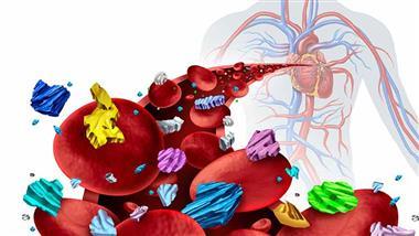
Story at-a-glance
- Microplastics and nanoplastics are now found in food, water, and air, and research suggests they accumulate in organs such as the liver, pancreas, kidneys, and brain. Laboratory and animal studies indicate they may contribute to oxidative stress, inflammation, and insulin resistance in these areas
- The same biological changes linked to microplastic exposure are also associated with Type 2 diabetes, including impaired blood sugar control, disrupted gut bacteria, and metabolic dysfunction that develops long before symptoms appear
- In animal studies, nanoplastics, which are microscopic, crossed the blood-brain barrier, where they triggered inflammation, interfered with how brain cells use glucose for energy, and contributed to memory and learning problems associated with cognitive decline
- Your body has a built-in defense system called Nrf2 that helps neutralize oxidative stress and repair cellular damage, and many nutrients found in colorful fruits, herbs, spices, and other plant foods help activate this protective pathway
- While eliminating all microplastic exposure is no longer realistic, reducing everyday plastic use, supporting healthy metabolism, tracking HOMA-IR, and eating a wide variety of nutrient-rich whole foods help strengthen your body’s resilience against plastic-related damage
You can’t see them, but the plastic you encounter every day is breaking down into fragments small enough to slip past your body’s defenses. These microplastics and nanoplastics arrive through the food you eat, the water you drink, and the air you breathe — and unlike the visible plastic waste we’ve been warned about for decades, these particles are tiny enough to interact directly with your cells.
Once inside, the smallest particles don’t just pass through and leave; the tiniest lodge in tissue and stay. Two recent reviews lay out what happens next, and read together, they trace a troubling path from your bloodstream to your brain. The first, published in Nutrients, examined how these particles may disrupt blood sugar regulation and contribute to the oxidative stress and insulin resistance associated with Type 2 diabetes.1
The second, published in the International Journal of Molecular Sciences, followed the same particles into the brain, where researchers believe they may feed the insulin resistance and cellular damage now linked to Alzheimer’s, a condition some scientists have begun calling “Type 3 diabetes.”2 Notably, this term is an informal label, not an officially recognized diagnosis.
What caught my attention was that neither review stopped at the damage. Both turned to the same question: can what you eat change how well your cells defend themselves? At the center of that defense sits a single protective pathway your body already has — one that certain food compounds appear able to switch on.
That shifts the conversation from simply avoiding exposure, which is no longer fully possible, to actively building resilience. What follows is how microplastics weaken those defenses, and which nutrients help restore them.
Your Diet Influences How Well Your Cells Fight Plastic-Related Damage
The Nutrients review examined how specific food-derived compounds interact with Nrf2, a protein that controls one of your body’s most important antioxidant defense systems.3 The researchers explored whether nutrients found in foods could strengthen cellular resilience and help counter the oxidative stress, inflammation, and metabolic disruption linked to plastic exposure.
Oxidative stress is what happens when damaging molecules called free radicals pile up faster than your cells can neutralize them, a kind of internal rust that wears down tissue over time.
Humans encounter microplastics and nanoplastics through contaminated food, beverages, household products, and airborne particles from textiles and plastics. Global plastic production was expected to reach roughly 400 million tons annually in 2025, while cumulative production could reach 33 billion tons by 2050.4 As exposure increases, scientists have become increasingly concerned about what happens when these particles accumulate inside the body.
• The concern extends far beyond simple pollution — Microplastics accumulate in organs involved in blood sugar regulation, including the liver, pancreas, and kidneys. In experimental studies, these particles triggered oxidative stress, inflammation, cellular barrier disruption, and metabolic dysfunction — processes that resemble those commonly seen in diabetes and its complications.
Several studies discussed in the review found that exposure to polystyrene microplastics and nanoplastics increased insulin resistance and worsened glucose tolerance in mice.5 Insulin resistance means your cells stop responding efficiently to insulin, forcing your body to keep blood sugar levels elevated for longer periods.
Researchers found that nanoplastics measuring just 100 nanometers produced greater toxicity than larger microplastic particles in diabetic mice. These tiny particles increased liver inflammation, disrupted gut bacteria, and intensified fat metabolism problems inside the liver. Their small size allows them to cross biological barriers more easily and reach tissues that larger particles struggle to enter.
• Your cells already have a built-in emergency response system — Researchers point to Nrf2 as the body’s master switch for antioxidant defense. Most of the time it sits idle, until cellular stress trips the alarm and it springs into action. When cells encounter stress, Nrf2 moves into the nucleus and switches on a large network of protective genes that help neutralize harmful molecules and repair damage.
Researchers emphasized that many plant-derived nutrients work by stimulating this same pathway, effectively helping your cells activate their own internal repair and defense programs.
• Several nutrient families activate the body’s defense genes — Polyphenols, flavonoids, phenolic acids, terpenoids, and related plant compounds are activators of the Nrf2 pathway. These naturally occurring compounds are found in a range of plant foods, including berries, herbs, spices, and colorful fruits and vegetables.
Once activated, Nrf2 switches on a network of antioxidant and detoxification enzymes that help cells neutralize oxidative stress, repair damage, and maintain normal metabolic function despite ongoing environmental exposures.
•The review connected environmental pollution with personalized nutrition — Researchers proposed that future dietary recommendations could be tailored not only to a person’s genetics, but also to their exposure to environmental pollutants such as microplastics. The goal would be to identify nutrients that activate protective pathways like Nrf2 and match those nutrients to individuals who are most vulnerable to oxidative stress, inflammation, and metabolic damage.
Microplastics Could Be Hitting Your Brain Too
A review published in the International Journal of Molecular Sciences examined how microplastics and nanoplastics affect the brain and whether specific nutrients help protect against that damage.6 The authors focused on a growing concern: the possibility that long-term plastic exposure contributes to brain insulin resistance, a condition increasingly linked to Alzheimer’s disease.
Unlike Type 2 diabetes, which affects how your body responds to insulin, brain insulin resistance affects how neurons use glucose for energy. Your brain consumes enormous amounts of energy every day. When brain cells struggle to access or use glucose efficiently, memory, learning, and cognitive function begin to suffer. Researchers explained that this process contributes to the buildup of beta-amyloid plaques and tau tangles, two hallmarks of Alzheimer’s disease.*
• Microplastics are capable of reaching the brain itself — Plastic particles have been detected in human tissues, including the lungs, liver, kidneys, and brain. Animal studies found that smaller nanoplastics cross the blood-brain barrier, the protective filter that normally shields the brain from harmful substances, more easily than larger particles. Once inside brain tissue, these particles trigger oxidative stress, inflammation, and cellular dysfunction.
• Brain inflammation creates a vicious cycle — Researchers described how plastic-induced oxidative stress damages neurons while simultaneously weakening the brain’s antioxidant defenses. As oxidative stress rises, inflammation increases. As inflammation increases, insulin signaling worsens. That decline in insulin signaling may promote additional oxidative stress and accelerate the formation of beta-amyloid plaques and tau protein abnormalities associated with cognitive decline.
Healthy brain cells rely on specialized glucose transporters that move glucose across the blood-brain barrier and into neurons. Aging, inflammation, and insulin resistance reduce the activity of key transporters, limiting glucose availability to brain cells. The result is an energy shortage that affects memory, concentration, and cognitive performance long before obvious symptoms appear.
• Several studies linked plastic exposure directly to memory problems — Researchers reviewed evidence showing that nanoplastics impaired learning and memory in animal models in a dose-dependent manner. One study found that exposure to 80-nanometer polystyrene nanoparticles disrupted learning and memory in mice after only seven days.7
Other experiments showed that microglia, the immune cells that protect the brain, became activated after exposure and shifted into a chronic inflammatory state that interfered with nearby neurons.8
• Researchers identified several nutrients that counteracted the same pathways disrupted by microplastics — The review highlighted a group of plant compounds that activate Nrf2 and related resilience pathways, which help defend brain cells against the oxidative stress, inflammation, and insulin-signaling problems linked to microplastic exposure.
Rather than acting through a single mechanism, these nutrients targeted multiple stages of the damage process, including antioxidant defenses, glucose metabolism, inflammation control, and neuronal survival. According to laboratory and animal research:
◦Ursolic acid, found in apple peels, berries, and herbs, improved memory, reduced beta-amyloid toxicity, and increased antioxidant enzyme activity.
◦Verbascoside, a plant compound found in species such as lilac, mullein, and lemon verbena, improved gut health, reduced inflammatory markers, and enhanced insulin sensitivity in the brain, supporting the growing connection between the gut-brain axis and cognitive health.
◦Diosmin, a flavonoid concentrated in the white pith of citrus fruit, which is exactly the part most people discard, boosted antioxidant defenses and improved both working and long-term memory.
◦Baicalein, a flavonoid extracted from the roots of Chinese skullcap (Scutellaria baicalensis), reduced neuroinflammation, lowered beta-amyloid plaque formation, and improved glucose metabolism and insulin signaling in brain tissue.
◦Cynarin, a major compound in artichokes, reduced inflammation, lowered beta-amyloid and tau protein accumulation, and improved cognitive performance in Alzheimer’s models.
• Many of these nutrients targeted brain insulin resistance directly — One of the review’s central themes was that Alzheimer’s disease shares many features with diabetes, including impaired insulin signaling inside the brain. In laboratory and animal studies, several of the highlighted nutrients improved key pathways involved in glucose uptake, insulin sensitivity, and cellular energy production, helping restore the ability of brain cells to access and use fuel efficiently.
Researchers argued that preserving these pathways could help interrupt the progression from oxidative stress and inflammation to cognitive decline. Rather than targeting a single symptom, the nutrients appeared to strengthen the underlying cellular defense network that keeps brain tissue functioning properly despite ongoing environmental stressors.
*These findings are from laboratory or animal research and may not directly apply to human health. To date, no long-term human studies have confirmed that microplastics cause Type 2 diabetes or Alzheimer’s disease in people.
Strengthen Your Defenses Against Microplastic Damage
The reality is that you can’t completely eliminate microplastic exposure. Plastic particles are already present in food, water, and the environment. That reality is why I wrote “Microplastics Cure,” available for preorder now. In it, I explain how these invisible particles enter and accumulate throughout the body, why they become increasingly difficult to remove once they lodge in tissues and blood vessels, and how everyday choices either increase or reduce that accumulation.
You’ll also find practical, science-based strategies that help lower ongoing exposure while supporting your body’s own protective systems. The goal is simple: understand what drives microplastic accumulation, identify the habits that contribute to it, and take meaningful steps to reduce the damage before the burden grows larger.
Avoidance is only part of the solution. A better strategy is to reduce your exposure wherever possible while strengthening the cellular defense systems that help your body handle oxidative stress and inflammation. Your daily choices influence how resilient your cells remain when they encounter environmental toxins.
1. Reduce the largest sources of plastic exposure — Start with the exposures you control every day. Avoid heating food in plastic containers, replace plastic water bottles with glass or stainless steel, and choose fresh foods instead of heavily packaged products whenever possible.
If you regularly drink bottled water, switching to filtered water stored in glass containers immediately reduces one of the most common sources of microplastic intake. Every source you eliminate lowers the amount of plastic your body has to process and store over time.
2. Build your meals around Nrf2-activating foods — The strongest nutritional message from both reviews is that certain plant compounds help activate Nrf2, one of your body’s most important cellular defense systems. Nrf2 helps switch on antioxidant and detoxification enzymes that protect tissues from the oxidative stress and inflammation triggered by microplastics.
Rather than focusing on a single food, build your meals around a variety of foods that naturally contain these protective compounds:
•Include apple peels, fresh herbs, and other minimally processed plant foods regularly, instead of relying on packaged products that often contribute additional microplastic exposure.
•Add artichokes to meals when available. They provide a unique mix of polyphenols that are largely absent from the modern processed-food diet.
•Choose whole citrus fruits regularly. They offer beneficial plant compounds along with fiber and nutrients that support metabolic health.
•Experiment with a wider variety of herbs, teas, and traditional plant foods rather than eating the same handful of foods every week. Greater plant diversity exposes you to a broader range of protective compounds.
•Build meals around colorful fruits, vegetables, herbs, and spices. Different colors signal different families of beneficial plant compounds, giving your body a wider range of nutritional tools to draw from.
3. Support cellular energy production — Oxidative stress becomes more damaging when your cells struggle to produce energy efficiently. Support mitochondrial function by getting regular sunlight, avoiding seed oils, eating adequate protein, and consuming enough carbohydrates — about 250 grams a day — to support healthy metabolism. Your cells repair damage more effectively when they have the energy required to run their antioxidant and detoxification systems.
4. Control blood sugar before metabolic damage develops — Microplastic exposure is linked to insulin resistance and impaired glucose control. One of the smartest steps you can take is to track your metabolic health before symptoms appear. I recommend monitoring your Homeostatic Model Assessment of Insulin Resistance (HOMA-IR) score, rather than relying solely on fasting glucose.
HOMA-IR is a calculation based on your fasting insulin and fasting glucose levels that provides one of the clearest pictures of how hard your body is working to keep blood sugar under control.
A higher score suggests greater insulin resistance, often years before obvious symptoms appear. Keeping your HOMA-IR low helps reduce oxidative stress and limits many of the same inflammatory pathways that microplastics appear to activate. For most people, if your HOMA-IR creeps above 1.0, it’s time to pay closer attention to factors like sugar intake, seed oils, plastic exposure, and gut health.
Talk to your health care provider about whether this testing is appropriate for you.
5. Create daily habits that activate your body’s resilience systems — The Nrf2 pathway responds to more than nutrition. Regular movement, quality sleep, healthy circadian rhythms, and consistent exposure to natural daylight all help maintain cellular resilience. Turn this into a simple daily challenge.
Get morning sunlight. Walk every day. Build meals around nutrient-dense whole foods. Reduce packaged products. Small actions repeated consistently strengthen your internal defense systems far more effectively than occasional bursts of effort.
FAQs About Food Nutrients and Microplastic Exposure
Q: How do microplastics affect my health?
A: Microplastics and nanoplastics can accumulate in tissues throughout your body, including the liver, pancreas, kidneys, and brain. Research — primarily in laboratory and animal models — suggests they may trigger oxidative stress, inflammation, and cellular dysfunction, which are linked to insulin resistance, metabolic disorders, and cognitive decline.
Q: What is Nrf2 and why is it important?
A: Nrf2 is one of your body’s most important cellular defense systems. When activated, it switches on genes that help neutralize oxidative stress, reduce inflammation, and support cellular repair. Researchers found that many beneficial plant compounds help activate this pathway, strengthening your body’s ability to cope with environmental stressors such as microplastic exposure.
Q: Can microplastics affect brain health and memory?
A: In animal studies, very small plastic particles crossed the blood-brain barrier and triggered inflammation, oxidative stress, and impaired insulin signaling inside the brain. These changes are associated with memory problems, reduced cognitive function, and biological processes linked to Alzheimer’s disease.
Q: Which foods contain nutrients that help protect against microplastic-related damage?
A: A variety of whole foods provide compounds that support your body’s antioxidant defenses. Apple peels, berries, herbs, and spices provide ursolic acid and other polyphenols. Artichokes contain cynarin, citrus fruits provide diosmin, Chinese skullcap contains baicalein, and plants such as lemon verbena and mullein contain verbascoside. These compounds help support antioxidant activity, healthy glucose metabolism, and inflammation control.
Q: What are the most effective ways to reduce the health risks associated with microplastics?
A: A practical approach combines reducing exposure and strengthening cellular resilience. Steps include avoiding heating food in plastic containers, reducing bottled water use, choosing fresh foods over heavily packaged products, supporting metabolic health, monitoring insulin resistance with HOMA-IR, and regularly eating nutrient-dense foods that activate protective pathways such as Nrf2.
Daily habits such as exercise, quality sleep, and natural daylight exposure also help maintain cellular defenses.
This article is for informational purposes only and does not constitute medical advice. Consult a qualified healthcare provider before making changes to your health regimen.
Why ignoring your lymphatic system is a fatal mistake
Reproduced from original article:
https://www.naturalhealth365.com/the-silent-system-most-people-ignore-and-the-damage-that-quietly-follows.html
by: July 6, 2026
 (NaturalHealth365) There is a network of vessels running through your entire body that most doctors never check. The lymphatic system moves fluid, clears waste, and keeps the immune system running properly. When that system stalls, the consequences reach far beyond a little swelling in your legs.
New research published in 2025 and 2026 is bringing the lymphatic system into a conversation that Western medicine has largely avoided. Researchers are finding direct links between poor lymphatic drainage and some of the world’s most common chronic diseases. Heart disease, chronic inflammation, and weakened immune function are all showing up in the data.
Your lymph system does more than most people realize
Think of the lymphatic system as the body’s slow but essential cleanup crew. Every day, lymph vessels collect excess fluid, cellular waste, and immune debris from your tissues and move them out. Without that clearance, the buildup creates exactly the conditions that disease needs to take hold.
A 2026 review from the University of Nevada Las Vegas confirmed that lymphatic vessels inside artery walls actively remove the cholesterol and inflammatory compounds that fuel plaque buildup. When those vessels stop draining properly, that material gets left behind.
The result is more inflammation, more plaque, and higher cardiovascular risk.
What happens when lymph fluid stops moving
Stagnant lymph does not just sit there quietly. Research published in Frontiers in Medicine in 2025 found that pooling lymph triggers inflammation, tissue scarring, and oxygen deprivation in the surrounding area.
What surprised researchers most was how far the damage spreads. People with lymphatic dysfunction showed immune and tissue changes in parts of the body far from the problem area. This is not a localized condition. When the lymphatic system struggles, the whole body feels the consequences.
Why sitting may be quietly stalling your lymph flow
Unlike the heart, the lymphatic system has no pump. Lymph moves only when muscles squeeze the surrounding vessels during physical movement. Prolonged sitting effectively shuts that movement down.
Research consistently links sedentary behavior to sluggish lymph flow and higher levels of systemic inflammation. This may partly explain why sitting too long raises disease risk across so many different organ systems. The lymphatic connection has been missing from that conversation for too long.
What you can do right now
Keep your body moving throughout the day, not just during a scheduled workout. Short walks, stretching, or even standing regularly throughout the day all contract the muscles that drive lymph flow.
Research suggests that frequent, brief movement is more effective for lymphatic drainage than a single long session. A mini-trampoline, or rebounder, is particularly effective because the up-and-down motion creates a pumping effect throughout the lymphatic network.
Feed your lymphatic system through your diet by reducing the inflammatory load the system has to carry. Organic leafy greens and fiber-rich whole foods reduce systemic inflammation and support the gut’s own lymphatic vessels, called lacteals. Processed seed oils, refined sugar, and heavily packaged foods increase the toxic burden flowing through lymph and make clearance harder.
What you eat directly determines how much work your lymph system has to do each day.
Support the body’s drainage pathways with targeted daily habits outside of eating and exercise. Dry brushing toward the heart before a shower stimulates the superficial lymph vessels just under the skin. Alternating warm and cold water during a shower causes the lymph vessels to contract and relax, which drives fluid movement.
In addition, staying well hydrated is essential, since lymph fluid is largely water, and dehydration slows the entire system.
The system your doctor is not checking
Cardiologists test cholesterol and blood pressure. Immunologists look at white blood cell counts. Almost nobody evaluates whether your lymphatic system is functioning properly.
Sources for this article include:
Frontiersin.org
Frontiersin.org
The forgotten organ that can predict your risk of infections and premature death by up to 50%
Reproduced from original article:
https://www.naturalhealth365.com/the-forgotten-organ-that-could-predict-how-long-you-live-researchers-now-confirm.html
by: Patrick Tims, staff writer | June 6, 2026
 (NaturalHealth365) Most adults have never given a thought to their thymus. Unfortunately, Western medicine rarely mentions this organ after childhood. On top of that, the conventional scientific community falsely believes that the thymus becomes largely irrelevant once the body matures. Now two landmark studies published in Nature are overturning that assumption entirely, and the numbers are impossible to dismiss.
Researchers at Mass General Brigham used artificial intelligence to analyze CT scans from more than 25,000 adults. The AI assessed each participant’s thymus and generated a thymic health score. People with the highest scores had a 50% lower risk of death from any cause and a 63% lower risk of dying from cardiovascular disease.
Remarkably, those adults also showed a 36% lower risk of developing lung cancer. A second study analyzed outcomes in 3,476 cancer patients receiving immunotherapy. Stronger thymic health was associated with a 37% lower risk of cancer progression and a 44% lower risk of death.
Your protection against infections and disease
The thymus sits in the chest, just behind the breastbone. During childhood, the organ trains T cells, the specialized immune defenders that protect the body from infection and disease.
Cancer defense and cellular repair both depend on these cells. After puberty, the thymus begins to shrink and produces fewer new T cells. Consequently, most physicians stopped paying attention to the thymus, assuming the work was finished.
However, research from Mass General Brigham reveals that the thymus continues to produce T cells throughout adulthood. The quality of that ongoing production matters enormously.
Moreover, adults with healthier thymuses maintain greater immune cell diversity. That diversity, the research shows, translates directly into lower rates of chronic disease and longer survival. “The thymus has been overlooked for decades and may be a missing piece in explaining why people age differently,” said lead researcher Hugo Aerts, PhD.
Why thymic decline happens faster in some people than others
The research did not simply confirm that thymic health predicts longevity. Importantly, the team identified specific factors associated with accelerated decline. Chronic inflammation, smoking, and high body weight each correlated with significantly poorer thymic health scores.
That finding matters because the research places a meaningful degree of control over immune aging in each individual’s hands. Rather than leaving the outcome entirely to genetics or time, lifestyle choices now emerge as direct determinants of thymic health.
A person managing chronic inflammation, reducing body weight, and avoiding tobacco is also protecting the immune system, which is responsible for long-term disease resistance. The research makes that connection direct and specific.
What the immunotherapy finding reveals about immune capacity
The second study’s finding deserves separate consideration. Researchers analyzed cancer patients receiving immune checkpoint inhibitors. These treatments work by removing molecular brakes that cancer cells place on T cells, allowing those immune cells to resume attacking tumors.
Patients with the strongest thymic health showed a 37% lower risk of cancer progression and a 44% lower risk of death. In fact, those results held even after accounting for tumor type, treatment protocol, and other patient variables.
These finding suggests the thymus functions as a reservoir of immune capacity. When that reservoir is depleted over decades of inflammation and poor metabolic health, the T cells available to respond to treatment become fewer and less diverse. As a result, the immune system’s ability to fight cancer may be substantially shaped years before a diagnosis is ever made.
Steps that protect thymic function starting now
Address chronic inflammation. The research confirms that systemic inflammation accelerates thymic decline. A diet rich in organic dark leafy greens, organic berries, and extra-virgin olive oil provides anti-inflammatory polyphenols, and antioxidants. Together, those compounds directly reduce the inflammatory burden on immune tissue.
Curcumin from turmeric has strong research support for reducing systemic inflammatory markers. In addition, high-quality omega-3 foods or supplementation can help to reduce the risk of inflammation at the cellular level.
Support metabolic health as inseparable from immune resilience. High body weight was directly linked to poorer thymic health in the research. Reducing visceral fat through consistent daily movement and a diet low in refined carbohydrates creates a metabolic environment that supports rather than burdens the immune system.
Targeted supplementation with berberine further improves insulin sensitivity through pathways that reduce systemic inflammation. And, finally, magnesium can help you with both blood sugar regulation and the stress response, reducing one of the least-recognized contributors to chronic inflammation.
Prioritize nutrients that support T cell development and immune diversity. Zinc plays an essential and largely underappreciated role in thymus function and T cell maturation.
Research consistently links zinc deficiency to accelerated thymic atrophy and reduced immune cell diversity. Plus, selenium supports the antioxidant defenses that protect thymic tissue from oxidative damage.
Vitamin D, maintained at functional blood levels between 50 and 70 ng/mL, supports immune genes critical for T cell activation. But, keep in mind, most people on standard supplement amounts will not reach that target without testing and adjustments, as needed.
Together, these targeted nutrients supply the biological inputs the thymus depends on to remain functional across the lifespan.
The immune system builds future resilience long before disease arrives
Overall, a 50% reduction in all-cause mortality linked to a single organ’s health status is a finding that should reshape how adults think about immune function. Most people manage symptoms as they appear. Very few consider the underlying immune infrastructure that determines whether those symptoms ever develop.
Sources for this article include:
Melatonin — the Missing Link Between Sleep Deprivation and Immune Dysregulation
Reproduced from original article:
https://articles.mercola.com/sites/articles/archive/2026/04/21/melatonin-sleep-deprivation-immune-dysregulation.aspx
Analysis by Dr. Joseph Mercola April 21, 2026

Story at-a-glance
- A 2025 review of 50 studies found that sleep deprivation lowers melatonin, which directly increases inflammatory signals and weakens immune cell performance
- When melatonin drops, your immune cells lose energy, struggle to coordinate their defense tasks and become less effective at fighting infections
- Chronic sleep disruption raises stress hormones like cortisol, shifts immune balance, and damages your gut barrier, compounding inflammation throughout your body
- Older adults, shift workers and people with insomnia face greater immune disruption because their melatonin levels are more easily suppressed
- Rebuilding melatonin through sunlight exposure, nighttime darkness, proper nutrition, and targeted nanoliposomal support strengthens immune timing, reduces inflammation and improves cellular resilience
You already know poor sleep makes you sick more easily — but you probably don’t know exactly why. The mechanism behind that vulnerability hasn’t been fully mapped — until now. A 2025 narrative review in the International Journal of Molecular Sciences pulled together two decades of research to answer a specific question: what actually breaks down between a short night of sleep and a weakened immune system?1 The answer centers on one hormone.
Melatonin does far more than make you drowsy. It coordinates immune cell activity, controls inflammatory signaling, and protects the energy systems inside every cell that mounts a defense against infection. When sleep loss suppresses melatonin night after night, those systems unravel — and the damage extends well beyond fatigue.
The review reveals how that unraveling plays out across your immune system, your gut, your stress hormones, and your cellular energy production, and it identifies who’s most at risk. Understanding the full chain of events clarifies why restoring melatonin isn’t optional but essential for immune resilience.
When Melatonin Drops, Sleep Loss Rewires Your Immune Defenses
For the study, researchers examined 50 studies published between 2000 and 2025 to determine how sleep deprivation alters melatonin and immune function.2 The goal was to identify whether reduced melatonin acts as the missing link between sleep loss and immune dysregulation.
• The findings consistently linked low melatonin to immune imbalance — Across the reviewed studies, consistent sleep loss lowered melatonin and coincided with elevated proinflammatory cytokines along with increased oxidative stress and reduced immune cell activity. Oxidative stress is essentially cellular rust — an accumulation of reactive molecules that damage the internal machinery your immune cells depend on.
Cytokines are chemical messengers that tell immune cells when to attack. When they stay elevated too long, your body shifts into a chronic inflammatory state. At the same time, natural killer cells and lymphocytes — white blood cells that destroy infected or abnormal cells — lost efficiency under sleep-deprived conditions. This translates into slower, weaker immune responses when your body needs them most.
• Certain groups showed stronger immune disruption — If you’re over 60, working night shifts or struggling with insomnia, you’re already starting with lower melatonin — and sleep loss compounds the deficit faster. Aging reduces melatonin production on its own, and night-shift light exposure suppresses it further.
In these groups, reduced melatonin aligned with higher infection risk and impaired immune regulation. If you fall into one of these categories, the biological stress from sleep loss compounds faster.
• Sleep deprivation amplified inflammatory markers — The review reported sustained increases in inflammatory markers even after sleep restriction ended in animal models, suggesting that immune disruption lingers beyond the night of poor sleep. One bad night doesn’t just cost you one day of weakened immunity — the inflammatory echo keeps reverberating even after you catch up on sleep.
• Immune balance shifted in measurable ways — Sleep deprivation revs up your hypothalamic-pituitary-adrenal (HPA) axis — the hormonal relay system that connects your brain to your adrenal glands and controls your stress response. The result: elevated cortisol, your body’s main stress hormone.
Elevated cortisol reduced lymphocyte counts in people with insomnia, shifting immune balance. In simple terms, your immune system changes gears and becomes less effective at fighting viruses and intracellular threats. The review also described activation of a genetic switch that increases proinflammatory cytokine production.
• Gut integrity and microbiota were directly affected — Animal models reviewed showed that sleep deprivation damaged the intestinal barrier, disrupted the gut microbiota, and reduced short-chain fatty acids (SCFAs).
SCFAs are beneficial compounds produced by gut bacteria that calm inflammation and support brain and immune signaling. Lower SCFA levels aligned with neuroinflammation and cognitive deficits following sleep loss. That means poor sleep affects not just immunity but also mental clarity and gut resilience.
How Melatonin Powers Immune Cells and Controls Inflammation
The review described melatonin as a protector of mitochondrial function, meaning it helps immune cells produce adenosine triphosphate (ATP) — the energy currency of your cells. Mounting an immune response is one of the most energy-demanding things your body does — a single activated T cell can burn through its ATP reserves in hours.
When melatonin levels fell, immune cells experienced higher oxidative stress and greater risk of cell death during immune activation. Without sufficient ATP, immune cells can’t mount coordinated responses. This connects sleep quality directly to cellular energy output.
• Melatonin directly modulated inflammatory signaling pathways — Melatonin interacts with immune cell receptors and inhibits macrophage activity, lowering nitric oxide production. Macrophages are immune cells that engulf pathogens but also drive inflammation when overstimulated.
By reducing these inflammatory outputs, melatonin helps maintain immune balance. The review also noted melatonin’s role in regulating signaling that governs expression of many proinflammatory genes.
• Circadian synchronization influenced immune timing — Melatonin, governed by the suprachiasmatic nucleus — your brain’s master clock — synchronized immune rhythms with the light-dark cycle. When sleep schedules were irregular, melatonin production dropped sharply, throwing off the timing of white blood cells and weakening their ability to carry out their core defense tasks. Desynchronization weakened coordinated immune action, increasing susceptibility to infection.
• Supplementation restored immune markers in certain contexts — The review cited experimental and clinical evidence showing that melatonin supplementation restored antioxidant capacity, enhanced natural killer cell activity, and improved immune markers in specific populations, including individuals undergoing chemotherapy and children with Down syndrome.
Nutritional strategies such as tryptophan-enriched foods also supported melatonin synthesis and improved sleep quality. These findings demonstrate that restoring nighttime melatonin aligns immune rhythm and improves resilience.
Rebuild Melatonin to Strengthen Immune Balance
If your immune system feels inflamed, reactive or easily overwhelmed, focus on the hormone that coordinates the entire repair cycle: melatonin. Sleep is important, but melatonin is the signal that tells your immune cells when to calm inflammation, repair tissue and restore balance. When melatonin production drops, inflammatory cytokines rise, oxidative stress increases and immune timing falls apart. Your goal is to rebuild melatonin naturally — at the mitochondrial level and at night.
Your body produces melatonin through two distinct pathways — and both need support. The first is your pineal gland, a small structure deep in your brain that releases melatonin into your bloodstream after dark. This is the melatonin that governs your sleep-wake cycle, synchronizes immune timing and signals your body to enter nighttime repair mode. It depends entirely on darkness.
The second pathway operates inside your mitochondria — the energy-producing structures within nearly every cell, including your immune cells. This mitochondrial melatonin acts as a localized antioxidant, neutralizing oxidative stress right where it’s generated and protecting the cellular machinery your immune system depends on. It doesn’t follow a circadian rhythm. Instead, it’s triggered by near-infrared light from the sun penetrating your skin and tissues during the day.
These two systems are complementary. Daytime sunlight builds your internal antioxidant defense. Nighttime darkness activates your circadian immune repair signal. When either pathway is neglected — too much time indoors during the day or too much artificial light at night — your total melatonin output drops and your immune system loses both its shield and its clock. The steps below address both pathways.
1. Use sunlight to stimulate mitochondrial melatonin — Most of your melatonin is produced inside your mitochondria as part of your antioxidant defense system. Near-infrared light from the sun penetrates your skin and activates signals that trigger this internal melatonin production. If you spend your days behind glass, under LED lights or indoors, you miss this signal.
Get outside daily. Allow natural sunlight to reach your skin gradually and safely. Avoid harsh midday sun until you have reduced seed oils, which are high in linoleic acid (LA), for at least six months — high LA levels increase sun sensitivity. Sunlight strengthens your cellular melatonin defense from the inside out.
2. Reinforce nighttime melatonin release with darkness — Your pineal gland releases melatonin only in darkness. Even small amounts of artificial light suppress that release. Dim your lights after sunset. Remove glowing electronics from your bedroom. Block outside light with blackout curtains if needed. Darkness isn’t optional — it’s the trigger that tells your body to enter immune repair mode.
3. Support melatonin synthesis with proper nutrient balance — Melatonin forms from serotonin, which forms from the amino acid tryptophan. You need adequate protein intake — roughly 0.8 grams per pound of lean body mass (or about 1.76 grams per kilogram), with one-third from collagen-rich sources like bone broth — to supply these building blocks.
Undereating protein or chronically restricting carbohydrates stresses your metabolism and suppresses hormonal balance. Most adults require 250 grams of carbohydrates daily to maintain strong mitochondrial energy production. When your metabolism is stable, melatonin synthesis improves.
4. Lower inflammatory burdens that suppress melatonin signaling — Chronic inflammation interferes with melatonin’s protective effects. Remove seed oils high in LA, including soybean oil, corn oil, canola oil, sunflower oil, and all processed foods that contain them.
Replace them with stable fats such as tallow, ghee or grass fed butter. Avoid ultraprocessed foods that disrupt your gut microbiome and increase endotoxin load. When oxidative stress drops, melatonin functions more efficiently inside your immune cells.
5. Optimize your sleep environment to amplify natural melatonin release — Your brain releases its strongest pulse of melatonin during deep, uninterrupted sleep in a cool, dark, and quiet environment. Keep your bedroom temperature slightly cool, ideally between 60 and 68 degrees Fahrenheit, to support deeper sleep cycles. Eliminate background noise or use steady pink noise to prevent stress-driven awakenings that blunt melatonin output.
Choose breathable bedding and avoid caffeine or alcohol, which fragment sleep and suppress nighttime melatonin signaling. When your sleep becomes deeper and more continuous, your melatonin curve strengthens, inflammatory signals calm down and your immune system regains its nighttime repair rhythm.
6. Use nanoliposomal melatonin strategically when needed — If your rhythm has been disrupted by travel, shift work or chronic stress, targeted support helps restore balance. Look for nanoliposomal (NLP) melatonin. NLP technology encloses melatonin in microscopic lipid particles that improve absorption and cellular delivery. This allows lower doses to achieve physiologic effects. Use it to reinforce your natural rhythm, not override it.
The objective is restoration of timing and immune coordination. When you rebuild melatonin at its source — through morning sunlight, evening darkness, the right nutrients and a lower inflammatory burden — your immune system doesn’t just recover. It regains the timing, precision and resilience it was designed to have.
FAQs About Melatonin and Immune Health
Q: How does sleep deprivation weaken your immune system?
A: Sleep deprivation suppresses melatonin, the hormone that coordinates immune timing and controls inflammation. When melatonin drops, inflammatory cytokines rise, oxidative stress increases and immune cells lose efficiency. Over time, this shift weakens your ability to fight infections and maintain immune balance.
Q: What role does melatonin play beyond sleep?
A: Melatonin does far more than make you sleepy. It protects mitochondrial function inside immune cells, helping them produce ATP — the energy required to mount a defense. It also regulates inflammatory signaling and keeps immune responses from becoming excessive or chaotic.
Q: Who is most vulnerable to low melatonin levels?
A: Older adults, people with insomnia and shift workers face greater risk because aging and nighttime light exposure reduce melatonin production. Chronic circadian disruption in these groups increases inflammation and raises susceptibility to infection.
Q: How does melatonin affect my gut and brain?
A: Sleep loss reduces melatonin and disrupts your gut microbiome, lowering SCFAs that help control inflammation. This weakens your intestinal barrier and contributes to neuroinflammation, which affects both immune resilience and cognitive clarity.
Q: What are the most effective ways to rebuild melatonin naturally?
A: Daily sunlight exposure, strict nighttime darkness, adequate protein and carbohydrate intake to support melatonin synthesis, elimination of inflammatory seed oils and strategic use of nanoliposomal melatonin all help restore proper melatonin rhythm. When melatonin timing is rebuilt, immune coordination and cellular energy improve.
Disease Categories with Strong Evidence for Molecular Hydrogen Therapy
Reproduced from original article:
https://articles.mercola.com/sites/articles/archive/2026/03/26/molecular-hydrogen-therapy.aspx
Analysis by Dr. Joseph Mercola March 26, 2026

Story at-a-glance
- Molecular hydrogen therapy is gaining scientific attention because it selectively neutralizes the most damaging reactive oxygen species that drive cellular damage, inflammation, and many chronic diseases
- Clinical research analyzing dozens of human trials found hydrogen therapy shows benefits across several major disease categories, including cardiovascular disease, cancer, respiratory disorders, and neurological injuries
- Hydrogen’s extremely small molecular size allows it to penetrate biological membranes and reach areas many drugs can’t access, including your brain and the mitochondria that produce your cellular energy
- Studies show hydrogen inhalation and hydrogen-rich water improve measurable health markers such as blood vessel function, inflammation levels, breathing performance, and neurological recovery after brain injury
- Using hydrogen therapy alongside lifestyle strategies that reduce oxidative stress — such as eliminating seed oils, avoiding alcohol, getting regular sunlight, and supporting your metabolism with adequate carbohydrates — strengthens cellular repair and improves overall resilience
More than 2,000 scientific papers now examine molecular hydrogen — a gas researchers once dismissed as biologically inert. That assumption held for decades until studies began revealing that hydrogen interacts directly with the reactive oxygen species responsible for cellular damage, inflammation, and chronic disease.
Your body produces reactive oxygen species — unstable molecules that damage your cells — naturally during energy metabolism. They’re a normal byproduct of being alive. But modern exposures accelerate their production far beyond what your defenses can handle. Pollution, ultraprocessed foods, toxins, and chronic illness all push your body toward oxidative stress — a state where damage outpaces repair.
This imbalance contributes to heart disease, cancer, neurodegenerative disorders, and metabolic dysfunction, making it one of the central problems in modern medicine. Most supplemental antioxidants work indiscriminately — they suppress harmful oxidants but also knock out the beneficial ones your cells use for signaling and immune defense.
Molecular hydrogen works differently, and the clinical research now emerging helps explain why scientists view it as one of the more promising therapeutic tools under investigation.
Molecular Hydrogen Therapy Influences Multiple Disease Systems
A scientific review published in the journal Molecules examined clinical research on molecular hydrogen therapy and evaluated its therapeutic effects across a wide range of diseases.1 The researchers analyzed 81 registered clinical trials and 64 peer-reviewed human studies investigating hydrogen administration through methods such as inhalation, hydrogen-rich water, and hydrogen-infused saline.
The goal of the review was to determine whether hydrogen gas could emerge as a viable medical therapy and identify which diseases respond most strongly to it. The researchers also explored the safety of hydrogen administration and how delivery methods influence outcomes in real-world clinical settings.
• Hydrogen therapy studies span heart disease, cancer, and brain disorders — Participants in the analyzed studies included patients with cardiovascular disease, cancer, respiratory illness, neurological disorders, autoimmune disease, metabolic conditions, and fatigue-related disorders. Researchers also examined hydrogen therapy in athletes, people with obesity, and patients recovering from severe infections.
Across these diverse groups, the researchers repeatedly observed improvements in markers of oxidative stress, inflammation, and metabolic function. Hydrogen’s benefits appear strongest in diseases driven by excessive oxidative stress — meaning conditions where unstable molecules damage tissues and disrupt normal cellular function.
• Cardiovascular studies revealed measurable improvements in heart and blood vessel function — In one early study, patients inhaled 2% hydrogen gas alongside standard temperature management therapy following cardiac arrest.2
The group receiving hydrogen experienced significantly higher 90-day survival rates compared to the control group receiving standard care alone. Researchers also recorded improvements in neurological outcomes, meaning patients retained better brain function after oxygen deprivation events associated with cardiac arrest.
• Other cardiovascular trials showed hydrogen improved blood vessel performance — Another clinical study examined how hydrogen-rich water affected endothelial function — the ability of blood vessels to expand and contract properly.
Participants who consumed water containing about 7 milligrams (mg) of dissolved hydrogen per liter showed significantly improved flow-mediated dilation compared to those receiving placebo water.3 Flow-mediated dilation measures how well arteries widen in response to blood flow, and impaired dilation is a warning sign of cardiovascular disease. Improved dilation indicates healthier blood vessel function and better circulation.
• Cancer research revealed hydrogen therapy influences immune activity and treatment tolerance — One clinical study involving 58 patients with advanced non-small cell lung cancer reported that patients receiving hydrogen inhalation experienced relief from pulmonary symptoms and longer progression-free survival compared to untreated patients.4
Progression-free survival refers to the length of time a patient lives without the cancer worsening. Researchers also observed that hydrogen therapy improved tolerance to chemotherapy, targeted therapy, and immunotherapy when used alongside conventional treatments.
• Hydrogen therapy improved quality of life during cancer treatments — Liver cancer patients undergoing radiation who drank hydrogen-rich water for six weeks reported meaningfully better quality of life — and importantly, the hydrogen didn’t weaken the radiation’s tumor-killing effects.5
The hydrogen therapy group showed reduced biological responses to radiation-induced oxidative stress without weakening the tumor-killing effects of radiation therapy. Researchers emphasized that hydrogen didn’t interfere with cancer treatment but instead reduced the damage to healthy tissue surrounding the tumors.
Hydrogen Therapy Improves Lung Inflammation and Brain Recovery
Hydrogen inhalation also demonstrated strong effects in respiratory disorders such as asthma and chronic obstructive pulmonary disease (COPD). In one clinical trial, patients inhaled a hydrogen-containing gas mixture for 45 minutes.6 Researchers observed significant reductions in inflammatory markers that signal inflammation in the lungs. Lower levels of these markers corresponded with reduced respiratory inflammation and improved breathing comfort.
• Neurological studies revealed hydrogen reaches the brain and improves recovery after injury — Because hydrogen molecules are extremely small and electrically neutral, they easily pass through biological membranes, including the blood-brain barrier that protects your brain from harmful substances.
Clinical trials investigating stroke and brain ischemia — meaning reduced blood flow to the brain — found that hydrogen inhalation or hydrogen-rich solutions reduced oxidative stress markers and improved neurological recovery scores in patients. Some patients also showed improvements in MRI imaging indicators that reflect reduced damage to brain tissue following stroke events.
• Hydrogen acts as a selective antioxidant — It neutralizes the most damaging reactive oxygen species, particularly hydroxyl radicals. These radicals are among the most destructive molecules produced inside cells because they attack DNA, proteins, and cellular membranes. By selectively neutralizing hydroxyl radicals while leaving beneficial signaling molecules intact, hydrogen preserves normal cellular communication while reducing oxidative damage.
• Hydrogen’s molecular size allows it to reach tissues other antioxidants can’t — Hydrogen has a molecular weight of only 2 Daltons, making it the smallest molecule in existence. Hydrogen is roughly 90 times smaller than vitamin C — small enough to slip through cell membranes the way air passes through a screen door.
Because of this extremely small size, hydrogen diffuses rapidly through biological membranes and distributes throughout the body, including inside mitochondria — the tiny structures that generate cellular energy. This rapid diffusion allows hydrogen to reach tissues that many drugs and antioxidants struggle to penetrate, including the brain and deep cellular compartments.
• Hydrogen therapy influences cellular signaling and inflammation regulation — Several clinical trials reported reductions in oxidative stress biomarkers and inflammatory cytokines after hydrogen therapy. Cytokines are small signaling proteins that coordinate immune responses. Think of cytokines as alarm signals your immune system sends — helpful in a crisis, but destructive when the alarm won’t stop ringing.
When cytokine levels become excessive, chronic inflammation develops and drives diseases such as heart disease, diabetes, and neurodegeneration. Hydrogen therapy helped bring these signaling molecules back to functional levels — enough to mount an immune response when needed, but not so much that inflammation becomes chronic and self-destructive.
Simple Ways to Use Molecular Hydrogen to Restore Cellular Balance
What makes these clinical findings so relevant is that hydrogen therapy isn’t locked behind a prescription pad or a hospital setting. Unlike many of the interventions studied in those trials — intravenous saline infusions, controlled gas inhalation under medical supervision — the core benefits of molecular hydrogen are accessible to you at home.
The same molecule that improved survival after cardiac arrest and reduced brain damage after stroke is available in forms you can use daily to protect your cells before disease takes hold. The question isn’t whether hydrogen works — the research increasingly confirms it does. The question is how to use it in a way that gives your body the greatest advantage.
Oxidative stress sits at the center of many chronic health problems. When damaging reactive oxygen species overwhelm your body’s defenses, mitochondria struggle to produce energy, inflammation rises, and recovery slows. Molecular hydrogen helps reset that system by supporting mitochondrial repair, improving cellular signaling, and reducing destructive oxidative stress.
When you combine hydrogen therapy with habits that protect your metabolism, you give your cells the conditions they need to recover and perform at their best. Here’s how to apply hydrogen therapy in a way that supports your body rather than working against it.
1. Start with hydrogen-rich water daily to restore cellular balance — If you want a simple place to begin, hydrogen-rich water offers one of the easiest entry points. Drop one hydrogen tablet into a glass of room-temperature water and drink it immediately after the tablet fully dissolves and the water turns cloudy. That cloudy appearance signals active hydrogen gas in the water.
Once the gas escapes, the therapeutic effect disappears. Drinking it right away ensures your body receives the hydrogen while it’s still active. If you deal with symptoms like brain fog, chronic fatigue, or persistent inflammation, taking hydrogen water two or three times daily — spaced at least an hour apart — creates a rhythmic pulse that strengthens your cellular defense systems.
2. Use the correct delivery method and timing — As noted in my interview with Tyler LeBaron, Ph.D., one of the world’s foremost experts on molecular hydrogen, hydrogen therapy works best when your cells receive it in short pulses rather than constant exposure. Hydrogen-rich water made from properly formulated tablets offers one of the most practical options for daily use. Drink it immediately after preparation so the hydrogen gas remains dissolved.
If you prefer inhalation, keep sessions short — about one to three hours — instead of continuous exposure. Intermittent exposure trains your cells to activate their own protective systems rather than becoming dependent on constant hydrogen supply.
3. Combine hydrogen therapy with habits that lower oxidative stress — Even the most powerful molecule can’t offset constant daily damage. If your goal is to restore cellular energy and reduce inflammation, the environment inside your body need to support that process.
Excessive intake of industrial seed oils, including soybean oil, corn oil, canola oil, or other vegetable oils, floods your cells with linoleic acid (LA), disrupting energy production and increasing oxidative stress. Replace seed oils with saturated fats such as grass fed butter, ghee, or tallow, and avoid ultraprocessed foods and restaurant meals, which are typically high in LA.
The goal is to get your LA intake below 5 grams, and ideally closer to 2 grams, daily. To track your intake, download the upcoming Mercola Health Coach app, which includes the Seed Oil Sleuth feature that calculates LA exposure with precise accuracy.
Alcohol also interferes directly with mitochondrial function and blocks recovery, so removing it from your routine protects the very systems hydrogen therapy strengthens. Daily sunlight exposure further amplifies the effect. Sunlight stimulates mitochondrial energy production and improves cellular signaling that hydrogen therapy supports.
If your body is full of LA from years of seed oil consumption, your skin is more prone to burning during midday sun. Avoid sunlight from 10 a.m. to 4 p.m. until you’ve reduced seed oils for at least six months, focusing instead on morning and late afternoon light. Once your tissues are free from these unstable fats, you’ll tolerate more sun safely.
4. Feed your metabolism with the carbohydrates your cells require — Starving your body with extreme dieting weakens mitochondrial energy production. When energy production drops, oxidative stress rises and your cells lose the ability to repair themselves. Your liver and muscles require adequate carbohydrates to maintain stable energy production.
Roughly 250 grams of carbohydrates per day supports metabolic function for most adults, with higher intake needed for people who exercise regularly. If your digestion struggles, begin with easy-to-digest foods like fruit and white rice before adding more complex carbohydrates. As your digestion improves, your body handles a wider range of foods and hydrogen therapy works more effectively.
5. Use hydrogen strategically before physical or mental stress — Timing hydrogen intake around stressful events strengthens your resilience. Drink hydrogen-rich water roughly 30 minutes before exercise, travel, intense workdays, or emotionally demanding situations. This pre-loads your cells with hydrogen right before the spike in oxidative stress that exercise, travel, or intense work normally triggers — giving your mitochondria a protective head start.
Many people notice sharper focus, better stamina, and faster recovery afterward. If you push hard physically or mentally, this timing strategy transforms hydrogen therapy from a passive supplement into a targeted tool that protects your cellular energy systems.
FAQs About Molecular Hydrogen Therapy
Q: What is molecular hydrogen therapy and why are scientists studying it?
A: Molecular hydrogen therapy uses hydrogen gas — the smallest molecule in existence — to help reduce oxidative stress inside the body. Oxidative stress occurs when reactive oxygen species build up and damage cells, contributing to diseases such as heart disease, cancer, neurological disorders, and metabolic dysfunction.
Research now includes more than 2,000 scientific papers and dozens of clinical trials investigating hydrogen therapy because it selectively neutralizes the most damaging free radicals without disrupting beneficial cellular signaling.
Q: What health conditions show the strongest evidence for hydrogen therapy?
A: Clinical studies reviewed in the journal Molecules report promising results across several major disease categories.7 These include cardiovascular diseases, cancer, respiratory illnesses such as asthma and COPD, neurological conditions including stroke and brain ischemia, metabolic disorders, autoimmune diseases, and chronic fatigue. Hydrogen therapy appears most effective in conditions driven by high oxidative stress and inflammation.
Q: How does molecular hydrogen work inside the body?
A: Hydrogen acts as a selective antioxidant that targets highly destructive molecules called hydroxyl radicals. These radicals damage DNA, proteins, and cellular membranes.
Because hydrogen has an extremely small molecular size, it easily diffuses through biological membranes and reaches tissues that many drugs can’t access, including your brain and mitochondria. This allows hydrogen to reduce oxidative stress, improve cellular signaling, and support mitochondrial energy production.
Q: What are the most common ways hydrogen therapy is used?
A: Hydrogen therapy is typically delivered in three ways: inhalation of hydrogen gas, drinking hydrogen-rich water, or receiving hydrogen-infused saline in clinical settings. Drinking hydrogen-rich water made with hydrogen-producing tablets is the simplest approach for daily use. In research studies, inhalation is also widely used, especially for respiratory and neurological conditions.
Q: How can you maximize the benefits of hydrogen therapy?
A: Hydrogen therapy works best when combined with lifestyle habits that lower oxidative stress. Limiting industrial seed oils that contain LA, avoiding alcohol, spending time in natural sunlight, and supporting mitochondrial energy production through adequate carbohydrate intake all help improve cellular health. Using hydrogen-rich water strategically — such as before physical or mental stress — also helps reduce oxidative stress and improve recovery.
Global Analysis Reveals Up to 4 in 10 Cancer Cases Could Be Prevented
Reproduced from original article:
https://articles.mercola.com/sites/articles/archive/2026/03/18/cancer-cases-prevention.aspx
Analysis by Dr. Joseph Mercola March 18, 2026

Story at-a-glance
- A sweeping global analysis found that nearly four in 10 cancer cases worldwide are linked to modifiable risk factors, meaning a large share of diagnoses are tied to exposures you can change
- Tobacco remains the single largest preventable driver of cancer, while infections such as HPV and H. pylori, and alcohol consumption account for millions of additional cases
- Lung, stomach, and cervical cancers make up nearly half of preventable cancers globally, highlighting how specific exposures translate into specific disease patterns
- Metabolic stressors — including excess body fat, poor mitochondrial function, and chronic inflammation — create an internal environment that supports tumor growth
- You can lower your long-term risk by eliminating smoking and alcohol, removing seed oils, balancing fat and carbohydrate intake, maintaining adequate protein, and committing to consistent daily movement such as walking
Cancer is often talked about as though it strikes at random — a matter of bad luck or inherited genes. But a sweeping new global analysis suggests otherwise. Published in Nature Medicine, the study from the World Health Organization and its International Agency for Research on Cancer (IARC) examined how much of the world’s cancer burden traces back to causes that are, in principle, avoidable.1
The findings reframe cancer less as an inevitability and more as a condition shaped by everyday exposures — what you breathe, drink, eat, and absorb over decades. The researchers’ conclusions carry enormous practical weight, because if a risk factor can be identified and measured, it can also be reduced. What follows is a breakdown of exactly how the analysis reached its conclusions, which exposures drive the most harm and what you can do to act on the data.
Global Data Show Where Cancer Risk Starts
For the study, researchers used data covering 36 cancer types across 185 countries and evaluated 30 risk factors that people and governments can address. They applied a statistical method that estimates how many cases would not occur if a risk factor were removed to determine how much of the global cancer burden traces back to specific exposures. This was not a small regional snapshot. It was a global audit of preventable cancer.
• More than one-third of new cancers were tied to modifiable causes — The investigators found that 7.1 million of 18.7 million new cancer cases in 2022 — 37.8% — were attributable to preventable risk factors. Among men, 45.4% of new cases were linked to these exposures, compared to 29.7% in women. That gap alone tells you something powerful: exposure patterns matter.
If nearly half of cancers in men connect to modifiable behaviors or environments, then your daily exposures shape long-term outcomes.
• Smoking dominated the preventable cancer landscape — Tobacco smoking accounted for 15.1% of all new cancer cases globally, making it the single largest contributor identified in the study. In men, smoking was responsible for an estimated 23% of new cancer cases, while in women it accounted for 6%.
This means smoking’s effect in men was nearly four times higher than in women. The numbers reflect cumulative DNA damage caused by carcinogens in tobacco smoke that trigger mutations and drive tumor formation. If smoking were removed, roughly 15% of global cancer cases would not occur.
• Infections and alcohol formed the next major tier of risk — Infections contributed 10.2% of global cancer cases, and alcohol consumption accounted for 3.2%. Among women, infections were responsible for 11% of new cancer cases — higher than smoking in that group.
That figure underscores how viruses and bacteria such as human papilloma virus (HPV) and Helicobacter pylori (H. pylori) reshape cancer risk through chronic inflammation and long-term cellular damage. Alcohol, by contrast, increases exposure to acetaldehyde, a toxic breakdown product that injures DNA and interferes with repair systems.
• Three cancer types made up nearly half of preventable cases — Lung, stomach and cervical cancers represented nearly 50% of preventable cancer cases globally. Lung cancer was linked primarily to smoking and air pollution. Stomach cancer largely traced back to H. pylori infection.
Cervical cancer was linked to HPV, but it’s important to note that, in most cases, only long-term, untreated HPV infections will trigger cervical cancer, and these are typically easily caught and treated with regular Pap smears.
• Regional differences exposed how environment shapes risk — Preventable cancers ranged from 24.6% to 38.2% in women depending on region and from 28.1% to 57.2% in men. East Asia showed the highest preventable cancer burden among men at 57%, while Latin America and the Caribbean showed the lowest at 28%.2
These differences reflect varying exposure to tobacco, infections, occupational hazards, and environmental pollutants. Where you live influences what you inhale, consume and encounter. The researchers concluded that “strengthening efforts to reduce modifiable exposures remains central to global cancer prevention.”
How to Reduce Your Preventable Cancer Risk
The data show that cancer risk is closely associated with what you inhale, drink, absorb, and store in your tissues. That means your strategy need to begin at the source. Focus first on removing metabolic stressors that damage mitochondria and drive inflammation, because cellular energy failure creates the environment where disease spreads. When you correct that terrain, you shift your long-term trajectory in a measurable way. Here are five steps you can take.
1. Shift your macronutrient balance toward carbs and away from excess fat — If your fat intake approaches 60% of daily calories, you force your body to rely heavily on fat for fuel. That metabolic state drives dysfunction and fuels disease spread, including cancer.3 Keep your fat intake between 30% and 40% of daily calories and aim for about 250 grams of healthy carbohydrates daily.
Build your meals around whole fruit, cooked root vegetables, white rice, healthy protein, and small amounts of well-tolerated whole grains if your gut handles them well. If your digestion struggles, begin with easier-to-digest carbohydrates like fruit and white rice and progress slowly. Your goal is steady glucose use, strong mitochondrial function and lower inflammatory signaling — chemical alarm signals that, when chronically elevated, create conditions favorable to tumor growth.
2. Eliminate vegetable oils and drive linoleic acid (LA) under 5 grams per day — The problem is not just fat. It’s LA, a polyunsaturated fat found in seed oils. High LA intake disrupts mitochondrial energy production, weakens your immune system’s ability to detect and destroy abnormal cells, and activates clotting factors that help tumors establish blood supply and spread.
Remove canola, corn, soybean, safflower, sunflower, and grapeseed oils from your kitchen. Replace them with grass fed butter, ghee or tallow.
For example, instead of sautéing vegetables in canola oil, cook them with ghee. Instead of store-bought salad dressing, use lemon juice and grass fed butter. Use olive oil and avocado oil sparingly, if at all, because they often contain hidden seed oils and high monounsaturated fat that stresses mitochondria.
You’re probably eating more LA than you think. When my Mercola Health Coach app launches, the Seed Oil Sleuth feature will help you track this down to the tenth of a gram. Aim for under 5 grams of LA daily, ideally under 2 grams.
3. Reduce inflammatory drivers by improving body composition and movement — Excess body fat releases inflammatory chemicals that support tumor growth. If you carry weight around your midsection, focus on restoring carbohydrate balance rather than crash dieting. Eat enough protein — about 0.8 grams per pound of ideal body weight, or 1.76 grams per kilogram — with one-third from collagen sources like bone broth.
This protects muscle mass while improving metabolic flexibility, your body’s ability to switch smoothly between burning glucose and fat for fuel. Commit to daily walking and avoid overdoing intense exercise. A sedentary body is a metabolically stagnant one — without regular movement, insulin sensitivity drops, inflammatory markers rise, and mitochondria lose their capacity to burn fuel efficiently.
At the same time, excessive high-intensity training spikes stress hormones and strains recovery. Working your way up to a one-hour brisk walk outdoors daily supports mitochondrial function, improves insulin sensitivity and reduces systemic inflammation.
If you’re starting from minimal activity, begin with 15 to 20 minutes a day and add five minutes per week. Add strength training two or three times per week, but don’t overdo intense sessions. If you feel wired, depleted or unable to recover, you’re pushing too hard. Daily movement builds resilience. Chronic overexertion breaks it down.
Making sure your vitamin D levels are optimized is also important. Multiple large-scale analyses link sufficient vitamin D to lower risk of colorectal, breast and other cancers.4
The mechanisms align with everything discussed in this article — vitamin D modulates immune function, supports your body’s ability to trigger programmed death in abnormal cells and helps regulate the inflammatory signaling that drives tumor growth. Test your blood levels twice a year and aim for 60 to 80 ng/mL (150 to 200 nmol/L).
Many people need supplemental vitamin D3, particularly during winter months or if they spend limited time outdoors. If you supplement, take it with a fat-containing meal to improve absorption, and pair it with magnesium and vitamin K2 to support proper calcium metabolism.
4. Lower chronic stress to protect metabolic function — Chronically high cortisol — your body’s main stress hormone — drives stubborn belly fat, suppresses immune function, and impairs the mitochondrial repair processes your body relies on to prevent abnormal cell growth. Lowering cortisol isn’t a luxury. It’s a metabolic intervention.
Start with slow, deep breathing several times a day and get early morning sunlight to reset your cortisol rhythm. Include healthy carbohydrates with your meals to stabilize energy and calm your nervous system.
Simple pleasures matter, too — laughter, music, time with pets, and doing something you genuinely enjoy all trigger measurable biochemical shifts that lower cortisol and signal safety to your brain. For deeper support, natural progesterone is one of the safest and most effective ways to block cortisol’s harmful effects, helping your body recover from stress overload and reestablish hormonal balance.
In addition, sleep is one of the most overlooked cancer-prevention tools. IARC classifies disrupted circadian rhythms as a probable carcinogen, and the reasons connect directly to the mechanisms discussed throughout this article — sleep deprivation impairs your body’s ability to repair damaged DNA, suppresses natural killer cells that hunt down abnormal cells and elevates the very cortisol you’re working to lower.
Prioritize seven to nine hours of uninterrupted sleep in a dark, cool room. Avoid screens for at least an hour before bed, dim lights after sunset, and keep a consistent sleep and wake time — even on weekends — to anchor your circadian rhythm.
5. Eliminate alcohol and smoking completely and remove other modifiable exposures — Alcohol acts as a metabolic poison and increases cancer burden, much like LA. When you drink, your liver converts ethanol into acetaldehyde, a toxic aldehyde that damages cell membranes and DNA. LA follows an almost identical route. As LA breaks down, it forms another toxic aldehyde called 4-hydroxynonenal (4-HNE).
Both acetaldehyde and 4-HNE are highly reactive molecules that attach themselves to proteins, phospholipids, and mitochondrial DNA, disrupting your body’s ability to generate adenosine triphosphate (ATP) — the energy currency that powers every function in your body, from heartbeat to DNA repair. Think of acetaldehyde and 4-HNE as molecular rust.
They corrode the very machinery your cells use to produce energy, leaving mitochondria damaged and inefficient. So, if you drink, stop. If you smoke, also stop. Address chronic infections where possible and limit exposure to environmental toxins like air pollution as much as possible. If you live near a busy road or in an area with poor air quality, run a HEPA air purifier in the rooms where you spend the most time, especially your bedroom.
When you walk or exercise outdoors, choose routes away from heavy traffic. Filter your drinking water with a high-quality system that removes chlorine, heavy metals, and industrial contaminants. Switch household cleaning and personal care products to versions free of synthetic fragrances, parabens, and phthalates. You don’t need to overhaul everything overnight — start with the exposures you encounter most frequently and work outward from there.
FAQs About Preventable Cancer Risk
Q: How much of cancer is actually preventable?
A: A large global analysis published in Nature Medicine found that roughly four in 10 new cancer cases worldwide are linked to modifiable risk factors.5 That means a significant share of the global cancer burden is tied to exposures such as smoking, alcohol, infections, excess body fat, and environmental pollutants rather than fate or genetics alone.
Q: What are the biggest preventable drivers of cancer?
A: Tobacco smoking ranks as the leading contributor worldwide. Infections such as HPV and H. pylori follow closely behind. Alcohol consumption also adds measurable risk. Lung, stomach, and cervical cancers account for nearly half of preventable cases globally, largely driven by these exposures.
Q: Why does body fat and metabolic health matter for cancer risk?
A: Excess body fat releases inflammatory chemicals that create a biological environment that supports tumor growth. Metabolic dysfunction also impairs mitochondrial function — your cells’ ability to produce energy efficiently. When your mitochondria can’t produce energy efficiently, your cells shift into a stressed, inflammatory state — and that’s precisely the environment where cancer cells thrive.
Q: How does diet influence cancer risk at the cellular level?
A: High intake of seed oils rich in LA and excess fat intake disrupt mitochondrial function and increase formation of toxic aldehydes that damage proteins, membranes and mitochondrial DNA. In contrast, balancing fat intake, prioritizing digestible carbohydrates and eliminating seed oils reduces metabolic stress and lowers inflammatory signaling.
Q: What practical steps reduce preventable cancer risk?
A: Stop smoking and eliminate alcohol. Remove seed oils to lower total LA intake. Maintain a balanced macronutrient profile with adequate carbohydrates and protein. Improve body composition through daily walking and moderate strength training rather than extreme dieting or overtraining. Address chronic infections and reduce exposure to environmental toxins whenever possible.
The Surprising Role of Cortisol in Alzheimer’s
Reproduced from original article:
https://articles.mercola.com/sites/articles/archive/2026/03/18/role-of-cortisol-in-alzheimers.aspx
Analysis by Dr. Joseph Mercola March 18, 2026

Story at-a-glance
- New research shows that people with Alzheimer’s have higher cortisol levels and a skewed cortisol-to-DHEA-S ratio, revealing that chronic stress drives early brain degeneration long before memory loss appears
- Cortisol, your body’s main stress hormone, damages neurons and disrupts energy metabolism when it stays elevated, while DHEA-S acts as a protective neurosteroid that helps your brain resist inflammation and oxidative stress
- Bioenergetic researcher Georgi Dinkov connected these findings to thyroid and metabolic health, showing that when your cells don’t produce enough energy, cortisol rises and accelerates cognitive decline
- Tracking your cortisol-to-DHEA-S ratio offers a powerful early warning sign for chronic stress and neurodegeneration, allowing you to take preventive action years before symptoms develop
- You can rebalance these hormones naturally by fueling your metabolism with adequate healthy carbohydrates, improving sleep and light exposure, reducing overtraining, practicing calming breathwork, and using natural progesterone to help quiet cortisol
Alzheimer’s disease doesn’t begin with memory loss — it begins years earlier with a slow, silent shift in your body’s stress chemistry. Long before neurons die, your brain’s hormonal balance starts to erode under constant pressure from everyday stress. The same hormones that once kept you alert and focused start working against you, wearing down your brain’s repair systems and disrupting the flow of energy your cells depend on.
Cortisol, the body’s main stress hormone, plays a central role in this process. When it stays high for too long, it drains your metabolic reserves and interferes with memory formation. Meanwhile, a second hormone called DHEA-S acts as cortisol’s natural counterbalance, helping protect neurons and stabilize brain function.
When the ratio between these two hormones tilts toward cortisol, your brain loses its resilience and becomes more vulnerable to aging and degeneration. This hormonal tug-of-war — shaped by stress, diet, and metabolism — has drawn new attention from researchers exploring why some people develop Alzheimer’s while others do not. The latest findings suggest that long-term hormonal imbalance, not just genetics or plaque buildup, could be one of the earliest warning signs of decline.
Understanding this relationship changes how you think about prevention. By strengthening your metabolism, restoring hormonal balance, and reducing chronic stress, you can support your brain’s ability to heal and adapt — long before symptoms appear. The new research provides a roadmap for how to start.
Stress Hormones Tip the Balance Toward Alzheimer’s
A clinical study published in Cureus examined 85 adults in Serbia — 45 with diagnosed Alzheimer’s disease and 40 healthy peers of similar age and sex — to determine how two hormones, cortisol and DHEA-S, relate to brain health.1 Cortisol is your body’s main stress hormone, while DHEA-S (dehydroepiandrosterone sulfate) acts as its built-in counterbalance — a neurosteroid that supports brain resilience and energy metabolism.
Unlike DHEA, which is the fast-acting, active form, DHEA-S is its sulfated storage form that circulates in your blood far longer and provides a more stable picture of long-term stress balance. By focusing on DHEA-S, the researchers could better gauge chronic stress effects on the brain rather than short-term fluctuations. The scientists wanted to know whether Alzheimer’s patients showed measurable differences in these hormones or in their ratio, which indicates how well your body manages prolonged stress.
• People with Alzheimer’s had higher cortisol levels but not lower DHEA-S — Those with Alzheimer’s showed cortisol levels averaging nearly 399 nanomoles per liter (nmol/L) — about 20% higher than healthy adults — yet their DHEA-S concentrations stayed roughly the same.
This imbalance means the stress response remains chronically activated without the brain’s natural protection. When cortisol dominates, neurons experience more inflammation and less regeneration. The study also noted that this skewed balance was strongest among participants aged 65 to 75, suggesting that middle-to-late adulthood is when stress hormones begin exerting their most damaging effects.
• The cortisol-to-DHEA-S ratio proved to be the real warning sign — Although each hormone alone tells part of the story, the researchers emphasized that their ratio — how much cortisol outweighs DHEA-S — offers a clearer window into chronic stress and brain decline.
In Alzheimer’s patients, that ratio climbed steeply, implying that the body’s defense system against cortisol’s toxicity was failing. This finding helps explain why some people with normal cortisol readings still experience cognitive decline: it’s the imbalance, not just the level, that matters.
• Men and women responded differently, revealing hormonal sensitivity — In healthy adults, men had significantly higher DHEA-S levels than women, meaning their brains could have greater protection from chronic stress. But that sex difference disappeared in those with Alzheimer’s.
The disease seemed to override normal hormonal patterns, flattening DHEA-S levels in both sexes. This means that once neurodegeneration begins, your brain’s ability to maintain hormonal balance — one of its self-defense tools — breaks down.
• Age changed the picture again, suggesting a nonlinear hormonal response — When researchers divided participants by age, they noticed that younger Alzheimer’s patients (60 to 65) had higher DHEA-S levels, which dropped sharply in the 66 to 75 group before rising again after age 75.
This unexpected curve points to a possible window of hormonal collapse, where midlife stress overwhelms the body’s compensatory systems. If you’re in this age range and facing chronic stress, that’s when intervention — stress reduction, adequate rest, and metabolic support — could be most protective for your brain.
Cortisol Acts Like an Overzealous Cleanup Crew That Damages What It’s Meant to Protect
Elevated cortisol increases inflammation and oxidative stress — chemical reactions that corrode neurons and disrupt communication between brain cells. It also suppresses the growth of new neurons in your hippocampus, the brain’s memory center, making it harder to store new information.
In contrast, DHEA-S supports neuronal survival, enhances energy metabolism, and shields brain tissue from the harmful effects of excessive cortisol. When cortisol wins this hormonal tug-of-war, brain networks lose their flexibility and begin to deteriorate.
• Why stress and memory loss are so tightly linked — Chronic cortisol elevation interferes with glucose uptake in brain cells, depriving them of the fuel needed to form memories. It also increases amyloid-beta and tau accumulation — the same proteins that define Alzheimer’s pathology.
Meanwhile, DHEA-S helps counter these effects by enhancing insulin sensitivity and calming overactive immune responses in your brain. In simple terms, one hormone burns your mental circuits, the other repairs them.
• A new biological marker for early intervention — Instead of waiting for memory loss or imaging changes, tracking your cortisol-to-DHEA-S ratio could signal early stress damage years before cognitive symptoms arise.
If your cortisol stays high while DHEA-S falls or stagnates, that’s a red flag. Supporting your metabolic health, prioritizing quality sleep, and restoring hormonal balance could help keep your brain’s internal environment stable long before Alzheimer’s develops.
Lowering Cortisol and Raising Metabolic Energy Could Reverse Brain Decline
In a commentary, bioenergetic researcher Georgi Dinkov analyzed the Cureus study showing that people with Alzheimer’s disease had significantly higher cortisol levels and a skewed cortisol-to-DHEA-S ratio compared to healthy adults.2 He explained that these results validate decades of bioenergetic research linking chronic stress, low metabolism, and neurodegeneration.
Dinkov emphasized that it’s not just elevated cortisol that drives decline — it’s the imbalance between cortisol and protective steroids such as DHEA, testosterone, and progesterone. When this ratio tips toward cortisol dominance, your body remains in a chronic “fight-or-flight” state that accelerates tissue breakdown and cognitive loss.
• Dinkov connected the findings to thyroid-driven metabolic stress — Building on the Cureus data, Dinkov explained that hypothyroidism — a sluggish thyroid that slows metabolic energy production — creates the same hormonal pattern seen in Alzheimer’s patients: high cortisol and suppressed DHEA-S.
When your metabolism slows, your body compensates by ramping up stress hormones to stay alert and energized. But this backfires over time, leading to chronic brain inflammation, poor glucose uptake, and reduced adenosine triphosphate (ATP) production — the energy currency your brain depends on.
• Your cortisol-to-DHEA-S ratio predicts long-term health better than any single hormone — According to Dinkov, this ratio — spotlighted by the Cureus research — is among the strongest predictors of all-cause mortality and neurodegenerative risk. Even when cortisol fluctuates throughout the day, the ratio reveals whether your stress and repair systems are balanced.
Dinkov suggested measuring cortisol and DHEA-S in hair or nails rather than blood, since these tissues reflect long-term hormonal patterns. For anyone trying to gauge chronic stress or cognitive risk, this offers a simple, objective biomarker that’s far more reliable than a one-time blood test.
• Natural compounds help restore hormonal balance and metabolic strength — Dinkov referenced several well-known substances — aspirin, niacinamide (vitamin B3), progesterone, pregnenolone, thyroid support, glycine, and emodin — that help correct the same imbalance observed in the Cureus study. These compounds work by lowering excess cortisol, improving mitochondrial energy output, and supporting the production of protective hormones.
Niacinamide, for instance, increases NAD+, which fuels cellular repair, while aspirin dampens inflammation and cortisol overproduction. Used together, these tools shift your body back into a “rest-and-repair” mode rather than the constant stress chemistry that drives brain aging.
• DHEA acts as a built-in cortisol regulator — Dinkov explained one of the key ways DHEA helps keep cortisol in check: it blocks the enzyme that turns inactive cortisol back “on” and boosts the one that clears excess cortisol from your body. This dual action makes DHEA a natural cortisol buffer that prevents the overactivation of stress pathways.
In other words, DHEA gives your brain a biochemical “cooling system,” stopping cortisol from overheating your neurons. Supporting DHEA through thyroid health, nutrition, and targeted supplementation helps restore this essential balance.
• Stress is a symptom of low energy, not just emotional strain — Dinkov described how the elevated cortisol levels observed in the Cureus Alzheimer’s cohort represent a deeper issue: energy failure. When your cells don’t make enough ATP — whether from poor thyroid output, nutrient deficiencies, or aging — they turn to cortisol to compensate.
The hormone breaks down tissue to release fuel, but that process worsens energy depletion over time. This self-reinforcing loop explains why chronic stress feels endless: it’s a metabolic, not psychological, trap. Dinkov concluded that maintaining a low cortisol-to-DHEA-S ratio protects more than memory — it sustains whole-body resilience.
People who keep this ratio balanced experience better sleep, stable mood, and slower biological aging. His message is practical: by restoring thyroid function, eating enough to prevent energy deficits, and lowering chronic inflammation, you directly influence the biochemical environment that determines whether your brain decays or endures.
Rebuild Your Energy System to Lower Cortisol and Protect Your Brain
If you wake up tired, crash midafternoon, or feel wired when you should be asleep, your body’s stress chemistry has taken over. The Cureus study3 and Dinkov’s review4 both point to the same conclusion: your brain suffers when your cells can’t make enough energy.
To fix that, you have to restore steady fuel, retrain your stress response, and help your body recognize that it’s no longer in survival mode. Here’s how to bring your hormones — and your energy — back into balance:
1. Feed your metabolism the fuel it’s been missing — Cutting carbs keeps your body trapped in a constant stress loop because cortisol spikes whenever blood sugar drops too low. Break that pattern by eating enough healthy carbohydrates — around 250 grams daily — to give your mitochondria a steady energy supply.
Start with gentle foods like fruit and white rice. When your digestion feels stable (no bloating or irregularity), add cooked root vegetables, then more vegetables, legumes, and well-tolerated whole grains. Once your body trusts it’s being fed regularly, cortisol naturally declines, and your energy and focus stabilize.
2. Move in ways that restore instead of deplete — Overdoing endurance exercise or high-intensity intervals keeps your body in fight-or-flight mode long after the workout ends. Cortisol stays elevated, recovery slows, and sleep suffers.
Replace long, punishing sessions with physical activities that build energy rather than drain it — strength training, walking outdoors, dancing, or swimming at an easy pace. Use how you feel afterward as your guide: if you finish feeling grounded and calm, you’ve helped your hormones, not hurt them.
3. Train your nervous system to shift out of stress — Your breath is the fastest lever you have to quiet cortisol and activate your parasympathetic, or “rest and digest,” system. Try rhythmic breathing patterns like 4-7-8 or 4-8 breathing — inhaling for four seconds, holding briefly, and exhaling slowly for seven to eight seconds.
The extended exhale stimulates your vagus nerve, lowering heart rate and cortisol while signaling safety to every organ. Practice before bed, after meals, or whenever tension rises. Over time, your body learns that it no longer needs to live in emergency mode.
4. Rebuild your circadian rhythm through light and sleep — Cortisol follows your light exposure, not your alarm clock. Get outside within an hour of waking to anchor your body’s circadian rhythm, and dim screens and overhead lighting at night so melatonin can rise naturally.
Keep your bedtime and wake-up times consistent — even on weekends — to lock in hormonal balance. Deep, regular sleep clears stress hormones, strengthens memory, and repairs brain tissue. If you’re dragging through the day, fix your light and sleep first instead of relying on caffeine.
5. Use natural progesterone to quiet the cortisol surge — Bioidentical progesterone acts as your body’s built-in cortisol brake, restoring calm where chronic stress has hijacked balance. Unlike synthetic versions, natural progesterone fits perfectly into your body’s own receptor system, lowering cortisol’s overstimulation and supporting deep rest.
FAQs About Cortisol and Alzheimer’s Disease
Q: What did the new Alzheimer’s study reveal about cortisol and DHEA-S?
A: Researchers found that people with Alzheimer’s disease had cortisol levels roughly 20% higher than healthy adults, while their DHEA-S levels stayed about the same. This created a skewed cortisol-to-DHEA-S ratio — meaning stress hormones were overpowering the brain’s natural defenses. That imbalance, not just genetics or amyloid buildup, appears to drive the early stages of brain decline.
Q: How are DHEA and DHEA-S different?
A: DHEA is the fast-acting form of the hormone, while DHEA-S is the stable, long-lasting form stored in your blood. Because DHEA-S changes slowly, it’s a better measure of long-term stress and brain resilience. It also acts as a neurosteroid, helping neurons resist inflammation and oxidative damage while buffering cortisol’s harmful effects.
Q: What did Georgi Dinkov’s analysis add to this research?
A: Dinkov explained that the Cureus study confirms a broader principle: high cortisol and low metabolic energy often go hand in hand. He connected these hormone shifts to thyroid sluggishness, nutrient depletion, and aging — all of which drain cellular energy and raise stress chemistry. He also noted that maintaining a low cortisol-to-DHEA-S ratio predicts not just better memory but longer life and greater overall resilience.
Q: What practical steps help lower cortisol and restore hormonal balance?
A: To calm your stress system, start by fueling your metabolism. Eat enough healthy carbohydrates — about 250 grams per day — to keep blood sugar stable. Cut back on overtraining, use rhythmic breathing to activate your vagus nerve, and rebuild your circadian rhythm by getting morning sunlight and sleeping on a consistent schedule. These changes lower cortisol naturally while improving energy and mental clarity.
Q: How does progesterone fit into this picture?
A: Natural progesterone acts as a built-in cortisol blocker. Your body recognizes it as a calming, balancing hormone that reduces overstimulation, helps you sleep deeply, and stabilizes mood. Natural progesterone effectively blocks cortisol by reducing blood concentrations, helping restore hormonal harmony, protecting your brain and body from the long-term effects of stress.
Vitamin E’s Therapeutic Benefits in Bone and Joint Disorders
Reproduced from original article:
https://articles.mercola.com/sites/articles/archive/2026/03/05/vitamin-e-bone-joint-health.aspx
Analysis by Dr. Joseph Mercola March 05, 2026

Story at-a-glance
- Vitamin E is gaining attention in orthopedics because it influences inflammation, oxidative stress, and immune signaling, all of which shape how your bones and joints maintain strength over time
- Osteoporosis develops when bone breakdown outpaces rebuilding, and studies show vitamin E helps suppress this activity, reduce inflammatory cytokines, and support antioxidant defenses involved in bone preservation
- Research also links vitamin E to improved outcomes in osteoarthritis, fracture healing, implant longevity, arthrofibrosis, and osteomyelitis, suggesting broader musculoskeletal relevance beyond bone density alone
- Many people fall short on vitamin E because the most common sources come packaged with high linoleic acid, which raises your antioxidant demand beyond what diet alone typically supplies
- Choosing the right vitamin E supplement matters, since natural full-spectrum forms provide greater biological activity than synthetic versions, especially for bone and joint support
Your bones and joints influence how comfortably you move, how well you recover from physical stress, and how resilient your body remains over time. Many orthopedic conditions develop gradually, shaped by more than calcium intake or bone density alone. Inside your tissues, inflammation, oxidative stress, and immune imbalance can slowly disrupt the processes that maintain bone strength and joint integrity.
That is where vitamin E has begun to draw more attention. Best known as an antioxidant, it is now being examined more closely for its role in supporting bone and joint tissue, with researchers exploring how it may help preserve the structural strength your musculoskeletal system relies on across many different stages of life.1
Vitamin E Helps Slow the Bone Breakdown That Drives Osteoporosis
Osteoporosis is the most common orthopedic condition affecting older adults, and it develops when bones gradually lose both mass and internal strength. This decline increases the risk of fragility fractures, especially in the hip, spine, and wrist. Among women over 65, about one in four is affected, and lifetime fracture risk in osteoporosis may reach as high as 40%, making it one of the most widespread and serious age-related skeletal conditions.2
• Your bones are not static structures — They are constantly being renewed through a process called bone remodeling. Old or damaged bones are broken down and replaced with new bone to preserve strength and structure over time.
This cycle depends on two main types of cells: osteoclasts, which remove old bone, and osteoblasts, which build new bone in its place. Osteoporosis develops when the breakdown side of this cycle becomes too active, leaving the bone thinner and more fragile.
• Vitamin E interferes with the molecular “on switch” that drives osteoclast formation — A review published in IntechOpen describes a central molecular signaling system that acts like a biological “on switch” for osteoclast formation. When this signaling becomes overactive, the body produces more osteoclasts, and bone loss accelerates.
Vitamin E has been highlighted as a nutrient that interferes with this process by reducing the signals that drive osteoclast development and increasing natural regulators that help keep bone breakdown in check.
• Inflammation also plays an important role in osteoporosis — Inflammatory cytokines act as chemical messengers that can stimulate osteoclast activity. The IntechOpen review reports that vitamin E inhibits cytokines such as TNF-α, IL-1, IL-6, and IL-27, all of which are linked to bone resorption.
• Oxidative stress accelerates osteoclast activity — Vitamin E decreases malondialdehyde, a marker of lipid damage caused by oxidative stress, while increasing antioxidant enzymes such as superoxide dismutase and glutathione peroxidase. These shifts matter because oxidative stress can promote osteoclast activity, and the antioxidant role of vitamin E is tied directly to suppressing bone-resorbing processes.
• Not all forms of vitamin E appear equally active in bone tissue — Vitamin E exists in different forms, including tocopherols and tocotrienols, and these do not always behave the same way in the body. In laboratory models where bone-building cells and bone-resorbing cells interact, tocotrienols reduced the signals that normally tell the body to form more osteoclasts.
Researchers also found that α-tocotrienols lowered c-Fos, an important molecular switch that helps immature cells develop into fully active osteoclasts, while alpha-tocopherol, the form used in most standard supplements, did not show the same effect.3
• Population data links higher vitamin E intake with lower osteoporosis risk — An analysis of data from the U.S. National Health and Nutrition Examination Survey (NHANES) involving 5,800 U.S. adults aged 50 and older found that higher dietary vitamin E intake was associated with lower osteoporosis risk.
Each additional 1 milligram per day corresponded to a 4% reduction in risk, and those in the highest intake group showed substantially reduced odds compared with the lowest intake group. Vitamin E intake also showed a stronger association with lower osteoporosis risk among participants with a history of hormone use, suggesting that hormonal context may shape skeletal outcomes in older adults.4
The rising risk of osteoporosis highlights the importance of maintaining adequate vitamin E levels as a beneficial strategy to support bone health. Learn more in “Hip Fractures from Osteoporosis Predicted to Double by 2050.”
Vitamin E in Other Orthopedic Conditions
Vitamin E’s relevance in orthopedics extends beyond osteoporosis, because many bone and joint problems involve the same internal stressors that wear down tissue over time. A narrative synthesis published in The Indian Journal of Orthopaedics highlights several orthopedic settings where vitamin E appears to influence tissue stability and healing:5,6
• Osteoarthritis — This involves the gradual degeneration of cartilage, the smooth tissue that cushions your joints. Vitamin E’s antioxidant and anti-inflammatory effects may help mitigate cartilage degeneration triggered by mechanical stress or free radical injury. Low vitamin E levels in blood or synovial fluid have also been correlated with more significant osteoarthritis progression.
One study cited in the review involved patients with late-stage knee osteoarthritis, where 400 IU of alpha-tocopherol daily for two months led to reduced pain, stiffness, and functional limitation. These improvements were accompanied by differences in oxidative stress markers in both blood and synovial fluid (the lubricating fluid inside your joints).
• Fracture healing — Bone healing occurs in stages, including callus formation, where temporary scaffolding stabilizes the fracture while new bone is built. A systematic review of animal studies reported that alpha-tocopherol demonstrated a significant positive influence on bone formation during late callus remodeling.
The review also summarizes findings from an osteoporotic fracture model where alpha-tocopherol improved the stage of fracture healing compared with untreated osteoporotic controls. This improvement was accompanied by significantly higher activity of antioxidant enzymes, such as superoxide dismutase and glutathione peroxidase during the early phase of repair.
• Joint replacement and implant longevity — In joint replacement surgery, implant materials slowly experience wear over time. Tiny debris particles can trigger inflammation and bone erosion around the prosthesis, contributing to loosening.
Meta-analyses cited in the review found that when vitamin E is incorporated into highly crosslinked polyethylene liners, a common implant material, it helps improve resistance to oxidative degradation and reduce wear-related bone loss around the implant.
• Post-surgical scar tissue — After orthopedic surgery, some patients develop excessive scar tissue inside the joint, which can restrict movement. This complication is called arthrofibrosis. Vitamin E’s local antioxidant and anti-inflammatory properties were reported to mitigate arthrofibrosis while improving final range of motion and weight-bearing symmetry.
• Osteomyelitis — Osteomyelitis is a serious bone infection, often caused by Staphylococcus aureus bacteria, that can destroy tissue and impair healing. The review highlights an animal study where titanium implants coated with a vitamin E phosphate layer showed lower bacterial levels, higher bone mineral density, and less bone tissue death, compared with uncoated implants.
• Tendon and ligament injuries — Some research suggests vitamin E may reduce swelling and tissue damage after musculoskeletal injury through its effects on inflammatory cytokines. The review notes preliminary suggestions that vitamin E could support healing after anterior cruciate ligament (ACL) tears or sprains by reducing excess scar buildup and protecting collagen fibers.
Across these orthopedic applications, the review presents vitamin E as a promising adjunct, with its consistent effects on inflammation, oxidative stress, and tissue healing.
The Role of Estrogen and Vitamin E in Postmenopausal Bone Health
Estrogen is often presented as a key protector of bone density, especially for women entering menopause. You’ve likely heard that once estrogen production declines, bone loss speeds up — and that’s true to a degree. After menopause, women typically lose bone mass at a faster rate, and this is commonly attributed to lower circulating estrogen levels.
What’s less widely recognized is how estrogen behaves in tissues compared to the bloodstream, and why vitamin E’s antiestrogenic activity may be especially relevant in this context.7,8,9
• Estrogen is not simply low across the board after menopause — Even when blood levels decline, estrogen can remain elevated in fat and other tissues, creating a hormonal environment that still favors estrogenic activity.
The use of hormone replacement therapy (HRT) intensifies this further by increasing estradiol and estrone concentrations within adipose tissue, while also reducing androgens like testosterone. The result is not a clean reversal of deficiency, but rather an increase in estrogen signaling where it already tends to accumulate.
• This is significant for bone health because estrogen affects more than bone density — In excess, estrogen contributes to higher intracellular calcium levels, increased oxidative stress, and suppressed mitochondrial function, which undermine bone regeneration and cellular energy metabolism.
It also fuels inflammatory processes that, over time, damage tissue and disrupt the balance of bone turnover. In this hormonal terrain, bone loss can continue or worsen despite estrogen being present in high amounts at the tissue level.
• Vitamin E helps rebalance the hormonal environment — It acts as a natural estrogen antagonist, blocking receptor activation, limiting estrogen’s stimulatory effects, and inhibiting aromatase, the enzyme that converts androgens into estrogens. These actions help blunt the excess estrogenic activity that accumulates in tissues, particularly in fat.
By doing so, vitamin E helps restore a more stable foundation for bone remodeling, not by increasing estrogen, but by limiting its dominance where it becomes counterproductive.
This perspective challenges the assumption that more estrogen is always better for bones. It also opens a path for targeted nutrient support that doesn’t carry the risks of pharmacological hormone manipulation. For women navigating midlife bone loss, and for anyone experiencing hormone-related disruptions in skeletal health, restoring hormonal balance through vitamin E may offer a safer and more sustainable foundation for long-term skeletal resilience.
Why Many People Don’t Get Enough Vitamin E from Their Diet Alone
Vitamin E exists in many foods, but that doesn’t mean you’re getting enough of it. One reason is that many common vitamin E sources, such as nuts, seeds, and vegetable oils, also come packaged with high amounts of linoleic acid (LA), an unstable omega-6 fat that increases oxidative stress. Instead of correcting a deficiency, these foods increase your demand for antioxidants like vitamin E, so you end up needing more just to keep up with the damage it generates.
• Some plant-based sources contain trade-offs that affect absorption — Beans and other legumes provide small amounts of vitamin E, but they also contain antinutrients such as lectins. In sensitive individuals, these compounds irritate the gut lining and interfere with nutrient absorption, making dietary vitamin E harder to utilize even when intake looks adequate.
• There are better options that provide vitamin E without the drawbacks — These include fresh fruits and vegetables such as pumpkin, asparagus, red bell peppers, tomatoes, kiwi, and mango. Pasture-raised, grass fed meats like beef and bison also contribute meaningful vitamin E, especially when you’re aiming to keep your LA intake low. These foods deliver vitamin E in a form your body absorbs more efficiently, especially when eaten alongside healthy fats.
• Modern diet is dominated by processed foods and industrial seed oils — For every gram of LA you eat, your vitamin E requirement goes up. This means someone eating 20 to 30 grams of LA per day, which is typical on a Western diet, would need far more vitamin E than what food alone can reasonably supply. This is why supplementing is a practical tool to counteract the effects of LA.
• Vitamin E supplementation becomes less necessary as LA burden declines — If you keep your LA intake below 5 grams per day for about three years, you may no longer need vitamin E supplements regularly, or you may only need them occasionally. If you do have a meal high in LA, taking a vitamin E capsule afterward helps protect your body from the effects of that specific meal.
Once your LA levels are lower, you’ll only need around 2 milligrams (mg) of vitamin E for every gram of LA you consume. With a recommended daily LA intake of 5 grams or less, that works out to about 10 mg of vitamin E per day, which is a relatively small amount. For best absorption, it’s ideal to take vitamin E with a healthy fat source, like coconut oil.
How to Choose the Right Vitamin E Supplement
Not all vitamin E supplements are created equal. Many products on the market use synthetic vitamin E, which is less biologically active than the natural form. Synthetic versions are often listed as alpha-tocopherol acetate, and the “acetate” label is a common sign that the product is not the same form of vitamin E found in food.
• Another issue is that many synthetic supplements are racemic — This means they contain a mix of mirror-image forms called isomers, like left and right hands. Your body strongly prefers one “handed” form of vitamin E. Only the “D” isomer provides health benefits, while the “L” isomer does not. In synthetic products, half of the vitamin E comes from this ineffective form, which significantly reduces the supplement’s overall potency.
• That is why label details matter — A high-quality supplement will list d-alpha-tocopherol, which indicates the natural D form. A label that says dl-alpha-tocopherol signals a synthetic mixture.
• Full-spectrum vitamin E provides broader benefits — For the best results, look for a supplement that provides the full range of vitamin E forms, including tocotrienols such as the beta, gamma, and delta types, in the natural D configuration. These different forms of vitamin E work synergistically to provide a wider range of health benefits.
• Choose a clean, food-based supplement without industrial additives — I recommend looking for a food-based supplement that contains all eight forms of vitamin E and avoids soy, soybean oil, and genetically engineered (GE) ingredients. Many lower-quality supplements use GE derivatives from corn, soy, or cottonseed, so choosing a clean, naturally sourced product helps ensure you are getting the highest-quality form available.
Frequently Asked Questions (FAQs) About Vitamin E and Bone Health
Q: What does vitamin E have to do with bone health?
A: Vitamin E influences the biological systems that control how your bones are maintained over time. Research shows it affects the activity of osteoclasts, the cells responsible for breaking down old bone, and helps regulate inflammatory and oxidative processes that accelerate skeletal weakening as you age.
Q: How might vitamin E help with osteoporosis?
A: Vitamin E has been shown to interfere with signals that drive excessive bone breakdown, including pathways involved in osteoclast formation. It also reduces inflammatory cytokines linked to bone resorption and supports antioxidant defenses that help protect bone tissue from oxidative damage, which plays a major role in osteoporosis progression.
Q: Why don’t I get enough vitamin E from food alone?
A: Many foods rich in vitamin E, such as nuts, seeds, and vegetable oils, are also high in linoleic acid (LA), which increases oxidative stress and raises your body’s demand for vitamin E. On a modern diet dominated by processed foods and seed oils, your requirement often exceeds what food alone can realistically supply.
Q: Are vitamin E supplements good for me?
A: Supplementing with vitamin E can be helpful, especially if you have a high LA burden or are trying to support bone and joint health during aging. Vitamin E is fat-soluble and provides antioxidant protection in tissues where oxidative stress contributes to degeneration, although the benefits depend heavily on using the right form.
Q: What should I look for in a high-quality vitamin E supplement?
A: Look for a natural form labeled d-alpha-tocopherol, not synthetic dl-alpha-tocopherol or acetate-based products. The best supplements include the full spectrum of vitamin E compounds, especially tocotrienols, and avoid soy, seed oils, and genetically engineered additives for cleaner, more effective support.
- 1, 5 JOIO 59, 1364–1380 (2025)
- 2, 3 IntechOpen, November 6, 2023
- 4 Front Endocrinol (Lausanne). 2024 Oct 21:15:1410581
- 6 Indian Journal of Orthopaedics 59(9) June 2025
- 7 J Clin Endocrinol Metab. 2024 Jul 10;110(2):511–522
- 8 Biomedicine & Pharmacotherapy June 2018, Volume 102, Pages 403-411 (Archived)
- 9 To Extract Knowledge from Matter February 9, 2025