Vitamins
now browsing by category
How to Conquer Fatigue and Low Energy
Reproduced from original article:
https://articles.mercola.com/sites/articles/archive/2022/05/22/eat-for-energy.aspx
The original Mercola article may not remain on the original site, but I will endeavor to keep it on this site as long as I deem it to be appropriate, and will not be bullied into removing it.
Analysis by Dr. Joseph Mercola Fact Checked May 22, 2022
STORY AT-A-GLANCE
- To address fatigue and low energy, you need to improve your mitochondrial energy production
- Mitochondria have a mutually exclusive dual function. They produce energy, but they’re also environmental sensors that detect threats inside the body. When a threat is detected, the mitochondria downshift energy production to focus on self-defense
- Common threats that can result in reduced mitochondrial energy production include oxidative stress, poor nutrition, environmental toxins, psychological stress and sleep deprivation
- Mitochondrial capacity declines by about 10% with each decade of life, but it’s not a natural function of aging. Rather, the loss of mitochondria is due to a lack of hormetic stress. Mitochondria have to be challenged and stimulated in order to stay big and strong
- Light deficiency is an extremely common cause for fatigue. Sun exposure is required for melatonin production in your mitochondria, which protects them from damage. Red and infrared light also stimulate tissue-specific growth factors, directly stimulate ATP production at the mitochondria level, and act as signals to encourage mitochondria to grow bigger and stronger
In this interview, Ari Whitten, a return guest, discusses his latest book, “Eat for Energy: How to Beat Fatigue, Supercharge Your Mitochondria, and Unlock All-Day Energy.”
Whitten has also written an excellent book about infrared light exposure or photobiomodulation as a healing modality, called “The Ultimate Guide to Red Light Therapy: How to Use Red and Near-Infrared Light Therapy for Anti-Aging, Fat Loss, Muscle Gain, Performance, and Brain Optimization.”
As you might guess, the focus of his latest book is fatigue, and the foundational core for addressing that is to improve mitochondrial energy production. Your circadian rhythm also plays an important role. Whitten explains the premise of the book:
“It’s a collection of science-based strategies as far as what to eat, how to eat and when to eat, that can be tied in with any particular dietary pattern that you’ve already adopted. So, I’m not asking if you’re paleo or vegan or keto or Mediterranean. I’m not asking you to change that.
It’s dozens of strategies that you can incorporate into the dietary pattern of your choosing. So, I feel like it’s a really key piece of the puzzle for a lot of people. They can just immediately plug in with pretty minimal effort and get big results …
In the last 100 years, we’ve seen massively accelerating risks of dozens of diseases, and that is not the result of everybody being a unique individual and responding randomly to what’s going on. It’s not that everybody’s genetics just decided to express lots of disease.
It’s because the modern world changed in very fundamental ways, as far as diet, the modern lifestyle, as far as being sedentary, being in climate-controlled offices, losing all these forms of hormetic stressors, sleeping less, disrupting our circadian rhythm — these are the main drivers of pretty much all of these different chronic diseases.
And the answer isn’t to treat everybody as a unique individual and pretend like we don’t know all of these universal factors that are the actual root cause drivers of these diseases. It’s to address the foundation of these root cause drivers of diseases. That’s where you have to start.”
A New Understanding of Mitochondria
In high school and college biology courses, we’re taught to think of mitochondria as mindless energy generators that take in the food we eat, mostly carbs and fats, and then pump out cellular energy in form of the ATP. However, in the past five to 10 years, we’ve gained a whole new understanding of mitochondria, largely thanks to the work of Dr. Robert Naviaux, who runs a mitochondrial medicine lab at the University of California, San Diego.
“Naviaux, I think, is one of the most brilliant scientists and has created one of the biggest breakthroughs in medicine in the last century, arguably. He figured out that mitochondria have a second role, other than energy production, and that is in cellular defense.
In his words, mitochondria are the central hub of the wheel of metabolism. They are not just energy producers, but also environmental sensors, and they are constantly sampling the environment around them, figuring out what’s going on in the body.
And, basically, they’re asking one fundamental question: Are we under attack? Is there something we need to defend against? And this is the big key — these dual roles of energy production and cellular defense are mutually exclusive. So, to the degree that mitochondria are picking up on some dangers that are present, they turn down the dial on energy production.
And if the mitochondria are turning down the dial on energy production, subjectively on a macro level, us as an organism, a collection of trillions of these cells filled with mitochondria, feel the symptom of fatigue. We can think of our energy levels as largely a function of the degree to which our mitochondria are detecting the presence of dangers or threats in the body.”
Common threats your mitochondria might be responding to if you feel fatigued include oxidative stress, poor nutrition, environmental toxins, psychological stress and sleep deprivation, just to name a few.
What’s Your Resilience Threshold?
Most if not all stressors can be boiled down to oxidative stress, reactive nitrogen species, inflammation and purinergic signaling (a situation in which energy molecules leak out of the cell). Even something like psychological stress can cause this kind of reaction. But whether the stressor is serious enough to cause fatigue depends on your resilience threshold. What wipes one person out might not affect another.
“I like to think of fatigue as having two fundamental causes,” Whitten says. “One is all of these different kinds of environmental and lifestyle stressors. The other thing that interplays with, and is often left out by a lot of people, is what is happening at the cellular level inside of your body.
And the key thing to understand here is that our cells can either be filled with big, strong mitochondria, and lots of them, or weak, fragile, shrunken, broken, dysfunctional mitochondria, and very few of them.
It’s been shown in research that mitochondrial capacity declines by about 10% with each decade of life. If you look at older people, they generally have somewhere between 50% to 75% lower mitochondrial capacity than a young person.
But it’s not a natural function of aging, because we know from other research that when they look at mitochondrial capacity of healthy 70-year-olds, who are lifelong athletes, they don’t have lower mitochondrial capacity than an adult at 40 years old.
What that tells us is the loss of mitochondria is not a function of aging, per se. It’s a function of lack of hormetic stress in your life. Mitochondria have to be challenged and stimulated in order to stay big and strong.”
Other Factors That Influence Your Energy Levels
While mitochondrial dysfunction is a central issue in fatigue, other factors also come into play, including:
- Low muscle mass — Greater muscle mass contributes to metabolic flexibility and health, because muscle acts as a sink for glucose, thereby reducing your risk of insulin resistance. Low muscle mass, on the other hand, is a leading contributor to early death, and it’s a major contributor to low energy and fatigue
- Elevated blood sugar and insulin resistance — This primarily goes back to a poor diet high in processed foods and constant grazing throughout the day. Eating a whole food diet and implementing TRE can go a long way toward normalizing your insulin and blood sugar
- Lack of hermetic stress such as insufficient amounts of exercise
- Stress
- Poor gut health
How Your Diet Influences Your Energy Level
Naturally, your diet has a central influence on your energy level. One key driver of low energy and ill health is excessive omega-6 linoleic acid (LA) intake. LA contributes to insulin resistance, obesity and chronic inflammation, and as mentioned earlier, when mitochondria detect inflammation, they dial down energy production to shift resources toward self-defense.
High LA consumption has also been implicated in neurodegenerative diseases, cancer and many other chronic diseases. Processed food, which is not only high in LA but also sugar, can also considerably impact your energy levels by impairing your hormone regulation.
Circadian Rhythm Disruption Is a Common Culprit
According to Whitten, arguably one of the most important things you can do to improve your energy level is to optimize your circadian rhythm by consistently going to bed and rising in the morning at the same times. Another crucial factor is to get plenty of daytime sunlight exposure and to minimize artificial light exposure at night. As explained by Whitten:
“The circadian clock in your brain learns to distinguish what is day and what is night based on the differences in light intensity, along with the color of the wavelengths of that light. When you start your morning in indoor environments, under indoor lighting, looking at screens, and end your day in indoor environments with indoor lighting, looking at screens, you don’t have a big [light intensity] differential.”
Your circadian rhythm is also influenced by nutrient sensors throughout your body. While you use light to optimize the central clock in your brain, you use nutrition to optimize the peripheral clocks and sync them with the central clock. One way to do this is through time-restricted eating (TRE), where you eat all your meals within a six- to 10-hour window.
The Importance of Sun Exposure
Light deficiency is another extremely common cause for fatigue. Sun exposure triggers vitamin D production, which is important, but it also has many other functions and benefits that can directly impact your energy level.
For example, the vast majority of melatonin, some 95%, is produced in your mitochondria in response to sunlight (specifically red near-infrared light, which is what provides warmth). Melatonin is a potent anti-inflammatory, so sunlight allows you to target oxidative stress right where it’s needed the most.
“Melatonin is absolutely vital for protecting your mitochondria from harm and preventing them from accumulating damage as you age,” Whitten says. But swallowing melatonin is useless for this, as oral melatonin cannot reach the mitochondria.
Red and infrared light directly stimulate ATP production at the mitochondria level. These wavelengths also create a transient increase in reactive oxygen species, which are signaling molecules that instruct the mitochondria to grow bigger and stronger.
Sunlight also allows for the conversion of retinol (vitamin A) to retinoids, which is crucial for the function of vitamin D, and interacts with your malanocortin system, which involves alpha-melanocyte-stimulating hormone that helps regulate inflammation and appetite.
Sunlight also creates a surge of nitric oxide, which helps normalize your blood pressure and reduce your cardiovascular disease risk. Whitten cites a Swedish study that showed women with the lowest sun exposure had a cardiovascular disease risk equivalent to smoking a pack of cigarettes per day. Red and infrared light also have a long list of other health benefits.
Importantly, red and infrared light directly stimulate ATP production at the mitochondria level. These wavelengths also create a transient increase in reactive oxygen species, which are signaling molecules that instruct the mitochondria to grow bigger and stronger.
Red and infrared wavelengths also stimulate tissue-specific growth factors. So, in muscle cells, it increases insulin-like growth factor 1, which is a key growth factor for muscle growth. In your thyroid, it stimulates growth factors that help regenerate thyroid gland tissue in the brain.
In your skin, fibroblasts are stimulated by red and near-infrared light to increase collagen production. So, essentially, red and infrared light act as signals that trigger growth and regeneration at the cellular level, throughout your whole body.
“Our biology has evolved for millions of years to require adequate sun exposure in order to express normal health,” Whitten says. “Just to function normally, we require these different bioactive wavelengths and red and infrared light …
We know that hermetic stress, that doing exercise, doesn’t just protect the cells and the mitochondria from harm from exercise. It protects against a broad range of other stressors.
So, having a body that does exercise, that’s fit, protects you from oxidative damage that might occur from psychological stress or sleep deprivation or environmental toxins — things that are totally unrelated to the initial source of that hermetic stress that led to those adaptations.
I think what we have with melatonin is probably something that’s very similar. This is something critical for protecting our mitochondria from a broad range of, basically, every type of stressor. You got to have those melatonin levels charged up, and that’s a function of exposing your body to light.”
The Antiaging, Energy-Boosting Benefits of Methylene Blue
Methylene blue is the parent molecule for hydroxychloroquine and chloroquine, an off-patent drug commonly used to treat not only malaria but also COVID-19. Interestingly enough, this molecule has been shown to have antiaging benefits when used topically. There’s even a new cosmetics brand that uses it in their formulas. While it has a blue tint, when mixed with a carrier oil it does not stain your skin blue. According to Whitten, methylene blue:
“… has profound antiaging effects, anti-wrinkle effects and protects that skin from damage. It has mitochondrial protective effects … it’s neuroprotective, combats neurological disease and improves long-term brain health. It can also help increase energy …
It’s worth mentioning that there are some contraindications, there are some interactions with certain drugs that can be dangerous. Definitely, SSRIs [are contraindicated]. There’s also a strange medical condition called G6PD [that it’s contraindicated for].”
Methylene blue also contains important copper enzymes called ceruloplasmin, which acts as a copper storage in your body. Copper is extremely important from mitochondrial energy production, and methylene blue forms a buffer to oxidation, which allows the copper to work better.
More Information
To learn more, be sure to pick up a copy of “Eat for Energy: How to Beat Fatigue, Supercharge Your Mitochondria, and Unlock All-Day Energy.” The book addresses several foundational nutritional causes of fatigue and how to fix it, including:
| The influence of your circadian rhythm and how to optimize it via nutrition |
| TRE and how to sync your eating window with your waking-sleeping cycle |
| Calorie stacking — how stacking more of your daily calories towards the earlier part of the day results in increased energy levels (in part by enhancing neurotransmitters and hormones that are synced with the circadian rhythm) |
| How to optimize your body composition |
| How to optimize your gut health |
| Superfoods and supplements to optimize energy levels |
You can also learn more by tuning into his popular podcast, “The Energy Blueprint,” where every week he delves into a wide variety of health promoting strategies
Higher Cholesterol Is Associated With Longer Life
Reproduced from original article:
https://articles.mercola.com/sites/articles/archive/2022/05/21/higher-cholesterol-is-associated-with-longer-life.aspx
Analysis by Dr. Joseph Mercola Fact Checked May 21, 2022
STORY AT-A-GLANCE
- Cholesterol, the soft, waxy substance found in every cell in your body, is used to produce several of your body’s vital functions, including hormones and vitamin D. High cholesterol is now recognized for its link to longer life
- Faulty science says your total cholesterol is not a gauge of your heart disease risk, because when your levels are measured, elevated levels called triglycerides are also taken into account
- Found in every cell of your body, cholesterol is used to produce several of your body’s vital functions, including those that involve hormones and vitamin D
- Studies show a contrary or inverse link between all-cause mortality and total cholesterol levels; in other words, mortality is highest when your cholesterol is low, without exception
- Considering the false information on cholesterol, continued prescriptions for statins, and the potentially devastating side effects they cause, it’s clear why researchers are calling for a paradigm shift in the way cholesterol is treated
For those who aren’t sure the conventional health community is wholly in support of individual health when it comes to cholesterol levels — which in some cases still adheres to the story line that too much cholesterol increases the risk of heart disease — you’re right to be concerned.
Taking a global view of what cholesterol is and how it affects your body is a smarter way to approach it than the simple “cholesterol kills” narrative that’s been the drum beat for so many years.
Cholesterol, the soft, waxy substance found in every cell in your body, is used to produce several of your body’s vital functions, including those that involve hormones and vitamin D. About 75% of it is made by your liver and the remainder is derived from the foods you eat; 25% is in your brain.
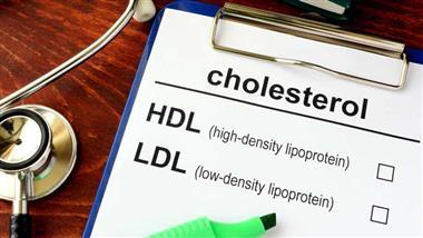
There are two types: High-density is the first. It is also known as HDL, or the “good” kind that keeps cholesterol away from your arteries and removes it from your arteries. The second type is low density lipoprotein, or LDL. LDL is the “bad” kind that can build up in your arteries, form plaque that narrows your arteries and form a clot. This can then make its way to your heart or brain and cause either a heart attack or stroke.
The American Heart Association (AHA)1 now recommends that you balance your levels at about 150 (milligrams per deciliter) (mg/dL). According to old, unfounded science, your total cholesterol — the sum of all the cholesterol in your body — is not a gauge of your heart disease risk. When your levels are measured, elevated levels of triglycerides are also taken into account. According to Børge Nordestgaard, from the University of Copenhagen and Copenhagen University Hospital:
“So far, both cardiologists and [physicians] have focused mostly on reducing LDL cholesterol, but in the future, the focus will also be on reducing triglycerides and remnant cholesterol.”2
Does It Matter What Kills You?
In a commentary posted on Mission.org, a rhetorical question is presented: With all the ways you can die, does it really matter what kills you? On one hand, “If you’re dead, you’re dead, no matter from what,” so it seems silly to “focus on changing something that lowers the risk of death from one cause only to raise that risk from another.”3
It’s an astute observation when you read studies showing conclusively that cholesterol has very little to do with heart disease. Even more importantly, cholesterol is crucial for your health. If it’s too low, then eventually, your hormones, disease risk, cell signaling pathways, and yes, your heart, will suffer. In fact, new research shows that a too-low LDL level could put you at higher risk for a stroke.4
The Dietary Guidelines Advisory Committee (DGAC), which reviews the Dietary Guidelines for Americans every five years, investigated the issue. On page 91 of the 572-page report, the 2015-2020 guidelines noted:5
“Previously, the Dietary Guidelines for Americans recommended that cholesterol intake be limited to no more than 300 mg/day.
The 2015 DGAC will not bring forward this recommendation because available evidence shows no appreciable relationship between consumption of dietary cholesterol and serum (blood) cholesterol, consistent with the AHA/ACC (American Heart Association / American College of Cardiology) report. Cholesterol is not a nutrient of concern for overconsumption.”
Yet, even though the government’s ChooseMyPlate doesn’t mention cholesterol, it’s obvious the cholesterol-is-evil myth is being pushed by physicians and medical agencies such as Harvard Health, which has proposed its own dietary guidelines called “The Healthy Eating Plate.”6
Chastising the USDA for remaining “silent on fat,” Harvard Health says this “could steer consumers toward the type of low-fat, high-carbohydrate diet that makes it harder to control weight and worsens blood cholesterol profiles.”
It’s no wonder people are confused about where cholesterol fits in your diet, when major health agencies send one message to the public and the government sends a completely different one in their dietary guidelines.
Interestingly, the 2015-2020 guidelines were created nearly seven years ago, but information on the importance of cholesterol is nearly two decades old.7 As the Honolulu Heart Program study published in The Lancet in 2001 states:
“Our data accord with previous findings of increased mortality in elderly people with low serum cholesterol, and show that long-term persistence of low cholesterol concentration actually increases risk of death.”
The narrative challenges those who insist that eating foods that contain fat of any kind — saturated fats and trans fats alike — are harmful. Many who make this claim also maintain that “Saturated fat is a bad fat because it raises your LDL level more than anything else in your diet.”8 However, as explained in the Mission.org article:
“While total cholesterol is a poor if not utterly worthless risk marker for heart disease, doctors have focused on it to the exclusion of how it might affect other causes of death. It does you little good to save yourself from heart disease if it means that you increase your risk of death from cancer. All-cause mortality — death from anything — is the most appropriate measure to use when looking at risk factors.”9
The Best Measure for Risk Factors
“Death from anything” may be a clearer term for the one so often used in clinical settings: All-cause mortality. Either way, as quoted by the following study, that’s what the latest research says is the best measure for the factors that increase the risk of what eventually and most likely will take someone’s life.
When it comes to your risk of death from heart disease, there’s much more evidence that inflammation is at the bottom of heart disease rather than high cholesterol, just as it is for a number of other serious diseases. If you want to find what might help you live longer, that’s the premise of a lengthy Japanese study published in the Annals of Nutrition & Metabolism. As the featured study, it notes that regardless of someone’s age, people with higher cholesterol live longer:
“Overall, an inverse trend is found between all-cause mortality and total (or low density lipoprotein [LDL]) cholesterol levels: mortality is highest in the lowest cholesterol group without exception. If limited to elderly people, this trend is universal. As discussed in Section 2, elderly people with the highest cholesterol levels have the highest survival rates irrespective of where they live in the world …
Based on data from Japan, we propose a new direction in the use of cholesterol medications for global health promotion; namely, recognizing that cholesterol is a negative risk factor for all-cause mortality and re-examining our use of cholesterol medications accordingly.”10
After showing that people of all ages with higher cholesterol levels live longer in Japan, in support of these conclusions, similar conclusions were made by a study based in the Netherlands and published in BMJ in 2016.11
The study adjusted for several heart risk factors like smoking, high blood pressure and a history of diabetes melllitus. Participants were placed in groups depending on whether their cholesterol levels were low, medium or high, and those with the highest cholesterol levels were found to have the lowest death rates.
The title of the BMJ review reveals the outcome — there was a “lack of an association or an inverse association between low-density-lipoprotein cholesterol and mortality in the elderly.”12
In short, older people with high LDL cholesterol generally live just as long as — and may even outlive — people with low LDL, which begs the question of how the current cholesterol theory was arrived at in the first place. At the very least, the study authors imply the cholesterol question should at least be re-evaluated. It concluded:
“Our review provides the basis for more research about the cause of atherosclerosis and (cardiovascular disease) and also for a re-evaluation of the guidelines for cardiovascular prevention, in particular because the benefits from statin treatment have been exaggerated.”13
One of the first studies published with information suggesting that high cholesterol is not as heart-damaging as once thought was the Honolulu Heart Program,14 offering further evidence that higher cholesterol levels may be heart protective. The authors concluded by questioning whether there is “scientific justification for attempts to lower cholesterol to concentrations below 4-65 mmol/L in elderly people,” adding that “prudence dictates a more conservative approach in this age group.”15
Can High Cholesterol Really Be Heart Protective?
Perhaps the question that would get to the point quicker is to ask why the Japanese study infers that people with low versus high cholesterol die sooner? The Mission offers referenced studies that show a few factors that could be weighing in:
• Cholesterol may protect against infections and atherosclerosis, as “the many observations that conflict with the LDL receptor hypothesis, may be explained by the idea that high serum cholesterol and/or high LDL is protective against infection and atherosclerosis.”16
• Cholesterol may protect against cancer, although in previous cases where low cholesterol was linked to cancer, exclusions were made to tip the scales, such as excluding possible prior drug treatment, namely clofibrate, a popular cholesterol-lowering drug before statins, by subjects — leaving the question open as to whether it was the low cholesterol that caused the cancer, or the drug treatments that contributed to it.17
• Low cholesterol (180 mg/dL and lower) and violence in psychiatric patients have been linked. When patients in a long-term psychiatric hospital with a history of seclusion or restraints were compared with other patients, there was a “highly significant and strong association between lower cholesterol levels and violent behavior.” The authors did caution, however, that cholesterol levels should not be used to predict violence.18
• There’s also an association between low cholesterol and suicide dating back more than a decade, as researchers found that people in the lowest quartile of cholesterol concentration had more than six times the suicide rates than those in the highest quartile.19
It should be noted that at least one study in Japan20 determined that high cholesterol and suicide were connected.
But, in fact, numerous recent studies have corroborated the earlier ones connecting low cholesterol to suicide,21 with various findings: For example, one found that low triglycerides, reduced BMI and waist circumference, specifically, but not total cholesterol, were connected to a higher risk of suicide;22 while a 2019 study23 found that “low cholesterol is associated with aggression in suicide attempters.”
So What About Statins?
Significantly, the authors of an Annals of Nutrition & Metabolism study didn’t hold back when drawing conclusions regarding why the cholesterol conundrum has gone on so long when the evidence is so clear: “For the side defending this so-called cholesterol theory, the amount of money at stake is too much to lose the fight.”24
The Annals of Nutrition & Metabolism study’s introduction mentions a medical practitioner who advocated statins to his patients to drive down cholesterol until he read the Scandinavian Simvastatin Survival Study,25 in which 4,444 patients with different types of heart disease were given simvastatin — which is touted to be a safe, long-term treatment to improve survival in cardiovascular heart disease patients.
As it turns out, the claim that high cholesterol causes heart disease and death is incorrect; it is, in fact, the opposite. Three reviews26,27,28 supporting the cholesterol hypothesis were found to contain altered data to support their conclusions, according to Expert Review of Clinical Pharmacology, in which it is noted that:
“Our search for falsifications of the cholesterol hypothesis confirms that … the conclusions of the authors of the three reviews are based on misleading statistics, exclusion of unsuccessful trials and by ignoring numerous contradictory observations.”29
Drugs.com30 contributors state that the 35 million people on statins often experience myriad side effects. Liver damage, for instance, is said to be “rare,” implying that ongoing liver tests while taking statins likely aren’t necessary. Some doctors, however, say you’ll need a baseline liver function test beforehand. The most common side effects of statins are:
- Headaches, muscle pain, lower back or side pain
- Nasal congestion or stuffiness, or a runny nose
- Difficulty sleeping
- Constipation
- Hoarseness
And, just in case you needed another source to implicate statins’ role in psychiatric problems, an April 2018 study found that lowering cholesterol levels in men could bring about changes in nerve cell membranes and behavior in men:
“Men seem to be more sensitive to low cholesterol levels as the association between low cholesterol levels and aggression is found mostly in men,” the authors said. “… Lowering cholesterol levels with statins brings about several changes in the serotonergic system, nerve cell membrane microviscosity and behaviour, and needs to be done with precaution in susceptible individuals.
Cholesterol levels could serve as a biological risk marker for violence and suicidal tendencies in psychiatric patients with depression and schizophrenia.”
More Studies and Reviews on Statin Use
Rather than pointing patients in the direction of finding dietary solutions, including eating both the whites and the yolks when having eggs, and ditching processed vegetable oils in favor of healthy cooking oils like coconut oil, olive oil and avocado oil, Harvard Health Medical School updated an article on how to “manage” muscle pain from taking statins, perpetuating the cholesterol myth. They stated:
“If you’re not taking a statin now, you may well be soon. These medications are commonly prescribed to lower ‘bad’ LDL cholesterol and have been shown to reduce the risk of heart attack, stroke, and death.
They are routinely recommended for people who have cardiovascular disease and for many people ages 40 to 75 who don’t have cardiovascular disease but have at least one risk factor (high blood pressure, high cholesterol, diabetes, or smoking) and a 7.5% or greater risk of a stroke or heart attack in the next decade.
Moreover, recent research indicates that they may benefit high risk individuals over age 75 as well … Taking a statin may give you some assurance that you’re doing all you can to avoid heart attack and stroke …”31
Exercising, losing weight, adopting healthy eating habits and including vitamin D and coenzyme Q10 (CoQ10) supplements in your diet are all good strategies for maintaining your cholesterol levels. Unfortunately, the common suggestion for alleviating muscle pain due to statin use is more of the same — just try taking a lower dose or switching to another statin prescription.32
As if that weren’t enough, experts say statins may impair your memory and cause amnesia, a possibility real enough to call for a warning on the labels of prescriptions.33 Statin use may precipitate a higher risk of developing cataracts34 and it “significantly” increases the likelihood of raising the fasting glucose levels of non-diabetics, as well as inducing high blood sugar.35
In another review of the adverse effects of statins, it was noted that “an array of additional risk factors for statin AEs [adverse events] are those that amplify (or reflect) mitochondrial or metabolic vulnerability, such as metabolic syndrome factors, thyroid disease, and genetic mutations linked to mitochondrial dysfunction.”36
Given the false information saying cholesterol is at fault for causing heart-related disease, the continued prescriptions being handed out to patients for statins, and the side effects they cause, it’s clear why, in their introduction, the authors of the Annals of Nutrition & Metabolism study stressed:37
“This, we believe, marks the starting point of a paradigm shift in not only how we understand the role cholesterol plays in health, but also how we provide cholesterol treatment …
Our purpose in writing this supplementary issue is to help everyone understand the issue of cholesterol better than before, and we hope that we lay out the case for why a paradigm shift in cholesterol treatment is needed, and sooner rather than later.”
- 1 AHA November 28, 2019
- 2 Medical News Today August 12, 2019
- 3, 9 Mission.org September 26, 2016
- 4 Neurology July 30, 2019
- 5 CNN. Cholesterol in Food Not a Concern, New Report Says. February 19, 2015
- 6 Harvard Health. Healthy Eating Plate vs. USDA’s MyPlate
- 7, 19 Epidemiology March 2001, Volume 12, Issue 2, pgs. 168-172
- 8 Medline Plus February 27, 2019
- 10, 24, 37 Annals of Nutrition & Metabolism April 30, 2015
- 11, 12, 13 BMJ June 12, 2016 Volume 6, Issue 6
- 14, 15 The Lancet August 4, 2001;358(9279):351-5
- 16 QJM: An International Journal of Medicine December 1, 2003
- 17 Q J Med 2012;105:383–388
- 18 Psychiatric Services February 1998;49(2):221-4
- 20 Acta Psychiatrica Scandanvica May 26, 2017 Volume 136 Issue 3
- 21 Annals of General Psychiatry April 17, 2017 Article number: 20 (2017)
- 22 Journal of Affective Disorders Volume 172, 1 February 2015, Pages 403-409
- 23 Psychiatry Research Volume 273 March 2019, Pages 430-434
- 25 The Lancet November 19, 1994; Volume 344, pgs. 1383-1389
- 26 The Lancet September 8, 2016; 388: 2532–61
- 27 JAMA September 27, 2016
- 28 European Heart Journal April 24, 2017; Volume 38, Issue 32, pgs. 2459–2472
- 29 Expert Review of Clinical Pharmacology October 11, 2018; Volume 11, Issue 10, pgs. 959-970
- 30 Drugs.com September 10, 2018
- 31 Harvard Health August 9, 2019
- 32 Medscape October 16, 2015
- 33 PLoS One September 10, 2013
- 34 JAMA November 2013
- 35 Cardiovascular Diabetology December 5, 2018
- 36 Am J Cardiovasc Drugs. April 6, 2008; 8(6):373-418
Two Remarkable Stress Relief Supplements; Take Them Together
Reproduced from original article:
https://articles.mercola.com/sites/articles/archive/2022/05/17/magnesium-and-vitamin-b6.aspx
The original Mercola article may not remain on the original site, but I will endeavor to keep it on this site as long as I deem it to be appropriate, and will not be bullied into removing it.
Analysis by Dr. Joseph Mercola Fact Checked May 17, 2022
STORY AT-A-GLANCE
- Vitamin B6 escorts magnesium to the cells that need it most, thus ensuring that the magnesium you’re getting, whether from foods or supplements, is being used as efficiently as possible. Vitamin B6 thus helps augment the many benefits of magnesium
- Research shows a combination of magnesium and vitamin B6 can lower perceived stress to a greater degree than magnesium alone in those with severe or extremely severe stress. Those taking magnesium and B6 in combination also experienced fewer side effects
- Magnesium and vitamin B6 are two nutrients commonly recommended for women with premenstrual syndrome. Magnesium deficiency may cause and intensify PMS symptoms, and magnesium appears to work because it has a calming effect on the neuromuscular system
- Researchers have found vitamin B6 and magnesium are most effective for lowering rates of depression, water retention and anxiety in women with PMS
- The recommended dietary allowance for magnesium ranges from 310 mg to 420 mg for adults over the age of 19, depending on age, gender and pregnancy status, and the adult RDA for vitamin B6 is between 1.2 mg and 2 mg per day, depending on age and gender; both nutrients are abundant in whole foods
This article was previously published June 10, 2019, and has been updated with new information.
You may be familiar with the connection between magnesium, calcium and vitamins K2 and D and how they work in tandem. But are you aware of the crucial link between magnesium and vitamin B6 (pyridoxine)? Individually, magnesium and vitamin B6 are both essential for heart and brain health. Both also play roles in the regulation of your blood sugar level.1,2
When you get insufficient amounts of magnesium from your diet, your body will leach magnesium from your bones, muscles and internal organs, which can lead to osteoporosis, kidney problems and liver damage.
Vitamin B6 can help ameliorate this by escorting magnesium to the cells that need it most, thus ensuring that the magnesium you’re getting, whether from foods or supplements, is being used as efficiently as possible. In so doing, vitamin B6 also helps augment the many benefits of magnesium.
Magnesium-B6 Combo Is Superior for Severe Stress
The importance of magnesium in combination with vitamin B6 was presented in a 2018 study3 in the journal PLOS ONE. Taken together, these two nutrients have been shown to have a complementary effect on stress reduction in animal studies.
In this randomized trial, they evaluated whether the combination of magnesium and B6 would improve perceived stress levels in 264 human subjects who also had low magnesium to start. Healthy adults with a depression anxiety stress scale score above 18 and a serum level of magnesium between 0.45 nanomoles per liter (mmol/L) and 0.85 mmol/L were randomized to receive either:
- 300 milligrams (mg) of magnesium in combination with 30 mg of vitamin B6
- 300 mg of magnesium only
The primary endpoint was a reduction in stress score from baseline to Week 8. While both treatment groups experienced similar reductions in their stress scores — the magnesium-B6 combo group reporting a 44.9% reduction in perceived stress and the magnesium-only group a 42.4% reduction — a more significant impact was shown in those with severe and/or extremely severe stress.
According to the authors, adults with a stress score at or above 25 had a 24% greater improvement with magnesium-vitamin B6 versus magnesium only at Week 8. Those taking magnesium and B6 in combination also experienced fewer side effects: 12.1% of those taking magnesium-vitamin B6 versus 17.4% of those taking magnesium only experienced some form of adverse event. As noted by the authors:4
“These findings suggest oral Mg supplementation alleviated stress in healthy adults with low magnesemia and the addition of vitamin B6 to Mg was not superior to Mg supplementation alone. With regard to subjects with severe/extremely severe stress, this study provides clinical support for greater benefit of Mg combined with vitamin B6.”
Magnesium and B6 May Ease Premenstrual Syndrome
Magnesium and vitamin B6 are two nutrients commonly recommended for women struggling with premenstrual syndrome. According to a research paper5 published in the Journal of Caring Sciences, magnesium deficiency has been proposed “as one of the factors causing and intensifying premenstrual syndrome symptoms,” and magnesium appears to work because it has a calming effect on the neuromuscular system.
“Vitamin B6 is another proposed treatment for this syndrome,” the paper notes.6 “On the one hand vitamin B6 increases serotonin and dopamine levels and improves premenstrual syndrome symptoms, and on the other, it has an essential role in the synthesis of prostaglandin and fatty acids, which are reduced in etiologies causing premenstrual syndrome.
Moreover, researchers believe that vitamin B6 deficiency decreases dopamine in the kidneys and therefore increase sodium excretion, which in turn causes water accumulation in the body and induces symptoms such as swelling in extremities, edema, and abdominal and chest discomfort. The administration of vitamin B6 can thus decrease these symptoms and improve premenstrual acne.”
To evaluate the effects of these two nutrients on premenstrual syndrome, 126 women diagnosed with premenstrual syndrome, based on American Psychiatric Association criteria, were divided into three groups, which received either 250 mg of magnesium oxide, 250 mg of vitamin B6, or a placebo, taken from the first day of the menstrual cycle until the beginning of the next cycle.
Magnesium and B6 Have Similar Rates of Effectiveness
Overall, magnesium and B6 had similar rates of effectiveness for premenstrual syndrome in this Journal of Caring Sciences study. Mean scores of premenstrual syndrome before and after intervention in the three groups were as follows:
| Magnesium | Vitamin B6 | Placebo | |
|---|---|---|---|
| Before intervention | 36.89% | 36.51% | 35.8% |
| After intervention | 22.22% | 22.84% | 28.41% |
As you can see, while the placebo also helped reduce premenstrual syndrome symptoms, magnesium and B6 did so more effectively, and at similar rates. When looking at specific symptoms, B6 and magnesium were found to be the most effective for lowering rates of depression, water retention and anxiety. In conclusion, the authors noted:7
“Considering the importance of premenstrual syndrome and the numerous effects it has on society and the lives of women, health groups should prioritize the diagnosis and treatment of this syndrome. Since there is no definitive etiology and treatment for this syndrome, many researchers have tried to find the best and most effective drug with the least side effects to prevent the occurrence of the syndrome …
The current study was also undertaken with the goal of finding an effective compound with no side effects to reduce the symptoms of this syndrome and its direct and indirect economic and social effects. All compounds used in the current study had no side effects, were effective, non-chemical, and acceptable by most groups of women in the society.
Hence, health groups, especially midwives, can compare the effectiveness the compound on their specific patients and select the most appropriate treatment for each individual. Moreover, in cases where the patient is prohibited from using chemical drugs to treat premenstrual syndrome, such as oral contraceptive pills and gonadotropin releasing hormone (GnRH) agonists, the use of these compounds seems effective …”
Unfortunately, a combination of magnesium and B6 was not evaluated in this study. It would have been interesting to see what their combined effect would have been. Considering the importance of both of these nutrients for health, I see no risk in combining them, though, should you struggle with premenstrual syndrome.
The Importance of Magnesium for Optimal Health
Magnesium8 is the fourth most abundant mineral in your body and the second most common intracellular cation9 (positively charged ion) after potassium. It’s required for the healthy function of most cells in your body, but is especially important for your heart, kidneys and muscles.
Low magnesium will impede your cellular metabolic function and deteriorate mitochondrial function, which can have far-reaching health consequences, seeing how loss of mitochondrial function is a foundational factor in most chronic diseases, including heart disease and cancer.
According to one scientific review,10 which included studies dating as far back as 1937, low magnesium actually appears to be the greatest predictor of heart disease, and other recent research shows even subclinical magnesium deficiency can compromise your cardiovascular health.11
Being one of the most abundant minerals in the human body, it’s not surprising that it has several hundred biological functions. To list just a few, magnesium helps:
- Relax your muscles as well as your blood vessels — Being deficient in it can cause muscle cramps and weakness
- Promote mental and physical relaxation — It’s a stress antidote that works by boosting GABA, an inhibitory neurotransmitter that relaxes your nervous system. Magnesium also helps boost your melatonin production
- Detoxification and reduces damage from electromagnetic fields
- Regulate blood sugar and improve insulin sensitivity, potentially protecting against Type 2 diabetes
Magnesium Is Required for Activation of Vitamin D
Magnesium is also a component necessary for the activation of vitamin D,12,13,14 and deficiency may hamper your ability to convert vitamin D from sun exposure and/or oral supplementation.
According to Mohammed Razzaque, professor of pathology at Lake Erie College of Osteopathic Medicine in Pennsylvania, coauthor of a study published in The Journal of the American Osteopathic Association (JAOA) in March 2018,15 “By consuming an optimal amount of magnesium, one may be able to lower the risks of vitamin D deficiency, and reduce the dependency on vitamin D supplements.”
Interestingly, the first paper I ever had published, back in 1985, was also in the JAOA. My paper was about the use of calcium to control hypertension, but if I had written the paper this century, it most certainly would have been about the use of magnesium for that purpose.16
A second study,17 published in The American Journal of Clinical Nutrition in December 2018 also concluded that your magnesium status plays an important role in your vitamin D status. Overall, people with high magnesium intake were less likely to have low vitamin D. They also had a lower mortality risk from cardiovascular disease and bowel cancer.
As explained by Dr. Qi Dai, professor of medicine at Vanderbilt University Medical Center and the lead author of this study, “Magnesium deficiency shuts down the vitamin D synthesis and metabolism pathway.” What’s more, magnesium was found to have a regulating effect, raising and lowering vitamin D based on baseline levels.
In people who had a baseline vitamin D level of 30 ng/mL (75 nmol/L) or below, magnesium supplementation raised their vitamin D level. However, in those who started out with higher vitamin D levels (50 ng/mL or 125 nmol/L), magnesium supplementation lowered their vitamin D.
Magnesium for Brain Health and Neurological Functioning
Magnesium is also crucial for optimal brain function, and is a common culprit in neurological ailments, including:
• Migraines18,19,20 — Researchers have noted that empiric treatment with a magnesium supplement is justified for all migraine sufferers.21
• Depression — Magnesium plays an important role in depression as it acts as a catalyst for mood-regulating neurotransmitters like serotonin. Research22 published in 2015 found a significant association between very low magnesium intake and depression, especially in younger adults.
Research23 published in PLOS ONE demonstrated magnesium supplementation improved mild-to-moderate depression in adults, with beneficial effects occurring within two weeks of treatment. In fact, the effects of magnesium were comparable to prescription SSRIs in terms of effectiveness, but without any of the side effects associated with these drugs.
Participants in the treatment group received a daily dose of 248 milligrams (mg) of elemental magnesium for six weeks, while controls received no treatment. According to the authors, “It works quickly and is well tolerated without the need for close monitoring for toxicity.”
• Memory problems and loss of brain plasticity — Memory impairment occurs when the connections (synapses) between brain cells diminish. While many factors can come into play, magnesium is an important one.
According to Dr. David Perlmutter, a neurologist and fellow of the American College of Nutrition, “magnesium is a critical player in the activation of nerve channels that are involved in synaptic plasticity.”24 Magnesium threonate, which most effectively permeates the blood-brain-barrier, is likely your best choice here.
The specific brain benefits of magnesium threonate were demonstrated in a 2010 study25 published in the journal Neuron, which found this form of magnesium enhanced “learning abilities, working memory, and short- and long-term memory in rats.”
Health Benefits of Vitamin B6
Like magnesium, vitamin B6 (as well as several other B vitamins) also plays an important role in heart and brain health. It is used in the creation of neurotransmitters, and is required for proper brain development during pregnancy and infancy.26
Vitamins B6, B9 (folate, or folic acid in its synthetic form) and B12 may be particularly important for supporting cognitive function as you age, and have been shown to play a major role in the development of dementia, including Alzheimer’s disease, which is the most serious and lethal form.
A primary mechanism of action here is the suppression of homocysteine,27 which tends to be elevated when you have brain degeneration. High homocysteine has also been implicated in the development of atherosclerosis.28,29
The good news is your body can eliminate homocysteine naturally, provided you’re getting enough B9 (folate), B6 and B12. One study confirming this was published in 2010.30 Participants received either a placebo or 800 micrograms (mcg) of folic acid (the synthetic form of B9), 500 mcg of B12 and 20 mg of B6.
The study was based on the presumption that by controlling homocysteine levels you might be able to reduce brain atrophy, thereby slowing the onset of Alzheimer’s. Indeed, after two years those who received the vitamin-B regimen had significantly less brain shrinkage compared to the placebo group.
A 2013 study31 took this research a step further, showing that not only do B vitamins slow brain shrinkage, but they specifically slow shrinkage in brain regions known to be most severely impacted by Alzheimer’s disease.
As in the previous study, participants taking high doses of folic acid and vitamins B6 and B12 lowered their blood levels of homocysteine, decreasing brain shrinkage by as much as 90%. High doses of vitamins B6, B8 (inositol) and B12 have also been shown to significantly reduce symptoms of schizophrenia, more so than standard drug treatments alone.32 Vitamin B6 is also important for healthy:
- Metabolism, by helping break down amino acids in the muscles to be used as energy and by converting lactic acid to glucose in your liver
- Immune system, as it helps create white blood cells that fight infections
- Hair and skin health, by reducing hair loss and alleviating dermatitis
How to Improve Your Magnesium and Vitamin B6 Status
The recommended dietary allowance (RDA) for magnesium ranges from 310 mg to 420 mg for adults over the age of 19, depending on age, gender and pregnancy status,33 and the adult RDA for vitamin B6 is between 1.2 mg and 2 mg per day, depending on age and gender.34
Both magnesium and vitamin B6 are abundant in whole foods. Good sources of magnesium include leafy greens, berries, avocado, seeds, nuts and raw cacao nibs. Eating a primarily processed food diet is the primary culprit in magnesium deficiency, and if you fall into this group, you’d be wise to take a magnesium supplement.
Vitamin B6 is abundant in animal foods such as beef and wild-caught salmon, as well as dark leafy greens, papaya, oranges, cantaloupe, sweet potatoes, avocados, bananas, spinach, pistachios and sunflower seeds.35 Nutritional yeast is another excellent source.
- 1 Diabetes Self Management, Magnesium, The Forgotten Healer
- 2 Michigan Medicine, Vitamin B6
- 3, 4 PLOS ONE 2018; 13(12): e0208454
- 5, 6, 7 Journal of Caring Sciences 2012 Dec; 1(4): 183–189
- 8 Medical News Today December 20, 2017
- 9, 11 Open Heart 2018:e000668 (PDF)
- 10 New Hope Network January 31, 2013
- 12 Live Science February 26, 2018
- 13 Medicalxpress.com February 27, 2018
- 14 News-Medical.net February 26, 2018
- 15 Eurekalert February 27, 2018
- 16 Journal of the American Osteopathic Association 1985; 85:104-107
- 17 American Journal of Clinical Nutrition December 1, 2018; 108(6): 1249-1258
- 18 Migraine.com, Magnesium
- 19, 21 J Neural Transm (Vienna). 2012 May;119(5):575-9
- 20 Cephalalgia 1996 Jun;16(4):257-63
- 22 Journal of the American Board of Family Medicine 2015 Mar-Apr;28(2):249-56
- 23 PLOS ONE June 27, 2017
- 24 Drperlmutter.com, Magnesium and Your Brain
- 25 Neuron January 27, 2010; 65(2): 165-177
- 26 Live Science August 20, 2015
- 27 Nutritionfacts.org January 18, 2019
- 28 American Journal of Pathology 1969 Jul;56(1):111-28
- 29, 35 Harvard TH Chan, The Nutrition Source: B Vitamins
- 30 PLoS ONE 5(9): e12244
- 31 PNAS 2013 Jun 4;110(23):9523-8
- 32 Psychological Medicine February 16, 2017; 47(9): 1515-1527
- 33 NIH.gov Magnesium
- 34 NIH.gov Vitamin B6
Is Ivermectin a Cancer Solution?
Reproduced from original article:
https://articles.mercola.com/sites/articles/archive/2022/05/12/ivermectin-antitumor-effects.aspx
The original Mercola article may not remain on the original site, but I will endeavor to keep it on this site as long as I deem it to be appropriate, and will not be bullied into removing it.
Analysis by Dr. Joseph Mercola Fact Checked May 12, 2022
STORY AT-A-GLANCE
- Ivermectin has notable antitumor effects, which include inhibiting proliferation, metastasis and angiogenic activity in cancer cells
- Ivermectin may target cancer in multiple ways, including inducing apoptosis and autophagy while also inhibiting tumor stem cells and reversing multidrug resistance
- Along with direct cytotoxic effects, it’s believed that ivermectin regulates the tumor microenvironment, mediating immunogenic cell death
- The development of an injectable form of ivermectin, or liposomal ivermectin, could help overcome some of its limitations regarding solubility, and open its use to a broader range of cancers
- Considering that the “war against cancer” has been ongoing for decades, with little to show in terms of lives saved, repurposing existing drugs with favorable safety profiles and notable anticancer effects — like ivermectin — makes sense
Ivermectin is a widely used antiparasitic drug that’s listed on the World Health Organization’s essential medicines list1 and approved by the U.S. Food and Drug Administration. In low- and middle-income countries, ivermectin is commonly used to treat parasitic diseases including onchocerciasis (river blindness), strongyloidiasis and other diseases caused by soil-transmitted helminthiasis, or parasitic worms.2
The drug is also used to treat scabies and lice. It’s estimated that the total number of ivermectin doses distributed is equal to one-third of the world’s population and, as such, “ivermectin at the usual doses (0.2–0.4 mg/kg) is considered extremely safe for use in humans.”3
Ivermectin also has demonstrated antiviral and anti-inflammatory properties and made headlines for its potential role in treating COVID-194 — although much of the positive press has been censored and falsely labeled misinformation.5 Now researchers are highlighting another potential use for ivermectin, which is equally as exciting as its potential role in COVID-19 — as an anticancer agent.
Ivermectin’s Powerful Antitumor Effects
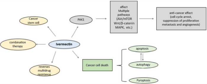
Ivermectin has notable antitumor effects, which include inhibiting proliferation, metastasis and angiogenic activity in cancer cells.6 It appears to inhibit tumor cells by regulating multiple signaling pathways, which researchers explained in the Pharmacological Research journal, “suggests that ivermectin may be an anticancer drug with great potential.”7
Their graphic, below, shows the multiple ways that ivermectin may target cancer, including inducing apoptosis and autophagy while also inhibiting tumor stem cells and reversing multidrug resistance. They stated that ivermectin “exerts the optimal effect when used in combination with other chemotherapy drugs.”8
Many may not be aware that scientists Satoshi ōmura and William C. Campbell won the Nobel Prize in Physiology or Medicine in 2015 for their discovery of ivermectin.9 The medicine is used to treat not only parasitic diseases like malaria but also shows promise for treating asthma and neurological diseases, in addition to cancer.
Along with direct cytotoxic effects, it’s believed that ivermectin regulates the tumor microenvironment, mediating immunogenic cell death — another reason for its promise as an anticancer agent.10 Research suggests the drug may be useful for the following cancers:11
| Breast cancer — The proliferation of multiple breast cancer cell lines was significantly reduced following treatment with ivermectin. |
| Digestive system cancer — Ivermectin significantly inhibited the proliferation of gastric cancer cells in vivo and in vitro. The drug also inhibited colorectal cancer cell lines and inhibited the development of hepatocellular carcinoma (liver cancer). |
| Urinary system cancer — Ivermectin significantly inhibited the proliferation of five renal (kidney) cell carcinoma lines without affecting normal kidney cells. It also had an inhibitory effect on prostate cancer cells. |
| Hematological cancer — In one study, ivermectin killed leukemia cells at low concentrations while leaving normal hematopoietic cells unharmed. |
| Reproductive system cancer — Ivermectin inhibited the proliferation of ovarian cancer cell lines and enhanced the efficacy of the conventional chemotherapy drug cisplatin, improving the treatment of epithelial ovarian cancer. |
| Brain glioma — Ivermectin inhibited the proliferation of human glioblastoma cells in a dose-dependent manner. |
| Respiratory system cancer — Ivermectin inhibited the development of nasopharyngeal carcinoma in mice, using doses that were not toxic to immune cells known as thymocytes. Ivermectin also significantly inhibited the proliferation of lung cancer cells and may reduce the metastasis of lung cancer cells. |
| Melanoma — When melanoma cells were treated with ivermectin, their activity was effectively inhibited. |
Ivermectin Shows Promise Against Colorectal Cancer
A study published in Frontiers in Pharmacology specifically highlighted ivermectin’s potential to fight colorectal cancer, which is the third most common cancer worldwide.12 The drug was found to inhibit colorectal cancer cell growth in a dose-dependent manner as well as promote cell apoptosis.
Further, even at low doses of 2.5 and 5 µM, ivermectin inducted cell arrest in colorectal cancer, leading researchers to state, “[I]vermectin might be a new potential anticancer drug therapy for human colorectal cancer and other cancers.”13 Considering that the “war against cancer” has been ongoing for decades, with little to show in terms of lives saved, repurposing existing drugs with favorable safety profiles and notable anticancer effects — like ivermectin — makes sense.
The Pharmacological Research scientists similarly noted, “Drug repositioning is a shortcut to accelerate the development of anticancer drugs.”14 Not only has ivermectin been shown to permeate tumor tissues effectively, but it has a long history of successful use in humans. They explained that even when doses were increased, no serious adverse effects were found:15
“[T]he broad-spectrum antiparasitic drug IVM (ivermectin), which is widely used in the field of parasitic control, has many advantages that suggest that it is worth developing as a potential new anticancer drug. IVM selectively inhibits the proliferation of tumors at a dose that is not toxic to normal cells and can reverse the MDR [multidrug resistance] of tumors.
Importantly, IVM is an established drug used for the treatment of parasitic diseases such as river blindness and elephantiasis. It has been widely used in humans for many years, and its various pharmacological properties, including long- and short-term toxicological effects and drug metabolism characteristics are very clear. In healthy volunteers, the dose was increased to 2 mg/Kg, and no serious adverse reactions were found …”
Is Liposomal Delivery a Game Changer?
The development of an injectable form of ivermectin, or liposomal ivermectin, could help overcome some of its limitations regarding solubility and open its use to a broader range of cancers. The cancer immunotherapy treatment pembrolizumab, for instance, is approved to treat PD-L1-positive, triple-negative breast cancer, which accounts for only about 20% of cases.
As an immune checkpoint inhibitor, it works best in so-called “hot” tumors, which are already infiltrated by T cells. If ivermectin could be injected into the tumor, inducing T-cell infiltration into the area and inducing immunogenic cancer cell death, it’s possible that it could turn a “cold” tumor into a “hot” one, thereby making it more effectively treated.16
Biotech company Mountain Valley MD has developed a liposomal delivery system for ivermectin that they believe could dramatically widen its treatment potential. In an interview with Medical Update Online, Dennis Hancock, Mountain Valley MD president and CEO, explained:17
“So the business value proposition really simply is, we take the best-selling and best-acting drugs and expand their ability to be used on … more types of cancer on a broader spectrum. So you still need the cancer drug and what our Ivectosol does is it enables it to be used in a broader universe …
What’s really exciting about the work that Mountain Valley MD is doing is we’re enabling drugs that have already been proven in their efficacy and safety to do better and do more faster — so we’re not asking people to ‘wait five years and see’…”
Most of the research involving ivermectin for cancer to date involves oral or in-vitro administration. Mountain Valley MD is conducting preclinical trials using liposomal ivermectin for metastatic melanoma, non-small cell lung cancer, triple-negative breast cancer and possibly bladder cancers. They also have plans to produce liposomal ivermectin for use in human trials.18 In a news release, Mike Farber, director of life sciences at Mountain Valley MD, stated:19
“The extensive research supporting the drug ivermectin as effective in the inhibition of proliferation, metastasis, and angiogenic activity in a variety of cancers, and as an initiator of immunogenic cell death, is overwhelming. Imagine what is possible when you have the world’s only human injectable form of ivermectin that can be directly injected into a tumor or provided through more bio-available forms such as intravenously.
We believe this will be groundbreaking research with near-immediate application to be able to proceed directly to human trials based on the safety and efficacy of ivermectin.”
What About Ivermectin for SARS-CoV-2?
In the U.S., ivermectin has been vilified as a treatment for SARS-CoV-2, despite its impressive inhibitory effects on the virus.20 Even the FDA has a dedicated webpage warning “why you should not use ivermectin to prevent COVID-19.”21
It’s interesting to note, however, that Africa has a lower number of cases, severity of disease, hospitalizations and deaths than other areas of the world,22 which may be due to using prophylactic medications for endemic infections — ivermectin and others, such as sweet wormwood — that have successfully treated COVID-19.
For instance, a study from Japan demonstrated that just 12 days after doctors were allowed to legally prescribe ivermectin to their COVID-19 patients, the cases dropped dramatically.23 The chairman of the Tokyo Medical Association24 noticed the low number of infections and deaths in Africa, where many use ivermectin prophylactically and as the core strategy to treat river blindness.25 More than 99% of people infected with river blindness live in 31 African countries.
Aside from these observations, a study published in the March 2022 issue of the International Journal of Infectious Diseases found that treatment with ivermectin reduced mortality in COVID-19 patients — and to a greater degree than remdesivir.26
Another recent investigation by Cornell University, posted on the University’s preprint server January 20, 2022, found ivermectin outperformed 10 other drugs against COVID-19, making it the most effective against the Omicron variant.27 It even outperformed nirmatrelvir (Paxlovid), which was granted emergency use authorization against COVID-19 in December 2021.
Remdesivir costs between $2,340 and $3,120,28 and nirmatrelvir costs $529 per treatment,29 while ivermectin’s average treatment cost is $58.30 Do you think this has anything to do with ivermectin’s vilification?
Dr. Pierre Kory, who is part of the group that formed the Front Line COVID-19 Critical Care Working Group (FLCCC) to advance early treatments for COVID-19, pleaded with the U.S. government early on in the pandemic to review the expansive data on ivermectin to prevent COVID-19, and to keep those with early symptoms from progressing and help critically ill patients recover — to no avail.31,32
However, if you’d like to learn more about its potential uses for SARS-CoV-2, FLCCC’s I-MASK+ protocol can be downloaded in full,33 giving you step-by-step instructions on how to prevent and treat the early symptoms of COVID-19.
FLCCC also has protocols for at-home prevention and early treatment, called I-MASS, which involves ivermectin, vitamin D3, a multivitamin and a digital thermometer to watch your body temperature in the prevention phase and ivermectin, melatonin, aspirin and antiseptic mouthwash for early at-home treatment.
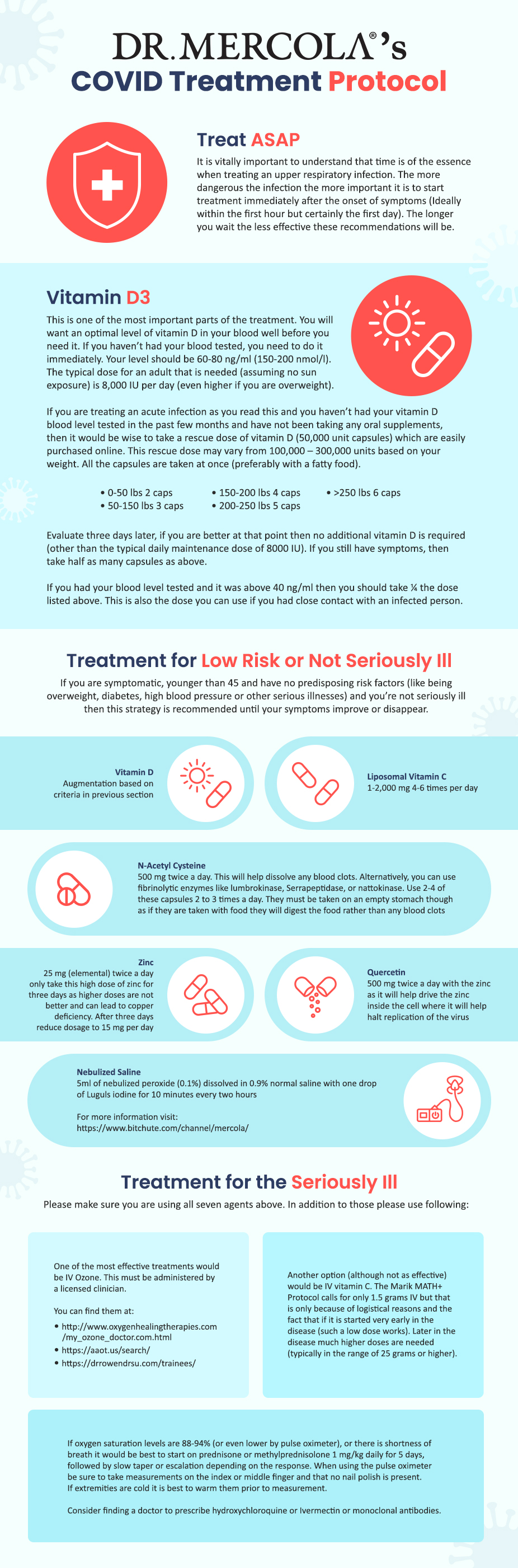
Household or close contacts of COVID-19 patients may take ivermectin (18 milligrams, then repeat the dose in 48 hours) for post-exposure prevention.34 Whether ivermectin’s potential as an anticancer agent will be stifled the same way it was for COVID-19 remains to be seen, but it appears to be a compound that’s worth watching as a potential powerful agent in the fight against cancer.
- 1, 2 WHO March 31, 2021
- 3 American Journal of Therapeutics: July/August 2021 – Volume 28 – Issue 4 – p e434-e460
- 4 In Vivo. Sep-Oct 2020;34(5):3023-3026. doi: 10.21873/invivo.12134
- 5 Trial Site News November 27, 2021, 3:52
- 6, 7, 8 Pharmacol Res. 2021 Jan; 163: 105207
- 9, 20 Pharmacol Res. 2021 Jan; 163: 105207., Introduction
- 10 Pharmacol Res. 2021 Jan; 163: 105207., 2.1 Breast cancer
- 11 Pharmacol Res. 2021 Jan; 163: 105207., 2. The role of IVM in different cancers
- 12, 13 Front Pharmacol. 2021; 12: 717529
- 14, 15 Pharmacol Res. 2021 Jan; 163: 105207., Summary
- 16, 17, 18 Medical Update Online June 19, 2021
- 19 Mountain Valley MD May 3, 2021
- 21 U.S. Food and Drug Administration, Why You Should Not Use Ivermetin to Prevent COVID-19
- 22 AP News, November 19, 2021
- 23 YouTube, November 23, 2021 Min 1:25
- 24 Tokyo Web, August 13, 2021
- 25 World Health Organization, Onchocerciasis
- 26 International Journal of Infectious Diseases March 2022; 116(Supplement): S40
- 27 Cornell University, January 20, 2022
- 28 AJMC June 29, 2020
- 29 Precision Vaccinations, November 19, 2021
- 30 JAMA 2022;327(6):584-587
- 31 FLCCC Alliance, Ivermectin & COVID-19
- 32 Mountain Home May 1, 2021
- 33 FLCCC Alliance, I-Mask+
- 34 FLCCC Alliance, I-MASS
The Restoration of Vitamin C Synthesis in Humans
Reproduced from original OMNS article (OrthoMolecular News Service):
https://orthomolecular.activehosted.com/index.php
Subscribe to the free Orthomolecular Newsletter: http://orthomolecular.org/subscribe.html
Go to the OMNS Archive: http://orthomolecular.org/resources/omns/index.shtml
Orthomolecular Medicine News Service, May 11, 2022
by Thomas E. Levy, MD, JD and Ron Hunninghake, MD
OMNS (May 11, 2022) The full importance of vitamin C in reaching and maintaining optimal health remains largely unappreciated by most traditional health care practitioners. Certainly, it is widely appreciated that vitamin C is required to sustain life, and that its severe deficiency in the body reliably results in the development of scurvy, with death following shortly thereafter in the absence of new vitamin C intake. However, it is not widely appreciated that optimal human metabolism requires the assimilation of new multigram (macronutrient) quantities of vitamin C on a daily basis, not just 50 to 100 milligram (micronutrient) quantities. Ironically, much of the reason for this misconception comes from vitamin C (ascorbic acid) being regarded solely as a “vitamin” in the first place, when it should be primarily regarded as the singularly most important nutrient in the body.
A vitamin is defined as an organic molecule needed in limited quantities for the proper functioning of the organism. Furthermore, a vitamin is considered to be an essential micronutrient available only through diet, unable to be synthesized in the organism in sufficient amounts. As a result, it is much better to regard vitamin C as follows:
Vitamin C is the primary macronutrient in the body, although it also possesses vitamin-like qualities, as very small amounts are needed to avoid the development of the associated deficiency disease (scurvy). And in the typical, but not necessarily “normal” human, it must be obtained on a daily basis through diet and supplementation.
Until most health care practitioners understand that vitamin C is a macronutrient with vitamin qualities and not just a vitamin micronutrient, the importance of restoring the ability of the human liver to synthesize and directly release vitamin C into the blood around the clock will remain unappreciated. After all, who cares if your body is making large amounts of something that you only need in tiny amounts?
Getting the blood levels of vitamin C to remain normal on a 24/7 basis has been shown to be an essential goal in order to maintain and/or restore optimal health. Many studies have clearly established that the higher the blood levels of vitamin C, the longer the life. Much of the literature addresses this in the context of reducing what is known as all-cause mortality. That is to say, sustaining high blood levels of vitamin C decreases the chances of dying from any disease, or cause. This is reflected consistently by studies that assess blood levels of vitamin C, as well as by studies that simply look at the health impact of higher dietary and supplemental intake of vitamin C. [1-11]
But once the primary “hurdle” of appreciating the enormous role of vitamin C in supporting the optimal function of every cell in the body is understood, it becomes apparent that restoring its endogenous synthesis in the liver “24/7” could well be considered the single most important clinical advancement in the history of medicine, at least to date. This advancement might finally now be available for many, and perhaps most, individuals. As Irwin Stone wrote about vitamin C in his ground-breaking book, The Healing Factor: “…the lack of this molecule in humans has contributed to more deaths, sickness, and just plain misery than any other factor in man’s long history.” [12] He referred to this genetic shortcoming of most people as hypoascorbemia. [13]
Mammalian Vitamin C Synthesis
In nearly all mammals, with the notable exceptions of guinea pigs and some bats, the liver is equipped with a sequence of four enzymes that progressively convert glucose into vitamin C, thereafter releasing it directly into the blood. [14] The formation of each enzyme starts with the precise transcription of a nucleotide sequence in the DNA into a segment of messenger RNA (mRNA). A structure known as a ribosome then attaches itself to this mRNA segment. Upon attachment, the ribosome proceeds linearly down the mRNA segment with the translation of this genetic information, somewhat like a train going down an extent of track. The fourth (last) enzyme in this sequence is L-gulonolactone oxidase (GULO). The actual metabolic pathway is as follows:
D-glucose–>D-glucuronic acid–>L-gulonic acid–>L-gulonolactone–>L-ascorbic acid [15]
It has long been felt that the human liver has an extensively mutated gene, or nucleotide sequence, in the DNA coding for the GULO enzyme, resulting in a genetic defect that is not only incapable of being overcome, but also shared by all humans. In fact, the human GULO gene has been documented to have accumulated a large number of mutations. [16] However, both the guinea pig and human vitamin C GULO genes have also been found to have significant compatibility with the gene of the vitamin C-producing rat. [17] This at least suggests that the guinea pig and human nucleotide sequence shortcomings might be partially overcome in the ribosome-reading translation part of the GULO-producing process.
The amounts of vitamin C per body weight vary widely among vitamin C-producing animals, with cats and dogs producing vitamin C at roughly 10% the degree that a goat can produce. [18] This could indicate that there is an incomplete production (quantity) of a normal vitamin C-producing GULO gene, or it could indicate that there is a larger production of that gene but with multiple incorrect amino acids and only partial functionality (quality). Factors that can impact how well the human vitamin C gene is expressed will be addressed below.
The normal ability of the mammalian liver to synthesize vitamin C from glucose is very dynamic, dramatically increasing in amount when significant new toxin and/or infection-related oxidative stress is encountered in the blood. The physiology in all diseased cells and tissues is ultimately due to the degree and extent to which new biomolecules are oxidized (depleted of electrons). This means that the triggered synthesis of large amounts of electron-donating vitamin C to repair (reduce) oxidized biomolecules while neutralizing new toxins and resolving ongoing oxidation-producing infections is the perfect natural response to promptly bring the organism back to a healthy status. When the redox (reduction-oxidation) status of the body is completely normal, there can be no disease. Of note, the acute presence of oxidative stress in the blood simultaneously triggers a large release of cortisol from the liver as well, the significance of which will be addressed below.
Among mammals who can synthesize vitamin C in their livers, goats in particular have been found to produce some of the largest amounts of vitamin C relative to body weight. In the absence of significant stress (new blood-borne toxins), a 150-pound goat can produce 13,000 mg or more of vitamin C daily. Nevertheless, the amount that the goat routinely produces by itself strongly supports the concept that what is commonly referred to a “megadosing” of vitamin C is woefully less that what should be naturally produced in the optimally-functioning liver. A great deal of the literature will routinely refer to the dosing of a few grams of vitamin C intravenously, or even orally, as a megadose. Such a dosing of vitamin C still falls far short of what the body needs to thrive, especially when faced with an acute oxidative challenge. Giving a sick patient 25 to 100 grams of vitamin C intravenously for an advanced infection, a life-threatening toxin exposure, or an advanced medical condition such as metastatic cancer is much more physiological in amount rather than excessive or extraordinary. Such physiological dosing vastly exceeds what the literature continues to mislabel as a mega-dose, even though that mega-dose vastly exceeds the 50 mg to 250 mg doses utilized in many “failed” clinical studies on the clinical benefits of vitamin C. Although largely ignored, the clinical success of this physiological dosing has been repeatedly established in the medical literature. [19-31]
The success (and safety) of this level of dosing is further supported by the evidence documenting that significant new toxic or infectious oxidative stresses can make baseline vitamin C production rapidly increase to a striking degree in animals depending on how much new oxidation is in need of reduction. [32,33] Different acute oxidative stresses have been shown to markedly increase the synthesis of vitamin C in the rat. [34-36] And, of course, this newly synthesized vitamin C goes directly into the blood, which would require a much higher oral vitamin C dosing to achieve the same result in the human incapable of new vitamin C production. This means that the regular multigram oral dosing of vitamin C is neither extraordinary nor inappropriate in the human making no vitamin C. Instead, such multigram dosing in the human will still be expected to fall short of optimal physiological dosing achieved with normal vitamin C synthesis in the liver.
While an acute and large toxic challenge will result in a large increase in vitamin C production in the vitamin C-producing animal, the effects of smaller but chronic toxin exposures can instead impair such production by oxidizing the critical molecule(s) involved in the production of GULO. [37] However, this will only occur with certain toxins that are chemically suited for attacking the vitamin C-producing apparatus, resulting in epigenetic translation modifications that decrease or stop the production of GULO. But when the toxin does not have such a biochemical specificity, the response of the animal liver will be just to increase vitamin C synthesis to counteract the toxin exposure.
Genetic and Epigenetic Interplay
Many consider genetic diseases or deficiencies to be due to irreparable and relatively large defects in the proper sequence of nucleotides in an extended segment of DNA coding for one or more proteins. When such large defects are present, it is then reasonably concluded that generating sequences of mRNA that are still capable of being translated into biologically active proteins is no longer possible. This is certainly the prevailing attitude toward the inability of the human liver to produce its own vitamin C. However, this does not appear to be correct.
Rather, it appears that the ability of the human liver to fully and accurately translate the mRNA produced by the gene coding for vitamin C is impaired and often completely blocked in most people. Such an inability to properly translate mRNA is one type of epigenetic defect. Oftentimes, such a defect cannot be overcome. Sometimes, however, this defect in translation can be secondary to modifiable factors that otherwise block or impair the ability of the ribosome to accurately translate that mRNA. Additionally, epigenetic defects can also involve certain molecules abnormally binding to the DNA, which can impact mRNA generation and/or integrity and its ultimate translation into a functional target protein. [38-42]
In a substantial number of genetic diseases, changing only a single base (adenine, cytosine, guanine, or thymine [A, C, G, or T]) in the nucleotide sequence of a single gene can cause a genetic disease by altering the makeup of the codon (sequence of three nucleotides) that corresponds to a specific amino acid. For example, sickle cell anemia results from a single point mutation in the beta hemoglobin gene, resulting in the GAG codon being converted to a GTG codon, which directs the insertion of the amino acid, valine, in place of the proper amino acid, glutamic acid. [43] Other genetic diseases that result from point mutations include color blindness, beta thalassemia, hemophilia, and Duchenne muscular dystrophy.
Of note, there are 64 different 3-base codon sequences in the DNA that can result from the four bases, and three of these serve to encode stop codons in order to end the translation of a segment of messenger RNA. These codons are always present to normally terminate the translation of mRNA by a ribosome, but these codons can also be abnormally present prematurely on the mRNA segment before a complete protein can be translated. Encountering a stop codon early in translation is one prominent example of an epigenetic factor stopping the production of a target protein. [44,45] However, a genetic disease with the transcription of an incorrect DNA sequence often works in concert with an epigenetic translation defect to prevent the proper formation of the target protein. In that sense, many inherited diseases have both genetic and epigenetic defects. However, generally speaking, the genetic defects (DNA nucleotide sequence) are fixed and the epigenetic defects are sometimes subject to biochemical modifications that can still result in the production of target proteins that have at least partial physiological function.
In the literature, a point mutation, known also as nonsense mutation, produces a single abnormal codon in the messenger RNA needing to be translated into the target protein. When this abnormal codon is one of the three stop codons, the translation of the mRNA is prematurely terminated. This results in an incomplete protein with either reduced or absent function. Sometimes the point mutation just encodes for the wrong amino acid and not a stop codon. This results in a full-length protein, but with the presence of an abnormal amino acid. Such a protein can have minimal to severe dysfunction physiologically, strictly depending on the placement and significance of that particular amino acid in the final protein. Point mutations have been found to account for at least 11% of all genetic defects in humans resulting in inherited disease. [46,47] Other authors consider such mutations to account for as much as 30% of genetic disorders. [48] Much larger defects in the genomes account for the rest of such diseases, greatly reducing the chances of finding treatment protocols that result in clinically significant improvements since any protein or peptide segments that result from the translation of the mRNA transcribed from such advanced DNA defects would not be expected to function at all.
It has also been documented that when a stop codon is prematurely placed on a segment of mRNA being translated, certain chemicals can result in a “skipping over” of such a stop codon. Such chemicals include the aminoglycoside antibiotics, which were documented to restore normal protein translation in a mouse model of muscular dystrophy. This was considered to be proof of concept information that the ribosome could continue translation through premature stop codons upon administration of the right agent. [49-51] This “skipping over” phenomenon is known as readthrough, resulting in target protein production in spite of the premature presence of the stop codon. The ability of such abnormally-placed stop codons to be passed over has resulted in a search for other agents that have this ability, preferably without the documented toxicity of the aminoglycosides, especially when taken for a prolonged period or even indefinitely. [52-56] The target protein can sometimes end up having normal to near-normal function in spite of the earlier presence of the premature stop codon if the final protein is not missing a critically-placed amino acid.
Nearly one-third of the defective genes causing genetic diseases encode for stop codons that prematurely terminate translation into a complete target protein. [57] However, many of these defective genes that encode for stop proteins are extensively mutated and involve far more than just a single point mutation. Such DNA sequences can produce mRNA with codons that encode for multiple abnormal amino acids without the presence of premature-placed stop codons. This can result in full-length target proteins, but containing multiple abnormal amino acids and having variable degrees of physiological activity. Other target proteins can be produced when a premature stop codon is in the mRNA sequence, and an agent that permits readthrough allows a full-length protein with only minimal abnormality in the amino acid sequence. Just like a train proceeding down a track, such agents permit the train (ribosome) to reach its destination after a track defect (stop codon) is repaired or bypassed.
The final protein can be almost completely normal in amino acid sequence, or it can be substantially adulterated with multiple abnormal amino acids in the target protein. This can result in a wide range of protein functionality, from effectively normal to completely nonfunctional, with many intermediate degrees of function. While the ultimate combination of genetic and epigenetic shortcomings in the loss of the ability in the human to make vitamin C remains to be clearly worked out, it nevertheless appears that GULO with sufficient integrity to convert L-gulonolactone into L-ascorbic acid can be achieved when the right epigenetic-modifying agents are ingested. This means that some individuals can regain near-normal GULO synthesis and function, with lesser degrees of synthesis and function regained by others.
Multiple natural and nontoxic molecules have been discovered which appear to readily facilitate the premature stop codon readthrough phenomenon noted above. [58-60] In cell, animal, and human studies, resveratrol, a nutrient polyphenol found in grapes and wine, was found to induce the production of hemoglobin in beta-thalassemia, a point mutation genetic disease. In 50% of thalassemia patients in one study, resveratrol administration completely eliminated the need for repeated transfusions, appearing to indicate the genetic defect in hemoglobin synthesis had been overcome for those patients, by whatever mechanism. [61-63]
Vitamin C Synthesis: Initially Present and Gradually Lost
Substantial evidence exists that indicates the ability of the human liver to make vitamin C is present at birth and for a variable amount of time thereafter. It would appear that some epigenetic factors are commonly acquired after birth which work to prevent an accurate and/or complete translation of the mRNA coding for vitamin C. In an early study, the authors asserted that “the amount of ascorbic acid in blood plasma from the umbilical cord blood of infants is from two to four times greater than that in maternal plasma taken at the time of delivery”. [64] In another study, the growing fetus appeared to make a large amount of vitamin C, with brain levels ranging between 400% and 1,100% higher than most adults. Furthermore, the umbilical blood was found to have 400% more vitamin C than the maternal blood. [65] Research on breast-fed babies showed that vitamin C blood levels persisted at a level 200% higher than that of the mother, and that there was no correlation with the vitamin C levels measured in the breast milk. The vitamin C blood levels in the breast-fed infants were also noted to maintain themselves at the same or higher concentrations than the vitamin C-supplemented infants fed by bottle. [66]
In Bantu community infants in South Africa, researchers demonstrated that despite extraordinarily minimal amounts of vitamin C intake (3 to 8 mg/day), the symptoms of scurvy or vitamin C deficiency were never seen. The investigators actually concluded that “the only alternative is to postulate an endogenous production of the vitamin”. [67] It is tempting to speculate that these babies in their relatively isolated “primitive” cultures were exposed to much lower amounts of environmental toxins (food, water, air) than their young peers living in the large cities. Just as with any other medical condition, it is the presence of pro-oxidant toxins that precipitates and maintains disease, and epigenetic defects appear to arise for the same reasons. In a study on mice, it has been shown that their ability to synthesize vitamin C declines markedly over time, likely indicating that the ability to make optimal amounts of vitamin C is not absolute, but directly related to an increased expression of epigenetic defect(s) due to the chronically increased toxin exposures seen with increased age. This reasoning also fits with the concept that decreased vitamin C blood levels play a prominent role in promoting the diseases associated with aging. [68]
Some adults also appear to make their own vitamin C, or at least maintain normal vitamin C blood levels through some other undefined mechanism, whatever that could possibly be. In one study, an adult female continued to show high blood levels of vitamin C as the vitamin C intake was progressively decreased. Another woman went 149 days without any significant dietary vitamin C intake, and she never developed any deficiency symptoms. [69] Similar findings were seen in some other humans and guinea pigs as well. [70-72]
At the very least, then, the preceding studies are clear that the inability of humans and guinea pigs to maintain normal vitamin C levels is not absolute. The precise mechanism by which some humans can keep vitamin C levels in the normal range without supplementation or very high dietary intake might remain open to speculation for some. However, vitamin C is simply not stored in sizeable amounts anywhere in the body, and the continued presence of normal vitamin C blood levels over an extended period of time in the absence of significant intake can only come from an ongoing internal source of vitamin C, since mobilization from various tissues in the body can simply never come close to supplying the amounts of vitamin C in question into the bloodstream. Internal vitamin C synthesis would appear to be the only logical conclusion.
Restoring Vitamin C Synthesis
Polyphenols are common plant metabolites found in many different fruits and vegetables, as well as coffee and tea. Over 8,000 different polyphenols have been identified, providing a defense against plant pathogens, largely because of their antioxidant properties. [73] Their presence in the human diet also confers antipathogen effects, along with protective effects against heart disease, diabetes, and cancer, as well as many other medical conditions. [74-79] These polyphenols are also considered to be key agents in modulating the epigenetic factors that promote age-related deterioration and cancer. [80-83]
In a landmark article, it was shown that the daily supplementation with 45 mg of the polyphenol hydroxytyrosol (HT) markedly increased vitamin C blood levels in 14 healthy volunteers. The primary goal of the study was to establish the safety of HT supplementation, and a wide range of blood parameters were examined, including vitamin C blood levels. While the degree of vitamin C response in all of the study subjects was not identical, the average vitamin C blood level fully doubled at both the 4-week and 8-week point of daily HT administration relative to initial baseline measurements. Furthermore, no negative impacts were seen in a wide array of other biochemical, hematological, vitamin, or mineral parameters that were also measured. The increased vitamin C levels singularly stood out. [84]
HT is found in significant amounts in both olive mill waste and virgin olive oil, and it is also a potent antioxidant by itself in addition to the effects it has on vitamin C blood levels. [85] Multiple studies have documented its positive effects along with other olive oil polyphenols on blood pressure, blood lipids, platelet aggregation, blood clotting, and inflammation in general. [86-89] A rabbit study looking at supplementation with HT alone documented an improvement in blood lipids, along with an improved antioxidant status and a reduction in the size of atherosclerotic lesions. [90] In vitro cell studies have also shown that HT can work as an iron chelator, decreasing its toxic impact. [91,92]
In a small unpublished study, a supplement with olive leaf extract containing 50 mg of HT administered daily increased vitamin C blood levels between 50% and 200% in five volunteers after taking it for only a week. Furthermore, in four of the five subjects a clear-cut elevation in those blood levels was seen the day after the first dose. And while the blood levels were not followed over an extensive period of time, significant increases in vitamin C blood levels were still seen 10 days after the stopping of the HT supplement, which had been given daily for a 2-week period.
Regardless of the underlying mechanism, the ability of a properly-dosed HT supplement to elevate blood vitamin C levels in only a day in four of five subjects certainly raises exciting clinical possibilities. It certainly suggests that the treatment of acute infectious and toxic conditions could be given a significant therapeutic boost for most individuals who promptly consume high doses of a quality olive leaf extract upon getting ill. As many patients simply do not want to be bothered with the inconvenience and expense of taking a supplement regularly on a long-term basis, the ability to quickly give a prompt assist to internal vitamin C levels appears to be another highly positive feature of such supplementation.
In a small experiment looking at the impact of a large toxin exposure on vitamin C levels in the authors who had been ingesting daily supplementation containing 50 mg of HT for an extended period of time, a striking outcome was realized. The toxin chosen was alcohol to the point of inebriation, with documentation of the peak alcohol blood levels achieved. The first vitamin C levels obtained upon the documentation of high blood alcohol showed a clear and prompt depression. However, the vitamin C levels then dramatically rose and stayed significantly higher for at least 18 hours after the alcohol ingestion. Symptomatically, both subjects felt exceptionally well the following morning, and there was a complete absence of malaise or any other symptoms associated with a hangover. Prior to this small experiment a less quantitative experiment was conducted on just one subject (TL), showing an equally dramatic rise in vitamin C blood levels following the stimulus of a large alcohol intake, except that the elevated blood levels were approximated by vitamin C urine dipstick testing. And as with the blood testing experiment, the urine dipstick indicated a persistent and striking vitamin C elevation for roughly 24 hours.


When a large infectious/toxin presence floods the circulation, an acute depression of vitamin C levels will always occur, as whatever circulating vitamin C is present will be rapidly consumed/oxidized. In the genetically deficient human liver, no new synthesis of vitamin C will ensue and the vitamin C levels will proceed to stay low or even become undetectable. However, upon detecting the presence of this acute oxidative presence in the blood not being quickly extinguished by the existing levels of vitamin C, the normally-functioning liver promptly starts making vitamin C in sufficient amounts to neutralize this oxidative presence in a compensatory manner. And the initial response has to be a dramatic drop in vitamin C levels in order to trigger the new vitamin C synthesis, since the liver can only respond and not anticipate. Furthermore, it appears that the liver continues to make increased amounts of vitamin C until the acute oxidative insult is resolved. It also appears that the resolution of that insult typically occurs with a natural “overshoot” of vitamin C production, resulting often in a sense of unusually good well-being rather than just a return to baseline of clinical “normalcy” after the acute toxin load has been neutralized.
While these small experiments are far from definitive due to size and limitations in the frequency of blood and/or urine testing, the results that were obtained strongly support the initial earlier published study on the impact of HT supplementation on vitamin C blood levels. Also, they clearly support the idea that not only can HT result in more vitamin C in the blood on a chronic basis, but that it can also result in massively larger amounts of vitamin C in the blood when a new large oxidative insult enters the blood. This makes the synthesis of new vitamin C in the liver a very likely reason for these results.
Further testing of the HT would certainly be appropriate and desirable in order to better establish how effective this polyphenol is in restoring vitamin C synthesis, along with establishing how many “nonresponders” exist. The limited data currently indicates that over 90% of supplementers have a significant increase in vitamin C blood levels following HT supplementation. But much detail remains to be worked out, including how sustained such a positive response would be over time. A very simple, proof of concept test would be to simply to look at pre- and post-supplementation GULO blood levels. This would also help quantify the degree of impact, with some subjects going from zero GULO levels in the blood to variable levels of GULO after supplementation. But the appearance of any GULO in the blood after being documented to be absent would be compelling evidence of vitamin C synthesis in the liver getting turned on.
Fight or Flight
The fight or flight response in vitamin C-producing animals is designed to quickly counteract the physiological impact of overwhelming stress, which includes the oxidative stress seen with large toxin exposures and significant infections. In fact, severe enough psychological stress quickly converts to oxidative stress that must be quenched to protect the animal in the same fashion as it is protected from a direct toxic or infectious insult. Practically speaking, both psychological stress and direct toxin/infectious stress serve to provoke a release of cortisol (and adrenaline) from the adrenals at the same time newly-synthesized vitamin C is being released from the liver. This natural coordination between the adrenals and liver of the vitamin C-producing animal rapidly works to increase the vitamin C (antioxidant) presence in the blood and, even more importantly, inside the cells of the body. This surge of new vitamin C, especially with the cortisol effectively pushing it inside the cells, gives the animal the most optimal physiological response to keep the cells from succumbing to the pro-oxidant insult encountered in the blood.
Addressed in greater detail in an earlier OMNS article, vitamin C and cortisol are the two most important anti-inflammatory agents, resolving oxidative stress even better than any prescription agents. The fight or flight response to increased oxidative stress clearly demonstrates that vitamin C and cortisol are actually designed by nature to interact together to optimize the antioxidant impact needed to resolve the disease-causing oxidation that always results from toxins, infections, and stress. [93] Cortisol works to effectively push vitamin C into the cells and rapidly optimize the antioxidant status of those cells. Because of this ability of cortisol to increase vitamin C uptake in the cells, the positive clinical impact of having vitamin C promptly appear in the blood at the same time cortisol is released into the blood cannot be overemphasized. When intracellular vitamin C levels are normalized in cells exposed to a large oxidative insult, the previously increased intracellular oxidative stress is returned to normal levels while any ongoing acute oxidative insult in the blood is neutralized and rendered nontoxic.
Furthermore, the natural design of vitamin C and cortisol working together so effectively underlines the importance of making sure a patient is producing normal amounts of cortisol at rest and upon facing oxidative stress. As discussed in another article, most adults today are not only dealing with acute infections and toxin exposures without the production of new vitamin C in the liver, they also are dealing with the release of suboptimal amounts of cortisol from adrenal glands that are minimally to severely deficient in function. [94]
Finding a physician who is willing to prescribe cortisol (hydrocortisone) to allow you to augment the impact of your daily vitamin C supplementation (or renewed synthesis?) is essential to optimize vitamin C intracellular delivery unless you have perfect cortisol-releasing adrenal function already, which would be very unusual in the older population. To have the optimal chances of restoring some vitamin C synthesis in your body, a quality olive leaf extract providing at least 50 mg of HT daily should be taken for life. However, if lifelong supplementation is not feasible, this supplement should still be taken in higher doses acutely when threatened with a toxic or infectious challenge, since vitamin C levels can increase very quickly in the face of such supplementation. And if your liver does not appear to respond with increased vitamin C synthesis when taking enough olive leaf extract, it is nevertheless an exceptionally good and nontoxic supplement that strongly supports the overall antioxidant status in the body.
Recap
The full importance of vitamin C remains unappreciated by most health care practitioners today, as it is the most important nutrient in the body, and daily intake must be multigram in amount to even approach the benefits that vitamin C affords the body when optimally present. It has been well-established that the higher the blood levels of vitamin C, the longer and healthier the life.
The inability of most human livers to make vitamin C from glucose appears to be a combination of genetic and epigenetic defects. However, it has been discovered that the intake of hydroxytyrosol (HT) in the form of a quality olive leaf extract allows most of the consumers to substantially increase their blood levels of vitamin C. It would appear that HT effectively overcomes an epigenetic translation defect allowing the formation of GULO which can then complete the synthesis of vitamin C in the liver. And while the underlying genetic details remain to be clarified and completely understood, multiple studies have indicated that many humans do make vitamin C in utero and after birth, clearly indicating that the ability to synthesize vitamin C is a lost ability, rather than one that was never present. This also indicates that epigenetic (acquired) defects likely play the major role in adults not having the ability to make vitamin C.
Limited and small experiments have also indicated that humans supplementing HT not only have the return of the ability to make vitamin C, but also the ability to make much larger amounts of vitamin C when faced with acute toxic and/or infectious oxidative stress in the blood. This ability would be profoundly synergistic with all other beneficial treatments for different medical conditions.
Finally, it appears that the human body should not only be making vitamin C, but it should be releasing it at the same time the adrenal glands release cortisol when faced with a substantial new oxidative insult in the blood. Proper supplementation of low-dose cortisol along with vitamin C supplementation can optimize this natural anti-inflammatory synergy.
References:
1. Kromhout D, Bloemberg B, Feskens E et al. (2000) Saturated fat, vitamin C and smoking predict long-term population all-cause mortality rates in the Seven Countries Study. International Journal of Epidemiology 29:260-265. PMID: 10817122
2. Khaw K, Bingham S, Welch A et al. (2001) Relation between plasma ascorbic acid and mortality in men and women in EPIC-Norfolk prospective study: a prospective population study. European Prospective Investigation into Cancer and Nutrition. Lancet 357:657-663. PMID: 11247548
3. Simon J, Hudes E, Tice J (2001) Relation of serum ascorbic acid to mortality among US adults. Journal of the American College of Nutrition 20:255-263. PMID: 11444422
4. Khaw K, Wareham N, Bingham S et al. (2008) Combined impact of health behaviors and mortality in men and women: the EPIC-Norfolk prospective population study. PLoS Medicine 5:e12. PMID: 18184033
5. Ford E, Li C, Cunningham T, Croft J (2014) Associations between antioxidants and all-cause mortality among US adults with obstructive lung disease. The British Journal of Nutrition 112:1662-1673. PMID: 25315508
6. Sotomayor C, Eisenga M, Neto A et al. (2017) Vitamin C depletion and all-cause mortality in renal transplant recipients. Nutrients 9:568. PMID: 28574431
7. Aune D, Keum N, Giovannucci E et al. (2018) Dietary intake and blood concentrations of antioxidants and the risk of cardiovascular disease, total cancer, and all-cause mortality: a systematic review and dose-response meta-analysis of prospective studies. The American Journal of Clinical Nutrition 108:1069-1091. PMID: 30475962
8. Jayedi A, Rashidy-Pour A, Parohan M et al. (2018) Dietary antioxidants, circulating antioxidant concentrations, total antioxidant capacity, and risk of all-cause mortality: a systematic review and dose-response meta-analysis of prospective observational studies. Advances in Nutrition 9:701-716. PMID: 30239557
9. Wang S, Fan J, Taylor P et al. (2018) Association of plasma vitamin C concentration to total and cause-specific mortality: a 16-year prospective study in China. Journal of Epidemiology and Community Health 72: 1076-1082. PMID: 30100578
10. Sheng L, Jiang Y, Pan A, Koh W (2022) Dietary total antioxidant capacity and mortality outcomes: the Singapore Chinese Health Study. European Journal of Nutrition Feb 5. Online ahead of print. PMID: 35122488
11. Xu K, Peng R, Zou Y et al. (2022) Vitamin C intake and multiple health outcomes: an umbrella review of systematic reviews and meta-analyses. International Journal of Food Sciences and Nutrition Mar 15. Online ahead of print. PMID: 35291895
12. Stone I (1972) The Healing Factor: “Vitamin C” Against Disease. New York, NY: Grosset & Dunlap
13. Stone I (1967) The genetic disease, hypoascorbemia. A fresh approach to an ancient disease and some of its medical implications. Acta Geneticae Medicae et Gemellologiae 16:52-62. PMID: 6063937
14. Chatterjee I (1973) Evolution and the biosynthesis of ascorbic acid. Science 182:1271-1272. PMID: 4752221
15. Chatterjee I (2009) The history of vitamin C research in India. Journal of Biosciences 34:185-194. PMID: 19550033
16. Nishikimi M, Yagi K (1991) Molecular basis for the deficiency in humans of gulonolactone oxidase, a key enzyme for ascorbic acid biosynthesis. The American Journal of Clinical Nutrition 54:1203S-1208S. PMID: 1962571
17. Nishikimi M, Koshisaka T, Ozawa T, Yagi K (1988) Occurrence in humans and guinea pigs of the gene related to their missing enzyme L-gulono-gamma-lactone oxidase. Archives of Biochemistry and Biophysics 267:842-846. PMID: 3214183
18. Chatterjee I, Majumder A, Nandi B, Subramanian N (1975) Synthesis and some major functions of vitamin C in animals. Annals of the New York Academy of Sciences 258:24-47. PMID: 1106297
19. Klenner F (1951) Massive doses of vitamin C and the virus diseases. Southern Medicine and Surgery 113:101-107. PMID: 14855098
20. Klenner F (1953) The use of vitamin C as an antibiotic. Journal of Applied Nutrition 6:274-278.
21. Klenner F (1971) Observations on the dose and administration of ascorbic acid when employed beyond the range of a vitamin in human pathology. Journal of Applied Nutrition Winter, pp. 61-88.
22. Klenner F (1974) Significance of high daily intake of ascorbic acid in preventive medicine. Journal of Preventive Medicine 1:45-69.
23. Cathcart R (1981) Vitamin C, titrating to bowel tolerance, anascorbemia, and acute induced scurvy. Medical Hypotheses 7:1359-1376. PMID: 7321921
24. Cathcart R. (1984) Vitamin C in the treatment of acquired immune deficiency syndrome (AIDS). Medical Hypotheses 14:423-433. PMID: 6238227
25. Cathcart R (1985) Vitamin C: the nontoxic, nonrate-limited, antioxidant free radical scavenger. Medical Hypotheses 18:61-77. PMID: 4069036
26. Landwehr R (1991) The origin of the 42-year stonewall of vitamin C. Journal of Orthomolecular Medicine 6:99-103. http://www.orthomolecular.org/library/jom/1991/pdf/1991-v06n02-p099.pdf
27. Jackson J, Riordan H, Bramhall N, Neathery S (2002) Sixteen-year history with high dose intravenous vitamin C treatment for various types of cancer and other diseases. Journal of Orthomolecular Medicine 17:117-119. http://orthomolecular.org/library/jom/2002/pdf/2002-v17n02-p117.pdf
28. Riordan H, Casciari J, Gonzalez M et al. (2005) A pilot clinical study of continuous intravenous ascorbate in terminal cancer patients. Puerto Rico Health Sciences Journal 24:269-276. PMID: 16570523
29. Padayatty S, Riordan H, Hewitt S et al. (2006) Intravenously administered vitamin C as cancer therapy: three cases. CMAJ 174:937-942. PMID: 16567755
30. Mikirova N, Jackson J, Riordan N (2007) The effect of high dose IV vitamin C on plasma antioxidant capacity and level of oxidative stress in cancer patients and healthy subjects. Journal of Orthomolecular Medicine 22:153-160. http://orthomolecular.org/library/jom/2007/pdf/2007-v22n03-p153.pdf
31. Marcial-Vega V, Gonzalez-Terron G, Levy T (2015) Intravenous ascorbic acid and hydrogen peroxide in the management of patients with Chikungunya. Boletin de la Asociacion Medica de Puerto Rico 107:20-24. PMID: 26035980
32. Subramanian N, Nandi B, Majumder A, Chatterjee I (1973) Role of L-ascorbic acid on detoxification of histamine. Biochemical Pharmacology 22:1671-1673. PMID: 4147115
33. Stone I (1979) Homo sapiens ascorbicus, a biochemically corrected robust human mutant. Medical Hypotheses 5:711-721. PMID: 491997
34. Conney A, Bray G, Evans C, Burns J (1961) Metabolic interactions between L-ascorbic acid and drugs. Annals of the New York Academy of Sciences 92:115-127. PMID: 13695066
35. Touster O, Hollmann S (1961) Nutritional and enzymatic studies on the mechanism of stimulation of ascorbic acid synthesis by drugs and carcinogenic hydrocarbons. Annals of the New York Academy of Sciences 92:318-323. PMID: 13777750
36. Aarts E (1966) Differentiation of the barbiturate stimulation of the glucuronic acid pathway from de novo enzyme synthesis. Biochemical Pharmacology 15:1469-1477. PMID: 4382013
37. Chatterjee I, Chatterjee G, Ghosh N et al. (1960) Biological synthesis of L-ascorbic acid in animal tissues: conversion of L-gulonolactone into L-ascorbic acid. The Biochemical Journal 74:193-203. PMID: 13809446
38. Srinivasan T (2011) Genetics, epigenetics, and pregenetics. International Journal of Yoga 4:47-48. PMID: 22022121
39. Kubota T, Miyake K, Hirasawa T (2012) Epigenetic understanding of gene-environment interactions in psychiatric disorders: a new concept of clinical genetics. Clinical Epigenetics 4:1. PMID: 22414323
40. Moosavi A, Ardekani A (2016) Role of epigenetics in biology and human diseases. Iranian Biomedical Journal 20:246-258. PMID: 27377127
41. Isac T, Isac S, Rababoc R et al. (2022) Epigenetics in inflammatory liver diseases: a clinical perspective (Review). Experimental and Therapeutic Medicine 23:366. PMID: 35481220
42. Zima L, West R, Smolen P et al. (2022) Epigenetic modifications and their potential contributions to traumatic brain injury pathobiology and outcome. Journal of Neurotrauma Apr 28. Online ahead of print. PMID: 35481812
43. Pace B, Starlard-Davenport A, Kutlar A (2021) Sickle cell disease: progress towards combination drug therapy. British Journal of Haematology 194:240-251. PMID: 33471938
44. Palma M, Lejeune F (2020) Deciphering the molecular mechanism of stop codon readthrough. Biological Reviews of the Cambridge Philosophical Society 96:310-329. PMID: 33089614
45. Wangen J, Green R (2020) Stop codon context influences genome-wide stimulation of termination codon readthrough by aminoglycosides. eLife 9:e52611. PMID: 31971508
46. Mort M, Ivanov D, Cooper D, Chuzhanova N (2008) A meta-analysis of nonsense mutations causing human genetic disease. Human Mutation 29:1037-1047. PMID: 18454449
47. Martins-Dias P, Romao L (2021) Nonsense suppression therapies in human genetic diseases. Cellular and Molecular Life Sciences 78:4677-4701. PMID: 33751142
48. Miller J, Pearce D (2014) Nonsense-medicated decay in genetic disease: friend or foe? Mutation Research. Reviews in Mutation Research 762:52-64. PMID: 25485595
49. Bidou L, Allamand V, Rousset J, Namy O (2012) Sense from nonsense: therapies for premature stop codons. Trends in Molecular Medicine 18:679-688. PMID: 23083810
50. Yesmin F, Bhuiyan R, Ohmi Y et al. (2020) Aminoglycosides are efficient reagents to induce readthrough of premature termination codon in mutant B4GALNT1 genes found in families of hereditary spastic paraplegia. Journal of Biochemistry 168:103-112. PMID: 32282910
51. Beryozkin A, Samanta A, Gopalakrishnan P et al. (2022) Translational read-through drugs (TRIDs) are able to restore protein expression and ciliogenesis in fibroblasts of patients with retinitis pigmentosa caused by a premature termination codon in FAM161A. International Journal of Molecular Sciences 23:3541. PMID: 35408898
52. Keeling K, Bedwell D (2011) Suppression of nonsense mutations as a therapeutic approach to treat genetic diseases. Wiley Interdisciplinary Reviews. RNA 2:837-852. PMID: 21976286
53. Lee H, Dougherty J (2012) Pharmaceutical therapies to recode nonsense mutations in inherited diseases. Pharmacology & Therapeutics 136:227-266. PMID: 22820013
54. Keeling K, Xue X, Gunn G, Bedwell D (2014) Therapeutics based on stop codon readthrough. Annual Review of Genomics and Human Genetics 15:371-394. PMID: 24773318
55. Lombardi S, Testa M, Pinotti M, Branchini A (2020) Molecular insights into determinants of translational readthrough and implications for nonsense suppression approaches. International Journal of Molecular Sciences 21:9449. PMID: 33322589
56. Politano L (2021) Read-through approach for stop mutations in Duchenne muscular dystrophy. An update. Acta Myologica 40:43-50. PMID: 33870095
57. Linde L, Kerem B (2008) Introducing sense into nonsense in treatments of human genetic diseases. Trends in Genetics 24:552-563. PMID: 18937996
58. Bianchi N, Zuccato C, Lampronti I et al. (2009) Fetal hemoglobin inducers from the Natural World: A novel approach for identification of drugs for the treatment of {beta}-thalassemia and sickle-cell anemia. Evidence-Based Complementary and Alternative Medicine 6:141-151. PMID: 18955291
59. Sermet-Gaudelus I, Namy O (2016) New pharmacological approaches to treat patients with cystic fibrosis with nonsense mutations. American Journal of Respiratory and Critical Care Medicine 194:1042-1044. PMID: 27797609
60. Mutyam V, Du M, Xue X et al. (2016) Discovery of clinically approved agents that promote suppression of cystic fibrosis transmembrane conductance regulator nonsense mutations. American Journal of Respiratory and Critical Care Medicine 194:1092-1103. PMID: 27104944
61. Fibach E, Prus E, Bianchi N et al. (2012) Resveratrol: antioxidant activity and induction of fetal hemoglobin in erythroid cells from normal donors and β-thalassemia patients. International Journal of Molecular Medicine 29:974-982. PMID: 22378234
62. Franco S, De Falco L, Ghaffari S et al. (2014) Resveratrol accelerates erythroid maturation by activation of FoxO3 and ameliorates anemia in beta-thalassemic mice. Haematologica 99:267-275. PMID: 23975182
63. Chowdhury et al. (2017) International Journal of Advanced Research 5:1816-1821.
64. Teel H, Burke B, Draper B (1938) Vitamin C in human pregnancy and lactation. American Journal of Diseases of Children 56:1004-1010.
65. Adlard B, De Souza S, Moon S (1974) Ascorbic acid in fetal human brain. Archives of Disease in Childhood 49:278-282. PMID: 4830116
66. Salmenpera L (1984) Vitamin C nutrition during prolonged lactation: optimal in infants while marginal in some mothers. The American Journal of Clinical Nutrition 40:1050-1056. PMID: 6496385
67. Andersson M, Walker A, Falcke H (1956) An investigation of the rarity of infantile scurvy among the South African Bantu. The British Journal of Nutrition 10:101-105. PMID: 13315928
68. Iwama M, Amano A, Shimokado K et al. (2012) Ascorbic acid levels in various tissues, plasma and urine of mice during aging. Journal of Nutritional Science and Vitaminology 58:169-174. PMID: 22878386
69. Cummings M (1981) Can some people synthesize ascorbic acid? The American Journal of Clinical Nutrition 34:297-298. PMID: 7211730
70. Williams R, Deason G (1967) Individuality in vitamin C needs. Proceedings of the National Academy of Sciences of the United States of America 57:1638-1641. PMID: 5231398
71. Odumosu A, Wilson C (1971) Metabolic availability of ascorbic acid in female guinea-pigs. British Journal of Pharmacology 42:637P-638P. PMID: 5116040
72. Ginter E (1976) Ascorbic acid synthesis in certain guinea pigs. International Journal for Vitamin and Nutrition Research 46:173-179. PMID: 1032629
73. Khan H, Sureda A, Belwal T et al. (2019) Polyphenols in the treatment of autoimmune diseases. Autoimmunity Reviews 18:647-657. PMID: 31059841
74. Mokni M, Limam F, Elkahoui S et al. (2007) Strong cardioprotective effect of resveratrol, a red wine polyphenol, on isolated rat hearts after ischemia/reperfusion injury. Archives of Biochemistry and Biophysics 457:1-6. PMID: 17125727
75. Du G, Zhang Z, Wen X et al. (2012) Epigallocatechin gallate (EGCG) is the most effective cancer chemopreventive polyphenol in green tea. Nutrients 4:1679-1691. PMID: 23201840
76. Steinmann J, Buer J, Pietschmann, Steinmann E (2013) Anti-infective properties of epigallocatechin-3-gallate (EGCG), a component of green tea. British Journal of Pharmacology 168:1059-1073. PMID: 23072320
77. Grootaert C, Kamiloglu S, Capanoglu E, Camp J (2015) Cell systems to investigate the impact of polyphenols on cardiovascular health. Nutrients 7:9229-9255. PMID: 26569293
78. Umeno A, Horie M, Murotomi K et al. (2016) Antioxidative and antidiabetic effects of natural polyphenols and isoflavones. Molecules 21:708. PMID: 27248987
79. Chu A (2022) Quarter-century explorations of bioactive polyphenols: diverse health benefits. Frontiers in Bioscience 27:134. PMID: 35468693
80. Arora I, Sharma M, Sun L, Tollefsbol T (2020) The epigenetic link between polyphenols, aging and age-related diseases. Genes 11:1094. PMID: 32962067
81. Selvakumar P, Badgeley A, Murphy P et al. (2020) Flavonoids and other polyphenols act as epigenetic modifiers in breast cancer. Nutrients 12:761. PMID: 32183060
82. Ganguly S, Arora I, Tollefsbol T (2021) Impact of stilbenes as epigenetic modulators of breast cancer risk and associated biomarkers. International Journal of Molecular Sciences 22:10033. PMID: 34576196
83. Kansal V, Agarwal A, Harbour A et al. (2022) Regular intake of green tea polyphenols suppresses the development of nonmelanoma skin cancer through miR-29-mediated epigenetic modifications. Journal of Clinical Medicine 11:398. PMID: 35054091
84. Lopez-Huertas E, Fonolla J (2017) Hydroxytyrosol supplementation increases vitamin C levels in vivo. A human volunteer trial. Redox Biology 11:384-389. PMID: 28063380
85. Tripoli E, Giammanco M, Tabacchi G et al. (2005) The phenolic compounds of olive oil: structure, biological activity and beneficial effects on human health. Nutrition Research Reviews 18:98-112. PMID: 19079898
86. Fito M, Gimeno E, Covas M et al. (2002) Postprandial and short-term effects of dietary virgin olive oil on oxidant/antioxidant status. Lipids 37:245-251. PMID: 11942474
87. Leger C, Carbonneau M, Michel F et al. (2005) A thromboxane effect of a hydroxytyrosol-rich olive oil wastewater extract in patients with uncomplicated type I diabetes. European Journal of Clinical Nutrition 59:727-730. PMID: 15798774
88. Dell’Agli M, Maschi O, Galli G et al. (2008) Inhibition of platelet aggregation by olive oil phenols via cAMP-phosphodiesterase. The British Journal of Nutrition 99:945-951. PMID: 17927845
89. Moreno-Luna R, Munoz-Hernandez R, Miranda M et al. (2012) Olive oil polyphenols decrease blood pressure and improve endothelial function in young women with mild hypertension. American Journal of Hypertension 25:1299-1304. PMID: 22914255
90. Gonzalez-Santiago M, Martin-Bautista E, Carrero J et al. (2006) One-month administration of hydroxytyrosol, a phenolic antioxidant present in olive oil, to hyperlipemic rabbits improves blood lipid profile, antioxidant status and reduces atherosclerosis development. Atherosclerosis 188:35-42. PMID: 16300770
91. Chimi H, Morel I, Lescoat G et al. (1995) Inhibition of iron toxicity in rat hepatocyte culture by natural phenolic compounds. Toxicology In Vitro 9:695-702. PMID: 20650146
92. Kitsati N, Mantzaris M, Galaris D (2016) Hydroxytyrosol inhibits hydrogen peroxide-induced apoptotic signaling via labile iron chelation. Redox Biology 10:233-242. PMID: 27810738
93. Levy T (2021) Vitamin C and cortisol: synergistic infection and toxin defense. http://orthomolecular.org/resources/omns/v17n28.shtml
94. Levy T (2022) How COVID helped me regain good health. http://orthomolecular.org/resources/omns/v18n06.shtml
Nutritional Medicine is Orthomolecular Medicine
Orthomolecular medicine uses safe, effective nutritional therapy to fight illness. For more information: http://www.orthomolecular.org
Find a Doctor
To locate an orthomolecular physician near you: http://orthomolecular.org/resources/omns/v06n09.shtml
The peer-reviewed Orthomolecular Medicine News Service is a non-profit and non-commercial informational resource.
Editorial Review Board:
Albert G. B. Amoa, MB.Ch.B, Ph.D. (Ghana)
Seth Ayettey, M.B., Ch.B., Ph.D. (Ghana)
Ilyès Baghli, M.D. (Algeria)
Ian Brighthope, MBBS, FACNEM (Australia)
Gilbert Henri Crussol, D.M.D. (Spain)
Carolyn Dean, M.D., N.D. (USA)
Ian Dettman, Ph.D. (Australia)
Susan R. Downs, M.D., M.P.H. (USA)
Ron Ehrlich, B.D.S. (Australia)
Hugo Galindo, M.D. (Colombia)
Martin P. Gallagher, M.D., D.C. (USA)
Michael J. Gonzalez, N.M.D., D.Sc., Ph.D. (Puerto Rico)
William B. Grant, Ph.D. (USA)
Claus Hancke, MD, FACAM (Denmark)
Tonya S. Heyman, M.D. (USA)
Patrick Holford, BSc (United Kingdom)
Suzanne Humphries, M.D. (USA)
Ron Hunninghake, M.D. (USA)
Bo H. Jonsson, M.D., Ph.D. (Sweden)
Dwight Kalita, Ph.D. (USA)
Felix I. D. Konotey-Ahulu, MD, FRCP, DTMH (Ghana)
Jeffrey J. Kotulski, D.O. (USA)
Peter H. Lauda, M.D. (Austria)
Alan Lien, Ph.D. (Taiwan)
Homer Lim, M.D. (Philippines)
Stuart Lindsey, Pharm.D. (USA)
Pedro Gonzalez Lombana, MD, MsC, PhD (Colombia)
Victor A. Marcial-Vega, M.D. (Puerto Rico)
Juan Manuel Martinez, M.D. (Colombia)
Mignonne Mary, M.D. (USA)
Jun Matsuyama, M.D., Ph.D. (Japan)
Joseph Mercola, D.O. (USA)
Jorge R. Miranda-Massari, Pharm.D. (Puerto Rico)
Karin Munsterhjelm-Ahumada, M.D. (Finland)
Tahar Naili, M.D. (Algeria)
W. Todd Penberthy, Ph.D. (USA)
Zhiyong Peng, M.D. (China)
Isabella Akyinbah Quakyi, Ph.D. (Ghana)
Selvam Rengasamy, MBBS, FRCOG (Malaysia)
Jeffrey A. Ruterbusch, D.O. (USA)
Gert E. Schuitemaker, Ph.D. (Netherlands)
T.E. Gabriel Stewart, M.B.B.CH. (Ireland)
Thomas L. Taxman, M.D. (USA)
Jagan Nathan Vamanan, M.D. (India)
Garry Vickar, M.D. (USA)
Ken Walker, M.D. (Canada)
Anne Zauderer, D.C. (USA)
Andrew W. Saul, Ph.D. (USA), Editor-In-Chief
Associate Editor: Robert G. Smith, Ph.D. (USA)
Editor, Japanese Edition: Atsuo Yanagisawa, M.D., Ph.D. (Japan)
Editor, Chinese Edition: Richard Cheng, M.D., Ph.D. (USA)
Editor, French Edition: Vladimir Arianoff, M.D. (Belgium)
Editor, Norwegian Edition: Dag Viljen Poleszynski, Ph.D. (Norway)
Editor, Arabic Edition: Moustafa Kamel, R.Ph, P.G.C.M (Egypt)
Editor, Korean Edition: Hyoungjoo Shin, M.D. (South Korea)
Editor, Spanish Edition: Sonia Rita Rial, PhD (Argentina)
Contributing Editor: Thomas E. Levy, M.D., J.D. (USA)
Contributing Editor: Damien Downing, M.B.B.S., M.R.S.B. (United Kingdom)
Assistant Editor: Helen Saul Case, M.S. (USA)
Technology Editor: Michael S. Stewart, B.Sc.C.S. (USA)
Associate Technology Editor: Robert C. Kennedy, M.S. (USA)
Legal Consultant: Jason M. Saul, JD (USA)
Comments and media contact: drsaul@doctoryourself.com OMNS welcomes but is unable to respond to individual reader emails. Reader comments become the property of OMNS and may or may not be used for publication.
This article may be reprinted free of charge provided 1) that there is clear attribution to the Orthomolecular Medicine News Service, and 2) that both the OMNS free subscription link http://orthomolecular.org/subscribe.html and also the OMNS archive link http://orthomolecular.org/resources/omns/index.shtml are included.
Riordan Clinic | Orthomolecular.org
3100 N Hillside Ave
Wichita, Kansas 67219
United States
Reversal of Cardiovascular Diseases with Orthomolecular Medicine
Reproduced from original OMNS article (OrthoMolecular News Service):
http://orthomolecular.org/
Subscribe to the free Orthomolecular Newsletter: http://orthomolecular.org/subscribe.html
Go to the OMNS Archive: http://orthomolecular.org/resources/omns/index.shtml
Orthomolecular Medicine News Service, May 1, 2022
New Free-Access Video:
Two Case Reports and Literature Review
https://youtu.be/0oeZeJRp0WY or https://www.brighteon.com/channels/OMES
Presented by Richard Z. Cheng, MD, PhD
OMNS (May 1, 2022) Cardiovascular diseases (CVDs) are the leading cause of death, killing some 18 million people globally, costing more than $320 billion annually in the USA alone. About 2/3 of these deaths are due to heart attacks and strokes, with atherosclerosis the key pathology. Although the causes of CVDs continue to be debated, it is generally accepted in the literature that atherosclerotic CVDs are inflammatory diseases with elevated oxidative stress. Oxidative stress is due to the imbalance of excessive oxidants (toxins) and deficient antioxidants. The resulting deficiency of vitamin C, a primary antioxidant, leads to impairment of collagen synthesis. Collagen plays a critical role in the integrity of arterial walls. Collagen deficiency results in arterial wall damage and an inflammatory response leading to atherosclerosis and associated pathologies including coronary artery stenosis and occlusion.
Based on the review and analysis of decades of research, we have previously proposed an integrative orthomolecular medicine approach that includes healthy lifestyle, nutritional supplementation with a focus on high dose vitamin C and other antioxidants, and toxin avoidance and removal (detox). We report here that with this protocol we have reversed two cases of atherosclerotic CVDs. Existing evidence and clinical experience suggest that atherosclerosis is preventable and reversible.
[The above 32 minute presentation inaugurates the new video arm of the Orthomolecular Medicine News Service. The Orthomolecular Medicine Educational Video Service is free-access, peer-reviewed, and non-commercial. https://www.youtube.com/channel/UC1dbpz2xB3jhAAyjvLn2yOQ/about
Videos will also be posted on Brighteon at https://www.brighteon.com/channels/OMES
This video is from Brighteon as YuoTube indiscriminately delete videos that may affect the profits of the big drug companies.
Your comments, corrections and suggestions are invited and may be made directly to <DrCheng@drwlc.com> .]
More Cardiovascular Disease articles from OMNS:
Spencer A, Saul AW (2010) Vitamin C and Cardiovascular Disease. Orthomolecular Medicine News Service. http://orthomolecular.org/resources/omns/v06n20.shtml
Smith RG, Saul AW (2011) Two Vitamin C Tablets Every Day Could Save 200,000 Lives Every Year: Ascorbate Supplementation Reduces Heart Failure. Orthomolecular Medicine News Service. http://orthomolecular.org/resources/omns/v07n14.shtml
Orthomolecular Medicine News Service (2005) Vitamin C Fights Heart Disease – Vitamin C Saves Lives. http://orthomolecular.org/resources/omns/v01n02.shtml
Nutritional Medicine is Orthomolecular Medicine
Orthomolecular medicine uses safe, effective nutritional therapy to fight illness. For more information: http://www.orthomolecular.org
Find a Doctor
To locate an orthomolecular physician near you: http://orthomolecular.org/resources/omns/v06n09.shtml
The peer-reviewed Orthomolecular Medicine News Service is a non-profit and non-commercial informational resource.
Editorial Review Board:
Albert G. B. Amoa, MB.Ch.B, Ph.D. (Ghana)
Seth Ayettey, M.B., Ch.B., Ph.D. (Ghana)
Ilyès Baghli, M.D. (Algeria)
Ian Brighthope, MBBS, FACNEM (Australia)
Gilbert Henri Crussol, D.M.D. (Spain)
Carolyn Dean, M.D., N.D. (USA)
Ian Dettman, Ph.D. (Australia)
Susan R. Downs, M.D., M.P.H. (USA)
Ron Ehrlich, B.D.S. (Australia)
Hugo Galindo, M.D. (Colombia)
Martin P. Gallagher, M.D., D.C. (USA)
Michael J. Gonzalez, N.M.D., D.Sc., Ph.D. (Puerto Rico)
William B. Grant, Ph.D. (USA)
Claus Hancke, MD, FACAM (Denmark)
Tonya S. Heyman, M.D. (USA)
Patrick Holford, BSc (United Kingdom)
Suzanne Humphries, M.D. (USA)
Ron Hunninghake, M.D. (USA)
Bo H. Jonsson, M.D., Ph.D. (Sweden)
Dwight Kalita, Ph.D. (USA)
Felix I. D. Konotey-Ahulu, MD, FRCP, DTMH (Ghana)
Jeffrey J. Kotulski, D.O. (USA)
Peter H. Lauda, M.D. (Austria)
Alan Lien, Ph.D. (Taiwan)
Homer Lim, M.D. (Philippines)
Stuart Lindsey, Pharm.D. (USA)
Pedro Gonzalez Lombana, MD, MsC, PhD (Colombia)
Victor A. Marcial-Vega, M.D. (Puerto Rico)
Juan Manuel Martinez, M.D. (Colombia)
Mignonne Mary, M.D. (USA)
Jun Matsuyama, M.D., Ph.D. (Japan)
Joseph Mercola, D.O. (USA)
Jorge R. Miranda-Massari, Pharm.D. (Puerto Rico)
Karin Munsterhjelm-Ahumada, M.D. (Finland)
Tahar Naili, M.D. (Algeria)
W. Todd Penberthy, Ph.D. (USA)
Zhiyong Peng, M.D. (China)
Isabella Akyinbah Quakyi, Ph.D. (Ghana)
Selvam Rengasamy, MBBS, FRCOG (Malaysia)
Jeffrey A. Ruterbusch, D.O. (USA)
Gert E. Schuitemaker, Ph.D. (Netherlands)
T.E. Gabriel Stewart, M.B.B.CH. (Ireland)
Thomas L. Taxman, M.D. (USA)
Jagan Nathan Vamanan, M.D. (India)
Garry Vickar, M.D. (USA)
Ken Walker, M.D. (Canada)
Anne Zauderer, D.C. (USA)
Andrew W. Saul, Ph.D. (USA), Editor-In-Chief
Associate Editor: Robert G. Smith, Ph.D. (USA)
Editor, Japanese Edition: Atsuo Yanagisawa, M.D., Ph.D. (Japan)
Editor, Chinese Edition: Richard Cheng, M.D., Ph.D. (USA)
Editor, French Edition: Vladimir Arianoff, M.D. (Belgium)
Editor, Norwegian Edition: Dag Viljen Poleszynski, Ph.D. (Norway)
Editor, Arabic Edition: Moustafa Kamel, R.Ph, P.G.C.M (Egypt)
Editor, Korean Edition: Hyoungjoo Shin, M.D. (South Korea)
Editor, Spanish Edition: Sonia Rita Rial, PhD (Argentina)
Contributing Editor: Thomas E. Levy, M.D., J.D. (USA)
Contributing Editor: Damien Downing, M.B.B.S., M.R.S.B. (United Kingdom)
Assistant Editor: Helen Saul Case, M.S. (USA)
Technology Editor: Michael S. Stewart, B.Sc.C.S. (USA)
Associate Technology Editor: Robert C. Kennedy, M.S. (USA)
Legal Consultant: Jason M. Saul, JD (USA)
Comments and media contact: drsaul@doctoryourself.com OMNS welcomes but is unable to respond to individual reader emails. Reader comments become the property of OMNS and may or may not be used for publication.
Click here to see a web copy of this news release: https://orthomolecular.acemlna.com/p_v.php?l=1&c=226&m=229&s=a8c8fe7bea3fdaa4efae896c7612b3de
This article may be reprinted free of charge provided 1) that there is clear attribution to the Orthomolecular Medicine News Service, and 2) that both the OMNS free subscription link http://orthomolecular.org/subscribe.html and also the OMNS archive link http://orthomolecular.org/resources/omns/index.shtml are included.
Riordan Clinic | Orthomolecular.org
3100 N Hillside Ave
Wichita, Kansas 67219
United States
India Supreme Court Supports Vaccination Choice
Reproduced from original OMNS article (OrthoMolecular News Service):
https://orthomolecular.activehosted.com/index.php
Subscribe to the free Orthomolecular Newsletter: http://orthomolecular.org/subscribe.html
Go to the OMNS Archive: http://orthomolecular.org/resources/omns/index.shtml
Landmark decision on May 2, 2022
by Shalini Mehta
OMNS (May 6, 2022) We are a family of three: myself, my husband, and our 17-year-old daughter. I started following a vitamin regimen in 2015 when I was desperately seeking answers for my husband’s health after he went through a surgery. I knew that medication alone would not allow him to fully recover. On the other hand, my daughter experienced some horrendous side effects of the MMR vaccine. Finally, God heard my prayers and I happened to hear Andrew Saul in the Food Matters documentary. [1] He was apparently a God-sent angel, and his knowledge helped me to understand the importance of supplements of essential nutrients. It turned around my and my family’s health.
Then came COVID-19 to India, where I live. On 20th March 2020 our country went for a complete lock-down. Just like everyone we didn’t know what this virus was and how it would impact the world health scenario. But I knew that my family and I were enthusiastic and devoted consumers of vitamins and minerals, even though it’s not easy to get supplements in India.
In the meantime, I heard Dr. Thomas Levy talk about how vitamin C and hydrogen peroxide are helpful against COVID-19. [2] Also I heard Dr. Saul mention that vitamin C, selenium, vitamin D, vitamin A and zinc are important supplements for preventing COVID, and how in some countries intravenous vitamin C is being used to treat COVID. [3-10] We had already been on these supplements since 2015. And we were also very particular about getting our vitamin D and zinc levels checked.
Apart from these supplements, all three of us have been very particular about our health and life choices. We wear a mask while going out to a market place, sanitize our hands and use hydrogen peroxide in diluted form on everything that comes from outside in our home. All our vegetables are washed either in salt water or baking soda. We have stopped going to crowded places. We put a shoe rack at the entrance of the house and to date we don’t use outside shoes inside the house. We use organic sesame oil in the inner lining of our nose twice a day. In Ayurvedic medicine, sesame oil is stated to be very beneficial to keep any air borne virus from going down the respiratory tract. We eat only organic, local, seasonal, home-cooked food and we are vegetarians. We exercise for half an hour, five times a week and are very active in our home chores. My husband is 53 years old and I am 49 years, and we don’t suffer from any co-morbidity. As a result of this simple yet effective protocol, none of us has been infected with COVID-19.
But a big concern was vaccination. In December 2021 we noticed that even though Government of India was encouraging public to get vaccinated, there was no compulsion. In my country the Government never forces people to do anything related to personal choice, allowing people to make their informed choices. But I was a little uneasy when I learned that vaccination was becoming compulsory in some countries. And in our country, some organizations without consent of the Government made it mandatory for people to be double vaccinated, and barred entry of unvaccinated people into their premises.
So, in March 2022, we were encouraged when a couple of good Samaritan lawyers filed various petitions in the Supreme Court of India requesting that vaccination should not be forced upon people and that the results of vaccine trials done in India should be made public. Based on this data, it should be a personal choice to get vaccinated, and using evidence, they proved in the court that vaccinated people were spreading infections just as much as non-vaccinated people. They also brought forth the argument that COVID vaccine had created some major challenges in people’s health because the vaccine trials were not extensive and Emergency Use Authorization (EUA) was given injudiciously.
So, on May 03, 2022 we were elated to wake up to the very positive judgement of the Supreme Court of India that vaccines should not be forced on people because Article 21 of our Indian constitution states ‘the right to life.’ [11] The Court clearly stated and I quote – “With respect to bodily integrity and personal autonomy of an individual in the light of vaccines and other health measures introduced to deal with the COVID-19 pandemic, we are of the opinion that bodily integrity is protected under Article 21 (Right to Life) of the constitution and no individual can be forced to be vaccinated.”
I wish other countries would follow suit.
References:
1. IMDB (2008) Food Matters. Movie, 1h 20m. https://www.imdb.com/title/tt1528734
2. Levy TE (2021) Hydrogen Peroxide Nebulization and COVID Resolution: Impressive Anecdotal Results. Orthomolecular Medicine News Service. http://orthomolecular.org/resources/omns/v17n13.shtml
3. Saul AW, Doctor X (2021) Vitamin C Treatment of COVID-19: Case Reports. Orthomolecular Medicine News Service. http://orthomolecular.org/resources/omns/v16n47.shtml
4. Holford P (2020) Vitamin C Cuts COVID Deaths by Two-Thirds. Orthomolecular Medicine News Service. http://orthomolecular.org/resources/omns/v16n50.shtml
5. Passwater M (2021) Vitamin C Levels in Critically Ill Covid-19 Patients. Orthomolecular Medicine News Service, http://orthomolecular.org/resources/omns/v17n17.shtml
6. Downing D (2020) How we can fix this pandemic in a month. Orthomolecular Medicine News Service. http://orthomolecular.org/resources/omns/v16n49.shtml
7. Grant WB (2021) Update on the beneficial effects of vitamin D for cancer, cardiovascular disease, type 2 diabetes and COVID-19. Orthomolecular Medicine News Service. http://orthomolecular.org/resources/omns/v17n21.shtml
8. Levy, TE (2021) Canceling the Spike Protein: Striking Visual Evidence. Orthomolecular Medicine News Service. http://orthomolecular.org/resources/omns/v17n24.shtml
9. Rasmussen MPF (2020) Vitamin C Evidence for Treating Complications of COVID-19 and other Viral Infections. Orthomolecular Medicine News Service. http://orthomolecular.org/resources/omns/v16n25.shtml
10. Gonzalez MJ (2020) Personalize Your COVID-19 Prevention: An Orthomolecular Protocol. Orthomolecular Medicine News Service. http://orthomolecular.org/resources/omns/v16n31.shtml
11. https://www.livelaw.in/top-stories/nobody-can-be-forced-to-get-vaccinated-vaccine-mandates-not-proprortionate-supreme-court-198032 and https://indianexpress.com/article/india/supreme-court-centre-covid-19-vaccine-policy-7897266/ and https://tv9telugu.com/national/supreme-court-rules-no-one-can-be-forced-to-get-vaccinated-for-covid-19-697450.html
Nutritional Medicine is Orthomolecular Medicine
Orthomolecular medicine uses safe, effective nutritional therapy to fight illness. For more information: http://www.orthomolecular.org
Find a Doctor
To locate an orthomolecular physician near you: http://orthomolecular.org/resources/omns/v06n09.shtml
The peer-reviewed Orthomolecular Medicine News Service is a non-profit and non-commercial informational resource.
Editorial Review Board:
Albert G. B. Amoa, MB.Ch.B, Ph.D. (Ghana)
Seth Ayettey, M.B., Ch.B., Ph.D. (Ghana)
Ilyès Baghli, M.D. (Algeria)
Ian Brighthope, MBBS, FACNEM (Australia)
Gilbert Henri Crussol, D.M.D. (Spain)
Carolyn Dean, M.D., N.D. (USA)
Ian Dettman, Ph.D. (Australia)
Susan R. Downs, M.D., M.P.H. (USA)
Ron Ehrlich, B.D.S. (Australia)
Hugo Galindo, M.D. (Colombia)
Martin P. Gallagher, M.D., D.C. (USA)
Michael J. Gonzalez, N.M.D., D.Sc., Ph.D. (Puerto Rico)
William B. Grant, Ph.D. (USA)
Claus Hancke, MD, FACAM (Denmark)
Tonya S. Heyman, M.D. (USA)
Patrick Holford, BSc (United Kingdom)
Suzanne Humphries, M.D. (USA)
Ron Hunninghake, M.D. (USA)
Bo H. Jonsson, M.D., Ph.D. (Sweden)
Dwight Kalita, Ph.D. (USA)
Felix I. D. Konotey-Ahulu, MD, FRCP, DTMH (Ghana)
Jeffrey J. Kotulski, D.O. (USA)
Peter H. Lauda, M.D. (Austria)
Alan Lien, Ph.D. (Taiwan)
Homer Lim, M.D. (Philippines)
Stuart Lindsey, Pharm.D. (USA)
Pedro Gonzalez Lombana, MD, MsC, PhD (Colombia)
Victor A. Marcial-Vega, M.D. (Puerto Rico)
Juan Manuel Martinez, M.D. (Colombia)
Mignonne Mary, M.D. (USA)
Jun Matsuyama, M.D., Ph.D. (Japan)
Joseph Mercola, D.O. (USA)
Jorge R. Miranda-Massari, Pharm.D. (Puerto Rico)
Karin Munsterhjelm-Ahumada, M.D. (Finland)
Tahar Naili, M.D. (Algeria)
W. Todd Penberthy, Ph.D. (USA)
Zhiyong Peng, M.D. (China)
Isabella Akyinbah Quakyi, Ph.D. (Ghana)
Selvam Rengasamy, MBBS, FRCOG (Malaysia)
Jeffrey A. Ruterbusch, D.O. (USA)
Gert E. Schuitemaker, Ph.D. (Netherlands)
T.E. Gabriel Stewart, M.B.B.CH. (Ireland)
Thomas L. Taxman, M.D. (USA)
Jagan Nathan Vamanan, M.D. (India)
Garry Vickar, M.D. (USA)
Ken Walker, M.D. (Canada)
Anne Zauderer, D.C. (USA)
Andrew W. Saul, Ph.D. (USA), Editor-In-Chief
Associate Editor: Robert G. Smith, Ph.D. (USA)
Editor, Japanese Edition: Atsuo Yanagisawa, M.D., Ph.D. (Japan)
Editor, Chinese Edition: Richard Cheng, M.D., Ph.D. (USA)
Editor, French Edition: Vladimir Arianoff, M.D. (Belgium)
Editor, Norwegian Edition: Dag Viljen Poleszynski, Ph.D. (Norway)
Editor, Arabic Edition: Moustafa Kamel, R.Ph, P.G.C.M (Egypt)
Editor, Korean Edition: Hyoungjoo Shin, M.D. (South Korea)
Editor, Spanish Edition: Sonia Rita Rial, PhD (Argentina)
Contributing Editor: Thomas E. Levy, M.D., J.D. (USA)
Contributing Editor: Damien Downing, M.B.B.S., M.R.S.B. (United Kingdom)
Assistant Editor: Helen Saul Case, M.S. (USA)
Technology Editor: Michael S. Stewart, B.Sc.C.S. (USA)
Associate Technology Editor: Robert C. Kennedy, M.S. (USA)
Legal Consultant: Jason M. Saul, JD (USA)
Comments and media contact: drsaul@doctoryourself.com OMNS welcomes but is unable to respond to individual reader emails. Reader comments become the property of OMNS and may or may not be used for publication.
This article may be reprinted free of charge provided 1) that there is clear attribution to the Orthomolecular Medicine News Service, and 2) that both the OMNS free subscription link http://orthomolecular.org/subscribe.html and also the OMNS archive link http://orthomolecular.org/resources/omns/index.shtml are included.
Riordan Clinic | Orthomolecular.org
3100 N Hillside Ave
Wichita, Kansas 67219
United States
Study links COVID-19 infection to excessive hair loss
Reproduced from original article:
https://www.naturalhealth365.com/study-links-covid-19-infection-to-excessive-hair-loss.html
by: April 30, 2022
 (NaturalHealth365) As the COVID-19 virus swept the globe, people got sick, then they got better, and then … they started losing their hair. That was a delayed aftereffect that no one saw coming. Stress levels were already running high with the lockdowns, illness, crazy mandates, medications, rushed vaxxes, and unrelenting controversy surrounding the pandemic. Add to that, the stress on the body from the virus itself, and you have a recipe for serious stress-related responses.
A study published in the Irish Journal of Medical Science in late August 2021 found a distinct link between acute telogen effluvium – a form of temporary hair loss – and COVID-19. Researchers wanted to examine the virus’s possible effects on the human hair growth cycle and establish a relationship between this condition and COVID.
Post COVID-19 infection associated with excessive hair loss, according to new study
The insanity of the COVID-19 pandemic has taken over our lives. Reports of overcrowded hospitals, deaths, job losses, closed businesses, and resource shortages have stressed the already fearful masses. Mainstream media did a fantastic job keeping the fear-mongering going, scaring people with news about newer variants, the ever-increasing death toll, etc.
So, when reports started coming in about people losing their hair months after having COVID, it was a symptom that no one saw coming.
Authors confirm relationship between COVID-19 infection and telogen effluvium
Researchers assembled 39 patients ranging from 22 to 67 years old. All of the patients had been diagnosed with COVID. Most only had mild and moderate symptoms, and none required hospitalization during their illness.
Two to three months after the illness, all patients in the trial began to experience excessive hair loss. In the study, the authors confirmed the link between COVID-19 infection and acute telogen effluvium. Furthermore, the drugs used to treat the virus were excluded as a cause of hair loss.
Therefore, the researchers believe it is specifically due to the so-called virus itself. But, now, let’s shift our focus to helping those in need.
Support hair growth with these nutrients
In addition to optimal nutrition, good hydration, and good quality sleep, several supplements can support natural hair growth.
Some of the top supplements include:
- Vitamin D: boosts immunity and combats vitamin D deficiency which can contribute to hair loss
- Vitamin E: a powerful antioxidant that is found in many hair growth oral supplements
- Biotin: vitamin B7 is a popular vitamin for hair growth and health
- B12: helps heal the central nervous system and helps the body better handle stress and increases blood flow
- Zinc: powerful promoter of hair follicle recovery
- Vitamin A: an essential vitamin needed by every part of the body
Some natural herbal supplements have also shown success:
- Saw Palmetto
- Curcuma aeruginosa
- Reishi Mushroom
- Pumpkin
- Red clover
- Panax ginseng
- Fish oil (or flaxseed oil)
- Gotu Kola
Finding the right combination for you may take some trial and error, but it is possible to regrow your hair after COVID.
Sources for this article include:
NIH.gov
The Atlantic
NIH.gov
NIH.gov
NIH.gov
Medical News Today
NIH.gov
Erectile Dysfunction and Niacin
Reproduced from original OMNS article (OrthoMolecular News Service)
Subscribe to the free Orthomolecular Newsletter: http://orthomolecular.org/subscribe.html
Go to the OMNS Archive: http://orthomolecular.org/resources/omns/index.shtml
Orthomolecular Medicine News Service, April 26, 2022
by W. Todd Penberthy, PhD and Andrew W. Saul
OMNS (Apr. 26, 2022) There is solid evidence that niacin (the standard form of vitamin B3) on its own can help effectively treat erectile dysfunction. [1] Niacin’s long-term circulatory benefits may include facilitating a male’s erection, which is dependent on blood supply to the penis, because niacin normalizes blood lipids. Niacinamide will not achieve this. [2,3]
It’s important to appreciate that an ED diagnosis likely denotes greater significance than just the loss of ability to have satisfactory sex. In fact, ED is considered to be one the earliest presentations prognostic of atherosclerotic cardiovascular disease – the most common age-related cause of death. [4] Hyperlipidemia, hypercholesterolemia, tobacco abuse, diabetes, or coronary artery disease are common risk factors present in ED. [5]
Fortunately, pennies a day of over-the-counter high-dose niacin is established as an effective treatment for directly addressing atherosclerosis, hyperlipidemia, hypercholesterolemia, and coronary disease and niacin has been used safely and effectively for over 60 years in clinical medicine. [6-8] Generally, the higher the dose of niacin, the more effective it is. [9] It is always best to take it with food and not in a fasted state.
Similar to popular ED drugs, niacin is also a vasodilator, but the vasodilation induced by ED drugs may be longer-lasting. For example, the niacin flush itself, commonly ends within half an hour or so. Still, it is not entirely known whether the flush pathway is the primary mechanism of action for how niacin works to provide benefits for ED patients as there are many therapeutic mechanisms for high-dose niacin treatment.
In short, the engorgement of the penis relies on a healthy circulation and niacin directly addresses these concerns in a safe fashion.
The biochemistry of a male erection initiates from a cascade nerve signals starting in the brain and running the length of the spinal cord down to the penis. In response to these signals, nerves within a spongy vasculature (corpus cavernosom) release nitric oxide gas molecules that drive production of cyclic guanosine monophosphate (CGMP) that ultimately exerts the relaxing expansive effect characteristic of an erection.
However, a permanent erection is of course unacceptable, so nature provided the phosphodiesterase type 5 (PDE5) enzyme to break down the CGMP and accordingly pharmaceutical companies developed drugs like Viagra to inhibit PDE5 to keep the cGMP levels elevated with stimulation of erection as desired. While Viagra is the most used approach, penile injections of prostaglandin (PGE1) mimetics were used prior to the introduction of Viagra and have been considered the gold standard for directly addressing ED, albeit with more side effects. [10] Niacin treatment increases nitric oxide synthase production and niacin causes a massive release of prostaglandins including PGE2 in the flush, but it is unknown whether niacin boosts PGE1 as well.
eNOS: Endothelial nitric oxide synthase, an enzyme that helps to make nitric oxide (NO),
PDE5: Phosphodiesterase type 5 (PDE5) breaks down cGMP (below)
cGMP: Cyclic guanosine monophosphate is involved in producing more NO
IIEF: International Index of Erectile Function, an accepted standard for measuring ED outcomes.
There is evidence that niacin alone does work for ED. The first niacin-only for treating ED study was “Effect of Niacin on Erectile Function in Men Suffering Erectile Dysfunction and Dyslipidemia.” [1] The authors wrote:
“[W]hen patients were stratified according to the baseline severity of ED, the patients with moderate and severe ED who received niacin showed a significant improvement in IIEF-Q3 scores and IIEF-Q4 scores compared with baseline values, but not for the placebo group. The improvement in IIEF-EF domain score for severe ED patients in the niacin group were 5.28 (p≤0.001) as compared to 2.65 (p < 0.04) for placebo respectively.”
The results had high statistical significance (meaning they were almost certainly not due to chance). The authors concluded that “Niacin alone can improve the erectile function in patients suffering from moderate to severe ED and dyslipidemia.”
Sildenafil (a common generic version of the better-known Viagra) is prescribed for pulmonary hypertension. It is a vasodilator. While niacin has not been commonly recognized as a treatment for hypertension, it can assist in lowering blood pressure. However, the effect of niacin is much more continuous than sildenafil, with a far greater number of clinically proven benefits than the PDE5 inhibitors.
This makes perfect sense, given that increased circulation is the very basis of the male erection and any dyslipidemia-associated blockage can be expected to interfere with this process.
While PDE5 inhibitors work ultimately by increasing nitric oxide (NO), niacin is also known to increase NO.
Severe ED most commonly involves poor circulation that can be effectively treated with high-dose niacin therapy. This comes with the side benefit of possibly saving your life from a cardiovascular event. Reduction in mortality by the most common cause of death has been proven for niacin in RCTs even 9 years after cessation of treatment (2.4g/d), but not for Viagra/sildenafil except in neonates, but not adults with pulmonary hypertension. [11-13]
One study has examined the PDE5 inhibitor vardenafil, with or without niacin, with propionyl-l-carnitine and l-arginine (NCA). Treatment with NCA alone increased IEFF5 by 2 points, vardenafil increased it by 4 points, NCA plus vardenafil increased it by 5 points, and placebo exerted no change at all. [14]
In addition to ED, sildenafil (Viagra) is used for pulmonary hypertension, high altitude edema, and Raynaud’s phenomena. We personally know people whose Raynaud’s disappeared after 2 weeks of treatment with high dose niacin. The vasodilation effect has been reported to be sufficient to noticeably help, and the warming sensation of the flush is welcomed by people with cold extremities. It is about dose: enough niacin must be taken to get the flush. This varies greatly from person to person. Remember that vasodilation occurs with standard niacin but not with other niacin forms. Niacinamide will not work, and inositol niacinate probably will not work. For comfort, niacin should be taken with food and not in a fasted state.
To the amazement of physicians and patients, high doses of niacin routinely correct dyslipidemia. Higher doses generally confer the greatest effect. This includes high-dose niacin-mediated routine correction of elevated total cholesterol, triglycerides, VLDL, and LDL, while simultaneously boosting HDL (“good cholesterol”) more than any known pharmaceutical.
It should also be noted that there are many different marketed laboratory tests for assessing the lipid contributions to the risk of atherosclerotic cardiovascular disease. Among the most significant contributors prognostic of future cardiovascular events is high Lp(a) and fibrinogen. [15,16] Niacin lowers Lp(a) and fibrinogen. [1,6,16] By contrast, statins in fact raise Lp(a), which is completely undesirable, and they well known to have many side effects, including some that can be serious. Still statins are commonly used and continue to be explored in new clinical trials given the great profit incentive.
Sildenafil (Viagra) is used for ED, pulmonary hypertension, high altitude edema, and Raynaud’s phenomena. We personally know people whose Raynaud’s disappeared after 2 weeks of treatment with high dose niacin.
PDE5 inhibitors (sildenafil/Viagra, vardenafil, tadalafil, and avanafil) do not directly cause penile erections but instead affect the response to sexual stimulation. Sildenafil was the first to be approved and is most effective in men with mild-to-moderate ED. Sexual stimulation is necessary to activate the response. The side effects of sildenafil include headache, flushing, indigestion, nasal congestion, and impaired vision, including photophobia and blurred vision. By comparison, niacin has been around much longer than sildenafil/Viagra. Since the 1930s, niacin has been examined in many more trials than sildenafil/Viagra, which was first marketed for ED in 1998.
Niacin’s dyslipidemia-correcting benefits are highly dose-dependent, with plain old original immediate release niacin exerting the greater benefits than timed release. [9] These studies have clearly shown that correction of dyslipidemic measures of HDL cholesterol, triglycerides, total cholesterol, and LDL-C are greater when administering the high dose of 3g/d as compared to 2g, 1g, or 500mg.
Niacin requirements and dosing is highly individual. Some individuals are more sensitive than others to the niacin flush response. First, one should always take high-dose niacin with food and not in a fasted state; Secondly, the more one takes a high dose of niacin, the more one can tolerate it. So, one may start their “high-dose” at 50mg three times a day and go for 1-2 weeks before increasing this to higher doses. Ideally an individual can eventually comfortably take 500mg or up to 2g per administration taken once or twice a day. Some are very sensitive to even 50mg, while others can take 2g and notice nothing. Generally, the latter is not a good sign.
To prevent ED, it will likely be most effective to correct deficiencies in ED-associated biochemical pathways, including the levels of niacin and arginine, because these pathways depend on niacin and arginine for nitric oxide production. Further, the simple fact that niacin costs less than three percent of the cost of ED drugs is in itself reason to investigate further.
(Research biochemist Dr. W. Todd Penberthy is a continuing medical education writer and consultant [ www.cmescribe.com ]. In addition to numerous journal publications, he has contributed chapters describing the latest vitamin research to textbooks including Present Knowledge in Nutrition and Biochemical, Physiological, and Molecular Aspects of Human Nutrition. Andrew Saul, along with Drs. Abram Hoffer and Harold Foster, is author of Niacin: The Real Story, which has been translated into German, Polish, and Chinese. In addition to authoring or coauthoring a dozen other books, Saul is editor of the textbook The Orthomolecular Treatment of Chronic Disease.)
References
1. Ng C-F, Lee C-P, Ho AL, et al. (2011) Effect of niacin on erectile function in men suffering erectile dysfunction and dyslipidemia. J Sex Med. 8:2883-2893. https://pubmed.ncbi.nlm.nih.gov/21810191
2. Hoffer A, Saul AW, Foster HD (2015) Niacin: The Real Story. Basic Health Pubs. ISBN-13: 978-1591202752. Summary and detailed contents at http://www.doctoryourself.com/niacinreviews.html .
3 Parsons W (2000) Cholesterol Control Without Diet. Lilac Press. ISBN-13: 978-0966256871
4. Nehra A, Jackson G, Miner M, et al. (2012) The Princeton III Consensus Recommendations for the Management of Erectile Dysfunction and Cardiovascular Disease. Mayo Clin Proc 87:766-778. https://pubmed.ncbi.nlm.nih.gov/22862865
5. Miner M, Seftel AD, Nehra A, et al. (2012) Prognostic utility of erectile dysfunction for cardiovascular disease in younger men and those with diabetes. Am Heart J. 164:21-28. https://pubmed.ncbi.nlm.nih.gov/22795278
6. Carlson LA (2005) Nicotinic acid: the broad-spectrum lipid drug. A 50th anniversary review. J Intern Med. 258:94-114. https://pubmed.ncbi.nlm.nih.gov/16018787
7. Creider JC, Hegele RA, Joy TR (2012) Niacin: another look at an underutilized lipid-lowering medication. Nat Rev Endocrinol. 8:517-528. https://pubmed.ncbi.nlm.nih.gov/22349076
8. Guyton JR, Bays HE (2007) Safety considerations with niacin therapy. Am J Cardiol. 99:22C-31C. https://pubmed.ncbi.nlm.nih.gov/17368274
9. Pieper JA (2002) Understanding niacin formulations. Am J Manag Care 8:S308-S314. https://pubmed.ncbi.nlm.nih.gov/12240702
10. Preckshot J (1999) Male impotency and the compounding pharmacist. Int J Pharm Compd. 3:80-83. https://pubmed.ncbi.nlm.nih.gov/23985547
11. Canner PL, Berge KG, Wenger NK, et al. (1986) Fifteen year mortality in Coronary Drug Project patients: long-term benefit with niacin. J Am Coll Cardiol. 8:1245-1255. https://pubmed.ncbi.nlm.nih.gov/3782631
12. Kelly LE, Ohlsson A, Shah PS. (2017) Sildenafil for pulmonary hypertension in neonates. Cochrane Database Syst Rev. 8:CD005494. https://pubmed.ncbi.nlm.nih.gov/28777888
13. Wang R, Jiang F, Zheng Q, et al. (2014) Efficacy and safety of sildenafil treatment in pulmonary arterial hypertension: a systematic review. Respir Med. 108:531-537. https://pubmed.ncbi.nlm.nih.gov/24462476
14. Gentile V, Antonini G, Antonella Bertozzi M, et al. (2009) Effect of propionyl-L-carnitine, L-arginine and nicotinic acid on the efficacy of vardenafil in the treatment of erectile dysfunction in diabetes. Curr Med Res Opin. 25:2223-2228. https://pubmed.ncbi.nlm.nih.gov/19624286
15. Tunstall-Pedoe H, Woodward M, Tavendale R, et al. (1997) Comparison of the prediction by 27 different factors of coronary heart disease and death in men and women of the Scottish Heart Health Study: cohort study. BMJ 315:722-729. https://pubmed.ncbi.nlm.nih.gov/9314758
16. Jacobson TA (2013) Lipoprotein(a), cardiovascular disease, and contemporary management. Mayo Clin Proc. 88:1294-1311. https://pubmed.ncbi.nlm.nih.gov/24182706
Nutritional Medicine is Orthomolecular Medicine
Orthomolecular medicine uses safe, effective nutritional therapy to fight illness. For more information: http://www.orthomolecular.org
Find a Doctor
To locate an orthomolecular physician near you: http://orthomolecular.org/resources/omns/v06n09.shtml
The peer-reviewed Orthomolecular Medicine News Service is a non-profit and non-commercial informational resource.
Editorial Review Board:
Albert G. B. Amoa, MB.Ch.B, Ph.D. (Ghana)
Seth Ayettey, M.B., Ch.B., Ph.D. (Ghana)
Ilyès Baghli, M.D. (Algeria)
Ian Brighthope, MBBS, FACNEM (Australia)
Gilbert Henri Crussol, D.M.D. (Spain)
Carolyn Dean, M.D., N.D. (USA)
Ian Dettman, Ph.D. (Australia)
Susan R. Downs, M.D., M.P.H. (USA)
Ron Ehrlich, B.D.S. (Australia)
Hugo Galindo, M.D. (Colombia)
Martin P. Gallagher, M.D., D.C. (USA)
Michael J. Gonzalez, N.M.D., D.Sc., Ph.D. (Puerto Rico)
William B. Grant, Ph.D. (USA)
Claus Hancke, MD, FACAM (Denmark)
Tonya S. Heyman, M.D. (USA)
Patrick Holford, BSc (United Kingdom)
Suzanne Humphries, M.D. (USA)
Ron Hunninghake, M.D. (USA)
Bo H. Jonsson, M.D., Ph.D. (Sweden)
Dwight Kalita, Ph.D. (USA)
Felix I. D. Konotey-Ahulu, MD, FRCP, DTMH (Ghana)
Jeffrey J. Kotulski, D.O. (USA)
Peter H. Lauda, M.D. (Austria)
Alan Lien, Ph.D. (Taiwan)
Homer Lim, M.D. (Philippines)
Stuart Lindsey, Pharm.D. (USA)
Pedro Gonzalez Lombana, MD, MsC, PhD (Colombia)
Victor A. Marcial-Vega, M.D. (Puerto Rico)
Juan Manuel Martinez, M.D. (Colombia)
Mignonne Mary, M.D. (USA)
Jun Matsuyama, M.D., Ph.D. (Japan)
Joseph Mercola, D.O. (USA)
Jorge R. Miranda-Massari, Pharm.D. (Puerto Rico)
Karin Munsterhjelm-Ahumada, M.D. (Finland)
Tahar Naili, M.D. (Algeria)
W. Todd Penberthy, Ph.D. (USA)
Zhiyong Peng, M.D. (China)
Isabella Akyinbah Quakyi, Ph.D. (Ghana)
Selvam Rengasamy, MBBS, FRCOG (Malaysia)
Jeffrey A. Ruterbusch, D.O. (USA)
Gert E. Schuitemaker, Ph.D. (Netherlands)
T.E. Gabriel Stewart, M.B.B.CH. (Ireland)
Thomas L. Taxman, M.D. (USA)
Jagan Nathan Vamanan, M.D. (India)
Garry Vickar, M.D. (USA)
Ken Walker, M.D. (Canada)
Anne Zauderer, D.C. (USA)
Andrew W. Saul, Ph.D. (USA), Editor-In-Chief
Associate Editor: Robert G. Smith, Ph.D. (USA)
Editor, Japanese Edition: Atsuo Yanagisawa, M.D., Ph.D. (Japan)
Editor, Chinese Edition: Richard Cheng, M.D., Ph.D. (USA)
Editor, French Edition: Vladimir Arianoff, M.D. (Belgium)
Editor, Norwegian Edition: Dag Viljen Poleszynski, Ph.D. (Norway)
Editor, Arabic Edition: Moustafa Kamel, R.Ph, P.G.C.M (Egypt)
Editor, Korean Edition: Hyoungjoo Shin, M.D. (South Korea)
Editor, Spanish Edition: Sonia Rita Rial, PhD (Argentina)
Contributing Editor: Thomas E. Levy, M.D., J.D. (USA)
Contributing Editor: Damien Downing, M.B.B.S., M.R.S.B. (United Kingdom)
Assistant Editor: Helen Saul Case, M.S. (USA)
Technology Editor: Michael S. Stewart, B.Sc.C.S. (USA)
Associate Technology Editor: Robert C. Kennedy, M.S. (USA)
Legal Consultant: Jason M. Saul, JD (USA)
Comments and media contact: drsaul@doctoryourself.com OMNS welcomes but is unable to respond to individual reader emails. Reader comments become the property of OMNS and may or may not be used for publication.
This article may be reprinted free of charge provided 1) that there is clear attribution to the Orthomolecular Medicine News Service, and 2) that both the OMNS free subscription link http://orthomolecular.org/subscribe.html and also the OMNS archive link http://orthomolecular.org/resources/omns/index.shtml are included.
Riordan Clinic | Orthomolecular.org
3100 N Hillside Ave
Wichita, Kansas 67219
United States
Lifesaving Vitamin Slashes Heart Disease Risk by 57%
Reproduced from original article:
https://articles.mercola.com/sites/articles/archive/2022/04/25/recommended-vitamin-k2-intake.aspx
The original Mercola article may not remain on the original site, but I will endeavor to keep it on this site as long as I deem it to be appropriate, and will not be bullied into removing it.
Analysis by Dr. Joseph Mercola Fact Checked April 25, 2022
STORY AT-A-GLANCE
- Vitamin K is a fat-soluble vitamin with significant health influence that many don’t get enough of. Your body stores very little of it, so it’s rapidly depleted without regular dietary intake. Common drugs such as statins and antibiotics can also deplete vitamin K
- Vitamin K1 is derived from green, leafy vegetables such as spinach, kale, broccoli and cabbage, and is best known for the role it plays in blood clotting
- Vitamin K2 plays a primary role in bone and heart health. Inside your body, vitamin K2 is synthesized by certain bacteria in your gut. The vitamin K2 MK-4 is found in animal products such as meat, eggs, liver and dairy, while MK-7 is found in fermented foods such as sauerkraut, certain cheeses and natto
- Recent research shows older individuals with low vitamin K1 status are more likely to have mobility problems and disability than those with higher levels
- Vitamin K2 inhibits arterial calcification and improves arterial flexibility, thereby reducing your risk of a cardiovascular event (such as a heart attack) or death. It’s also important for the prevention of osteoporosis
This article was previously published November 11, 2019, and has been updated with new information.
Vitamin K is a fat-soluble vitamin with significant health influence that many don’t get enough of. Your body stores very little of it, so it’s rapidly depleted without regular dietary intake. Common drugs can also deplete vitamin K, making elevated intake even more important.
There are many vitamin K-dependent proteins in your body, which are involved in things like blood coagulation, bone metabolism and mineralization of your blood vessels. As you’d suspect, that means vitamin K plays a particularly important role in the prevention of bleeding disorders, osteoporosis and heart disease.
There are two main forms of vitamin K, along with several subtypes, all of which can make matters a bit confusing. Here, I’ll do my best to break down the basics, starting with the naturally-occurring forms of vitamin K and their basic functions and sources:1,2
1. Vitamin K1 (phylloquinone) — Vitamin K1 is derived from green, leafy vegetables such as spinach, kale, broccoli and cabbage, and is best known for the role it plays in blood clotting.With insufficient K1, your blood cannot clot properly, which can have life-threatening consequences, as you could bleed to death. Excessive intake of dietary or supplemental K1 however, can overcome the anticoagulant effects of blood thinning medications.
As noted by vitamin K expert Leon Schurgers, Ph.D., absorption of vitamin K1 from food is low; only 10% of the vitamin K found in vegetables is absorbed, and there’s no way to significantly increase that absorption.
2. Vitamin K2 (menaquinones) — The menaquinones play a primary role in bone and heart health. Inside your body, vitamin K2 is synthesized by certain bacteria in your gut. There are several subtypes of K2, two of the most common of which are:3
a. Menaquinone-4 (MK-4) — A short-chain form of vitamin K2 found in animal products such as meat, eggs, liver and dairy.4,5 Source matters, however. For example, pasteurized dairy and products from factory farmed animals are not high in MK-4 and should be avoided. Only grass fed animals (not grain fed) will develop naturally high levels.
MK-4 has a short biological half-life — about 2.5 hours — making it a poor candidate as a dietary supplement. That said, natural MK-4 from food is important for good health as it plays a role in gene expression. For example, research6 has found it may lower your risk of liver cancer.
b. Menaquinone-7 (MK-7) — Longer-chained vitamin K2 found in fermented foods such as sauerkraut, certain cheeses and natto.7 There’s a variety of these long-chain forms, but the most common one is MK-7. This is the one you’ll want to look for in supplements, as this form is extracted from real food, specifically natto,8 a fermented soy product.
MK-7 is produced by specific bacteria during the fermentation process. However, not all strains of bacteria make it,9 so not all fermented foods will provide it. Most commercial yogurts, for example, are virtually devoid of vitamin K2, and while certain types of cheeses, such as Gouda, Brie and Edam, are high in K2, others are not.
One of the best ways to ensure a good source of vitamin K2 is to ferment your own vegetables using a special starter culture with bacterial strains that produce vitamin K2. While K1 in vegetables is poorly absorbed, virtually all of the K2 in fermented foods is readily available to your body.
The MK-7 formed in the fermentation process has two major advantages: It stays in your body longer and has a longer half-life than MK-4,10 which means you can take it just once a day.
Research11 has shown MK-7 helps prevent inflammation by inhibiting proinflammatory markers that can cause autoimmune diseases like rheumatoid arthritis. And, while vitamin K1 has been found to moderately reduce the risk of bone fractures,12 MK-7 is more effective than vitamin K1 at reaching (and protecting) your bone.13,14
Vitamin K2 Plays Key Role in Heart Health
The difference between vitamins K1 and K2 was clearly established in The Rotterdam Study,15 published in 2004, which focused on their effects on the heart. As it turns out, K1 has no effect on the heart at all, whereas K2 does.
That doesn’t mean K1 is useless — far from it. According to a 2019 study16 in the Journal of Gerontology, older individuals with low vitamin K1 status are more likely to have mobility problems and disability than those with higher levels. In reality, you need the whole suite of vitamin K, but vitamin K2 does have important benefits that K1 does not.
One of the reasons why vitamin K2 is so important for both bone and heart health in particular has to do with a complex biochemistry involving the enzymes matrix gla-protein (MGP, found in your vascular system17), and osteocalcin, found in your bone.
“Gla” stands for glutamic acid, which binds to calcium in the cells of your arterial wall and removes it from the lining of your blood vessels. Once removed from your blood vessel lining, vitamin K2 then facilitates the integration of that calcium into your bone matrix by handing it over to osteocalcin, which in turn helps “cement” the calcium in place inside your bone.
Vitamin K2 activates these two proteins, so without it, this transfer process of calcium from your arteries to your bone cannot occur, which raises your risk of arterial calcification.18
Calcium deposits in the arteries are responsible for atherosclerosis, hardening and narrowing of the arteries, slowly blocking blood flow. It’s a common cause of heart attacks, strokes and peripheral vascular disease.
Indeed, studies19,20 have demonstrated vitamin K2 inhibits arterial calcification and improves arterial flexibility, thereby reducing your risk of a cardiovascular event (such as a heart attack) or death.21
One 2019 study22 proposed therapeutic supplementation with vitamin K2 could prevent or halt progression of vascular calcification that may occur with excessive calcium intake, and previous research has demonstrated those eating the highest amount of vitamin K2 have the lowest risk of developing cardiovascular disease.
In the Rotterdam study,23 those who had the highest amount of vitamin K2 were 52% less likely to experience severe calcification in their arteries and 57% less likely to die from heart disease over a seven- to 10-year period.
People who consumed 45 micrograms (mcg) of K2 daily lived on average seven years longer than people getting just 12 mcg per day. This was a profound discovery, because such a correlation did not exist for K1 intake.
In a subsequent trial called the Prospect Study,24 16,057 women were followed for an average of 8.1 years. Here, they found that each additional 10 mcg of K2 in the diet resulted in 9% fewer cardiac events. Again, vitamin K1 had no apparent impact on heart disease risk.
Vitamin K2 Is an Important Ally Against Osteoporosis
To explain the importance of vitamin K2 for bone health and prevention of osteoporosis a bit further, this goes back to its influence over osteocalcin.25 Osteocalcin is a protein produced by your osteoblasts (cells responsible for bone formation), and is utilized within the bone as an integral part of the bone-forming process.
However, osteocalcin must be “carboxylated” before it can be effective. Vitamin K functions as a cofactor for the enzyme that catalyzes the carboxylation of osteocalcin.
A systematic review and meta-analysis26 published in 2006 found vitamin K2, in this case MK-4, reduced bone loss and had “a strong effect on incident fractures.” MK-4 supplementation was linked to a 60% reduction in vertebral fractures, a 77% reduction in hip fractures and an 81% lower risk for all nonvertebral fractures.
Another three-year-long placebo-controlled study27 done in the Netherlands found that postmenopausal women taking 180 mcg of MK-7 per day increased their bone strength, decreased the rate of age-related bone mineral decline and reduced bone density loss, compared to placebo.
Vitamin K2 ‘Kills Two Birds With One Stone’
Oftentimes, osteoporosis (precipitated by insufficient calcium in your bones) goes hand in hand with heart disease, and vitamin K2’s interrelationship with MGP and osteocalcin is one of the reasons for that.
In short, both osteoporosis and heart disease are related to vitamin K2 deficiency, and can be prevented by ensuring sufficient amounts.
As noted in a 2015 paper,28 “By striking the right balance in intake of calcium and K2, it may be possible to fight osteoporosis and simultaneously prevent the calcification and stiffening of the arteries.”
The following graphic from the 2014 paper,29 “Vitamin K: An Old Vitamin in a New Perspective,” illustrates the effect of vitamin K on bone and vascular health.
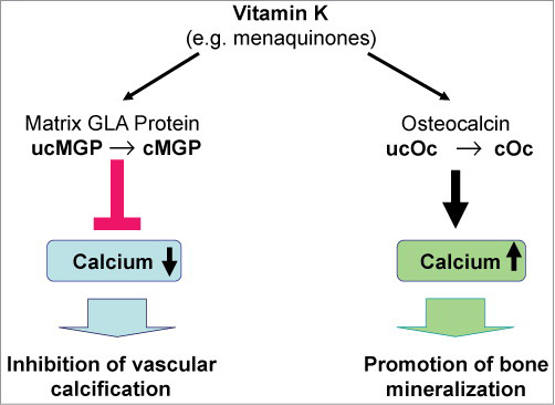
Keep in mind that vitamin K2 also works in tandem with vitamin D and magnesium. So, it’s important to remember that vitamin K2 needs to be considered in combination with calcium, vitamin D and magnesium, as these four all have a synergistic relationship that impacts your bone and heart health.30
Other Health Benefits of Vitamin K2
Vitamin K2 is also important for a variety of other health reasons and conditions, including the following:
| Dental health — Preserving enamel helps reduce cavity formation. Your enamel is made of hollow tubes that extend into the dentin. The enamel is made of nonliving cells, while the dentin has live cells called odontoblasts.
These cells release proteins using vitamin K2 to help prevent tooth decay through an immune response.31 Research32 has demonstrated osteocalcin may be found in inflamed tissue, and researchers consider the protein to have a reparative role in dental pulp. You teeth, like your bones, are storehouses of calcium, which supports the structure and hardness of the teeth.33 The way calcium is deposited in your teeth will either increase the hardness of your tooth or make it more brittle. Vitamin K2 once again acts like the traffic cop, telling calcium where and how to be used in your teeth. Working together with vitamin D, it also promotes a reduction in tooth decay or cavities.34 |
| Cancer — Cancer prevention is another health benefit of vitamin K2. As noted in the 2010 European Prospective Investigation into Cancer and Nutrition (EPIC) study,35 high intake of vitamin K2 — not K1 — reduces cancer risk and lowers cancer mortality by 28%. Findings published in 2008 showed men who consumed the greatest amounts of K2 had a 63% reduced incidence of advanced prostate cancer.36
The evidence also suggests vitamin K2 may reduce your risk for Non-Hodgkin lymphoma. Mayo Clinic researchers discovered that people with the highest intake of vitamin K2 (108 mcg/day or higher) had a 45% lower risk for this type of cancer, compared to those with the lowest vitamin K2 intake (39 mcg/day or less).37 They attribute this effect to vitamin K2’s ability to inhibit inflammatory cytokines, which are related to this type of lymphoma, and its role the life cycle of your cells. |
| Inflammatory conditions — The inflammatory response is a crucial component in many chronic diseases and diseases of aging. Scientific evidence suggests both vitamin K1 and K2 have anti-inflammatory activity, in part by inhibiting nuclear factor kappa-B.38
In one meta-analysis,39 researchers found evidence the family of vitamin K plays a protective role in chronic aging conditions, inflammation and cardiovascular disease. Another study40 confirmed the role the MK-7 form specifically has in the modulation of inflammatory biomarkers. The researchers evaluated natural vitamin K2 for its potential to inhibit gene expression and production of pro-inflammatory markers in vitro, finding MK-7 inhibited gene expression in a dose-dependent manner. Other research41 has demonstrated a reduction in C-reactive protein, a common biomarker used to evaluate inflammation in patients with rheumatoid arthritis. C-reactive protein is a risk marker for a wide range of diseases and the researchers concluded higher intake of vitamin K2 may lower overall risk. |
| Kidney health — There is also accumulating evidence suggesting vitamin K2 may play a role in chronic kidney disease.
A pre- and post-intervention clinical trial,42 which included patients with a diagnosis of chronic kidney disease who took 360 mcg of MK-7 for four weeks, found it significantly reduced calcification in the patients’ blood vessels. Patients with chronic kidney disease with a subclinical vitamin K deficiency may also benefit from a greater intake of the vitamin.43 As noted in a 2016 study44 in EBioMedicine, “MGP not only inhibits calcification in larger arteries but also may be renoprotective.” |
| Testosterone — Yet another role vitamin K2 plays in your body is the optimal production of sex hormones. At least two studies45,46 have shown MK-4, found in animal foods such as meat and dairy products, can help balance testosterone production. |
| Polycystic ovarian syndrome (PCOS) — Vitamin K2 has an effect in women as well, as the vitamin may be helpful in the management of PCOS. This is now the most common hormonal disorders in young women, causing enlarged ovaries and a heightened risk for fertility problems.47
In one randomized study,48 60 vitamin D deficient women with PCOS took either a placebo or a combination of calcium, vitamin D and vitamin K for eight weeks. Those in the treatment group enjoyed greater decrease in testosterone and symptoms than the placebo group. |
| Insulin sensitivity — An unexpected discovery was made when researchers found bone was an endocrine organ, contributing to the regulation of a number of physiological processes, including glucose homeostasis.49
There’s evidence to suggest vitamin K2 supplementation may improve insulin sensitivity via osteocalcin metabolism,50 and may help prevent the development of Type 2 diabetes.51 The evidence is not conclusive, however, as a systematic review52 of controlled trials found K2 supplementation had no effect on insulin sensitivity. Part of the problem may have something to do with the form of vitamin given. Numerous studies have demonstrated synthetic vitamin K2 supplements are not as effective as natto-derived K2 supplements. |
| Mitochondrial health — Research53 published in the journal Science in 2012 found vitamin K2 serves as a mitochondrial electron carrier, thereby helping maintain normal ATP production in mitochondrial dysfunction, such as that found in Parkinson’s disease. |
| Macular degeneration — According to a 2014 paper54 on vitamin K, MK-7 in particular may offer benefits for age-related macular degeneration. |
Signs and Symptoms of Vitamin K2 Deficiency
The major problem we face when it comes to optimizing vitamin K2 is that there’s no easy way to screen or test for vitamin K2 sufficiency. Vitamin K2 cannot at present be measured directly, so it’s measured through an indirect assessment of undercarboxylated osteocalcin. This test is still not commercially available, however.
Without testing, we’re left with looking at various lifestyle factors that predispose you to deficiency. As a general rule, if you have osteoporosis, heart disease or diabetes, you’re likely deficient in vitamin K2.
That said, it’s believed that the vast majority of people are in fact deficient these days and would benefit from more K2. One reason for this is because very few (Americans in particular) eat enough vitamin K2-rich foods.
So, if you do not have any of the health conditions listed, but do not regularly eat high amounts of the following foods, then your likelihood of being vitamin K2 deficient is still very high:
- Certain fermented foods such as natto, or vegetables fermented using a starter culture of vitamin K2-producing bacteria
- Certain cheeses such as Brie and Gouda (these two are particularly high in K2)
- Grass fed organic animal products such as egg yolks, liver, butter and dairy
Use of certain drugs may also affect your vitamin K2 status and make you more prone to deficiency. For example, research55 shows statins deplete vitamin K2, and there’s scientific evidence56 suggesting statin drugs may indeed increase calcification in the arteries.
Vitamin K2 depletion may be an explanation for that. So, if you’re on a statin, you’ll want to make sure you’re taking extra vitamin K2, as well as ubiquinol or coenzyme Q10, which is also being depleted by the drug.
As discussed in “Vitamin K2 Deficiency Is a Significant Cause of Cardiovascular Disease,” warfarin, antibiotics, anti-tuberculosis drugs, anticonvulsants can also raise your risk of vitamin K deficiency.
Dosage Recommendations
As for a clinically useful dosage of vitamin K2, some studies — including the Rotterdam study57 — have shown as little as 45 mcg per day may be sufficient. As a general guideline, I recommend getting around 150 mcg of vitamin K2 per day.
Others recommend slightly higher amounts; upward of 180 to 200 mcg. Fortunately, you don’t need to worry about overdosing on K2, as it appears to be completely nontoxic.
If you opt for a vitamin K2 supplement, make sure it’s MK-7. (The exception is if you’re on vitamin K antagonists, i.e., drugs that reduce blood clotting by reducing the action of vitamin K. If so, you should avoid MK-7 supplements.58) Also remember to take it with fat since it’s fat-soluble and won’t be properly absorbed otherwise, and to balance it with calcium, vitamin D and magnesium.
As a last tip, keep in mind that vitamin K2 may not necessarily make you “feel better” per se. Its internal workings are such that you’re not likely to feel the difference physically.
Compliance can therefore be a problem, as people are more likely to take something that has a noticeable effect. This may not happen with vitamin K2, but that certainly does not mean it’s not doing anything.
– Sources and References
- 1 Oregon State University, Vitamin K
- 2 Journal of Biological Chemistry, December 14, 2007
- 3, 10 Nutrition Journal November 12, 2012; 11, Article Number 93, Introduction
- 4 Journal of Agricultural and Food Chemistry 2006; 54: 483-468 (PDF)
- 5, 7 Haemostasis July 27, 2000; 30: 298-307
- 6 Journal of Hepatology, July 2007; 47(1): 83-92
- 8, 14 Nutraceutical Business Review, February 23, 2016
- 9 J Food Sci Technol. 2015 Aug; 52(8): 5212–5219
- 11 European Journal of Pharmacology, August 15, 2015; 761: 273-278
- 12 Medicine 2017;96(17): e6725
- 13 Osteoporosis International March 2013, DOI: 10.1007/s00198-013-2325-4
- 15, 23, 57 The Journal of Nutrition November 1, 2004: 134(11); 3100-3105 (The Rotterdam Study)
- 16 The Journals of Gerontology May 6, 2019, DOI: 10.1093/Gerona/glz108
- 17, 18 Thrombosis and Haemostasis, 2008; 100(4):393
- 19, 28 Integrative Medicine, 2015;14(1)
- 20 Oman Medical Journal, 2014;29(3)
- 21 Vasc Health Risk Manag. 2009;5(1):185-97
- 22 Frontiers in Cardiovascular Medicine February 5, 2019, DOI: 10.3389/fcvm.2019.00006
- 24 Nutr Metab Cardiovasc Dis. 2009 Sep;19(7):504-10
- 25 American Bone Health October 15, 2019
- 26 Archives of Internal Medicine 2006; 166: 1256-1261
- 27 Osteoporosis International September 2013: 24(9); 2499–2507
- 29, 58 Dermo-Endocrinology 2014: 6(1), Figure 3
- 30 Nutritional Outlook September 24, 2015
- 31 Steven Lin, Use Vitamin K2 to Remineralize Your Teeth
- 32 Journal of Endodontics, 2013;39(7)
- 33 NIH Office of Dietary Supplements, Calcium
- 34 Weston A Price, Vitamin K2 and Dental Health
- 35 Am J Clin Nutr. 2010 May;91(5):1348-58
- 36 Am J Clin Nutr. 2008 Apr;87(4):985-92
- 37 Medical News Today April 20, 2010
- 38 Anti-inflammatory Actions of Vitamin K March 22, 2017, DOI: 10.5771/63891
- 39 Current Nutrition Reports, 2016;5(2)
- 40 Journal of Medicinal Food, 2016;19(7)
- 41 Modern Rheumatology, 2012;23(5)
- 42 BMC Nephrology, 2017;18(191)
- 43 Nutrients, 2013;5(11), 3.1. Vitamin K Status in Patients With CKD Not Yet Requiring Dialysis
- 44 EBioMedicine 2016 Feb; 4: 16–17
- 45 Biochimica et Biophysica Acta, 2006;1760(10)
- 46 Lipids in Health and Disease, 2011; 10:158
- 47 BMC Medicine, 2010; 8:41
- 48 Hormone and Metabolic Research, 2016;48(7):446
- 49 Reviews of Endocrine and Metabolic Disorders, 2015;16(2)
- 50 Diabetes Care, 2011;43(9)
- 51 Diabetes Research and Clinical Practice, 2018; 136:39
- 52 Diabetes, Metabolic Syndrome and Obesity, 2017;10:169
- 53 Science 2012 Jun 8;336(6086):1306-10.
- 54 Dermo-Endocrinology 2014: 6(1)
- 55 Expert Review of Clinical Pharmacology 2015 Mar;8(2):189-99
- 56 Journal of the American College of Cardiology 2015;65(13):1273-1282