Vitamins
now browsing by category
Fitness Strategies That Are Creating World-Class Champions
Reproduced from original article:
https://articles.mercola.com/sites/articles/archive/2023/02/12/fitness-strategies-world-class-champions.aspx
The original Mercola article may not remain on the original site, but I will endeavor to keep it on this site as long as I deem it to be appropriate.
Analysis by Dr. Joseph Mercola Fact Checked February 12, 2023
STORY AT-A-GLANCE
- Sam Calavitta, aka Coach Cal, founder of The Treigning Lab, has put together much of the health science I’ve been teaching over the years into a unique training program that is getting extraordinary results
- The most central strategies are those that optimize ATP production (energy production) and mitochondrial function
- ATP (the energy currency of your cells) can be produced in three ways: aerobically in the mitochondria, anaerobically in the cytosol, or through phosphocreatine. Of these, the phosphocreatine pathway is the most effective in terms of boosting athletic performance, as it allows your body to use D-ribose, adenosine and phosphate to form ATP within your body at a higher level. Near-infrared light also boosts ATP production
- Mitochondrial function must also be optimized, or energy production cannot be elevated. Effective strategies include near-infrared sauna therapy and intermittent fasting
- Other strategies that help optimize fitness and athletic performance include sun exposure, boosting NAD+ with low-dose niacinamide, a cyclical ketogenic diet, intermittent fasting or time-restricted eating, addressing elevated iron levels, methylene blue and molecular hydrogen
In the video above, I interview Sam Calavitta, aka Coach Cal, founder of The Treigning Lab. We met through a biohacking conference in the fall of 2022. One of the organizers of the event introduced me to Miesha Tate, a professional and former Ultimate Fighting Championship (UFC Women’s Bantamweight champion) and mixed martial artist who in turn introduced me to Coach Cal.
He’s taken much of the health science I’ve been teaching over the years and put it together into a revolutionary training program that is producing extraordinary results. Here, we’ll review some of the strategies that can take your fitness to the next level.
Coach Cal’s Unique Approach Gets Results
Calavitta himself wrestled until he turned 40, at which point he switched to Ironman competitions, which is an entirely different paradigm in terms of energy expenditure.
“So, I had to figure out my energy systems,” Calavitta says. “I used a lot of the things that you have been preaching … in particular [strategies for] mitochondrial optimization.
I brought a lot of those practices into the sports, I brought them into Ironman, and I was able to hybridize Ironman training along with combat sports and wrestling, and came up with a very unique way of training athletes …
There’s a plethora of athletes who are seeking to get in to train with us … A huge portion of the UFC and Bellator athletes are my athletes … Almost the entire Olympic wrestling team, which this year [2022], won the first world title in wrestling in many years, beating out Russia [and] Iran.
Wrestling and combat sports are very weight-specific programs which tie right into many of your practices with intermittent fasting, with your meticulous directions on cyclic ketogenic diet, not overdoing it and things like that …
The Treigning Lab is known for some of the most magnificent body transformations ever seen. And … I appreciate your approach, [which is] ‘Treat the body the way God intended, and then sit back and watch the miracle take over.’ And it’s been really, really neat …
These athletes are the toughest athletes in the world, but they’re very cerebral and they appreciate being guided by expertise. And a lot of that expertise is directed by many of the things that you’ve taught me.
This is a young gal who fights in an organization overseas [see video for image]. We were able to bring her down from about 154 pounds to 125 pounds without ever bastardizing her hydration status or her caloric intake, all through many of the principles that were found in your book, ‘Fat for Fuel.’”
‘Proof of the Pudding’
Calavitta shows several other pictures of amazing body transformations in the video.
“That’s the influence that you’ve had on a huge plethora of different athletes,” he says. “You do a lot of health and wellness for the general population, but I don’t think you really understand the effect you’ve had on the world-class athletes … those who are truly listening to and appreciate the educational pieces that you put out daily, and your steadfast fight to let people facilitate the means to allow the human body to do what it can do.”
Frankly, I was unaware that so many elite athletes were using these principles that I’ve been teaching, and it was a real treat to see the kinds of results a person can get if they’re dedicated.
The good news is, you don’t have to be a professional athlete to get similar results — you just have to have a willingness to implement and follow through. If you’ve ever doubted or wondered what the strategies we discuss here can actually do for you, I think you’ll be inspired by Calavitta’s testimony, and the before-and-after pictures of his roster of athletes.

Download this Article Before it Disappears
Optimizing Energy Production
So, what are some of the most effective strategies in Calavitta’s toolbox? The most central strategies are those that optimize ATP production (energy production) in the mitochondria. This is a no-brainer, since you need optimal energy production in your cells order to perform at an optimal level physically.
“Basically, everything came down to ‘How can I optimize ATP production in these athletes, to allow their bodies to overtake that of an opponent?’” Calavitta says. “It came down to understanding that ATP could be produced in three main ways. One, aerobically in the mitochondria. Two, anaerobically in the cytosol. Or three, through phosphocreatine.”
Phosphocreatine is the phosphorylated form of creatine, which rapidly mobilizes reserves of high-energy phosphates in your skeletal muscle, heart and brain. Those phosphates, in turn, recycle adenosine triphosphate, which is the energy currency of your cells.
Of these three ATP producing pathways, the phosphocreatine pathway is the most effective in terms of boosting athletic performance, as it allows your body to use D-ribose, adenosine and phosphate to form ATP within your body at a higher level. That said, your mitochondrial function must also be optimized, or this energy boost cannot occur.
Near-Infrared Sauna Therapy for Mitochondrial Optimization
There are several ways to optimize mitochondrial function, but one of the most effective, as it provides more than one benefit, is near-infrared sauna therapy. First of all, it’s an effective detox strategy. Most people have toxins in their system — plasticizing chemicals, pesticides, flame retardants and heavy metals — and they need to be expelled.
Calavitta uses the Sauna Space near-infrared sauna bulbs with a wavelength of 700 to 960 nanometers. He also appreciates the science of electromagnetic field (EMF) damage, so he uses an EMF-free setup. He explains:
“I didn’t do that until I read your book ‘EMF*D.’ But when I understood what actually happens — the EMF damage, due to the voltage-gated calcium channels in the body, and the [subsequent] necrosis and breakdown of the cell, I thought ‘I can’t be using this with my athletes and having their cells break down.’
I want them in a state of autophagy, mitophagy and whatnot, to be able to clean out, replicate and rebuild. So, I took the far-infrared sauna, as you suggested, and outfitted it with seven of the Sauna Space bulbs, so I can get it up to 150 degrees.”
Autophagy is a really important built-in function your body has that most people fail to optimize. “Auto” means “self” and “phago” means “eat.” So, autophagy is the process by which your body removes damaged cellular parts so that they can then be replaced. Autophagy is activated through fasting, exercise and near-infrared sauna, and when you’re doing all three, you end up with a very efficient self-healing process.
Optimizing ATP Output With Photobiomodulation and Nutrition
Near-infrared sauna also has powerful photobiomodulation effects. As explained by Calavitta:
“Near-infrared light works in conjunction with chlorophyll to … convert ubiquinone to ubiquinol in the body, thereby allowing the ubiquinol to go through the bloodstream and be used in a very effective manner in the electron transport chain …
So, what I did is, I took the near-infrared light and I developed a protocol to use diindolylmethane — which is a metabolite of indole-3-carbinol, sulforaphane and chlorophyll [all of which are found in cruciferous vegetables] — and to administer the red [near-infrared] light in a very calculated fashion to put out a larger ATP production.
People say, ‘How do you know that?’ The thing is, I’m a mathematician, so I took your interview with Dr. Shallenberger, and I wrote the algorithm with a metabolic cart, to actually be able to calculate mitochondrial function and discreet ATP output within the human body. So, I’m not just telling you what we do with the red light, I’m telling you we can measure it, because what is measurable can be improved.”
Low-Dose Niacinamide Boosts NAD+
Calavitta also uses low-dose niacinamide (not niacin), 50 milligrams three times a day (or four times a day for bigger athletes), to boost nicotinamide adenine dinucleotide (NAD+), a crucial signaling molecule believed to play an important role in longevity.
It’s also important in the building and maintenance of muscle mass. When the NAD+ salvage pathways in muscle are impaired, mitochondrial dysfunction and decreased muscle mass ensue.
Many athletes get sold on intravenous NAD, which is extremely expensive, but oral supplementation with niacinamide has also been shown to significantly increase NAD, and at a fraction of the cost. Results doing this, he says, has been nothing short of spectacular.
“I implemented a very strict protocol, as per your suggestion, of one-sixty-fourth teaspoon, three times a day for the athletes,” Calavitta says.
“Their energy levels, which I have measured on the metabolic carts — in the way of resting metabolic rate, resting energy expenditure, metabolic efficiency crossover, as well as fat substrate and carbohydrate substrate use for fuels — have all been skyrocketing as a result of using this practice. My athletes call it the fountain of youth, and I myself use it.”
Sun Exposure for Vitamin D Optimization
Calavitta’s athletes also get an hour of sun exposure every day, which is the easiest and most effective way to optimize your vitamin D level, and then some. I think it’s one of the top two or three things everyone needs to do.
“I understand completely how this is going to be a game changer in my guys. I can’t have them going out there with low vitamin D. For my athletes, I want their vitamin D [level] between 80 ng/mL and 100 ng/mL.
I’ve actually seen athletes whose vitamin D levels were 21 ng/mL, and when we raised them up to the 80s and 90s, their performance absolutely went through the roof and were completely optimized. We try to steer clear of the supplementation with the D3-K2-magnesium cascade, if we can … So many people don’t understand that the D3 without the K2 is not going to end up, possibly, in the right tissues.”
Putting It All Together for Optimal Fuel Synthesis
Another practice Calavitta’s athletes have embraced is the cyclical ketogenic diet detailed in my book, “Fat for Fuel.”
“High-level athletes, with the amount of energy expenditure they have, there are certain things that have to be cycled in, otherwise we’re going to get a down-regulation in thyroid hormone, and I can follow that directly,” Calavitta says.
“By using the cyclical ketogenic diet and the intermittent fasting protocol, along with fasted training, the red-light therapy and the nutrients, we’re able to get some really fantastic pathways of fuel synthesis.
A lot of your fuels don’t just come from carbohydrates or glycolysis, but through … thiamine. Basically, what people don’t understand is that through many different pathways, even to be able to work within the mitochondria … you have to have thiamine in there …
So, all of these principles, the supplementation, [it’s about] reaching the mitochondria from an optimized performance of the facilities that are available to it …
One of the first things that I do with my athletes is reduce the linoleic acid (LA) [and] balance the ratios between omega-3s and omega-6s … We’re supposed to have, at best, a 3-to-1 [omega-3 to omega-6] profile. An outlier would be 6-to-1, and Americans are on average, anywhere from a 25-to-1 to 50-to-1 ratio. So, that’s a big one.”
He also checks iron levels to make sure they’re not elevated and he understands the important interplay and symbiotic relationship between iron and copper. As detailed in previous articles, more often than not, if you’re told you have low iron because your ferritin level is low, the real problem is copper deficiency, as copper deficiency prevents the proper recycling of iron.
Depending on their individual situation, an athlete may be placed on a blood donation schedule if iron is high, and if low on copper, copper-rich foods are added to their diet. Whole food vitamin C is an excellent choice if your copper needs a boost, such as acerola cherry and amla berry.
“So, being able to put all these pieces together, at the level that we have here … with all of the means that we’ve mentioned on this podcast, we’re able to see a complete picture of an individual, to enable their God-given potential to go to a level that is basically unsurpassable,” Calavitta says.
Other Tips and Tricks
Two additional strategies Calavitta has embraced are:
• Supplementing with methylene blue, which I think is one of the most exciting products available, with respect to mitochondrial function, especially for those who are tired all the time.
While it does not address the foundational cause of the problem, it’s a phenomenal crutch when you’re in a pinch or just need to get over a hump, as results are felt very rapidly, typically well within a couple of hours. As noted by Calavitta, its main benefit is its ability to bridge the gap when there’s a breakdown in the electron transport chain.
• Supplementing with molecular hydrogen tablets, to neutralize the free radical damage from ionized radiation during flights. It also helps athletes attenuate jet lag and water retention or edema. “People don’t understand how much oxidative damage actually happens when you’re flying,” he says. “I’m in a unique position because I measure everything and I’m able to see it. And I’m able to see the [decline in] performance that comes from it.”
More Information
To learn more about Coach Cal and all the performance and recovery tools he uses, check out TreigningLab.com and sign up for his newsletter. He also has a Treigning Lab phone app, available for both Apple and Android.
The Treigning Lab Recovery Center, located in Placentia, California, offers a variety of services, including hyperbaric oxygen therapy, full body cryotherapy, infrared light therapy, compression boots, hair and tissue mineral analysis and percutaneous hydrotomy.
Resolving Colds to Advanced COVID with Methylene Blue
Reproduced from original OMNS article (OrthoMolecular News Service):
http://orthomolecular.org/
Subscribe to the free Orthomolecular Newsletter: http://orthomolecular.org/subscribe.html
Go to the OMNS Archive: http://orthomolecular.org/resources/omns/index.shtml
Orthomolecular Medicine News Service, Feb 4, 2023
Editorial by Thomas E. Levy, MD, JD Contributing Editor, OMNS
OMNS (Feb 4, 2023) Linus Pauling first coined the word “orthomolecular” in an article he wrote in the journal Science in 1968. Quite simply, ortho means correct or right, and molecular refers to the simplest building block of any substance. Dr. Pauling started the whole concept of orthomolecular medicine by pointing out that the only true way to prevent or treat any medical condition is to restore whatever important natural substances (nutrients, vitamins, minerals) that are depleted. And the corollary of this approach to healthcare is that no patient is sick because they have low levels of any synthesized agents (prescription drugs) that are not found in nature.
As a rule, a pharmaceutical agent impacts some metabolic pathway in such a manner that helps to alleviate a symptom without lessening the pathology causing that symptom to be present. With the rarest of exceptions, pharmaceutical agents reliably allow a disease to continue to evolve while one or more of the associated symptoms are chronically suppressed. However, when deficiencies of important natural substances are corrected, positive clinical results not known to (or acknowledged by) traditional medicine are consistently seen. Clinical improvement following the resolution of such deficiencies typically indicates that the underlying disease is no longer progressing and possibly even reversing. And depending on how chronic the condition was, an eventual return to physiological normalcy might also result.
Probably the most noteworthy of chronically depleted nutrients in a large majority of people around the world is vitamin C. The literature supporting its positive health impact when properly administered is massive and extending over 80 years now. [1] The benefits of vitamin C and many other important orthomolecular substances continue to be suppressed and even belittled in biased and sometimes frankly fraudulent medical articles. The patented drugs that are promoted relentlessly make knowing the benefits of an inexpensive natural remedy difficult for even sincere healthcare practitioners who want the best for their patients.
Such “education” on the “essential” role of pharmaceutical drugs begins in medical and osteopathic schools, and it never stops. Furthermore, without the endorsement of traditional medicine, many healthcare practitioners are reluctant to use such agents even when they are fully aware of the benefits. Also, as evidenced by the past three years of the COVID pandemic, it appears that a majority of pharmaceutical companies, along with too many hospitals and far too many physicians, place profits far ahead of patient welfare. The application of scientifically-based treatment protocols, many based on orthomolecular principles, continues to be ignored and even suppressed.
Methylene blue (MB), a powerful antioxidant with a clinical impact comparable to even vitamin C, is a major exception to the principles of orthomolecular medicine. It is not produced in the body, and it is not naturally present in any animal or plant. Nevertheless, its documented beneficial health effects rival that of any other known substance, whether normally found in nature or coming out of a laboratory. Just like vitamin C, the true benefits of MB in so many different diseases remain unappreciated and unused by most clinicians, even though it has been safely utilized in many patients for a much longer time than even vitamin C.
The parallels between vitamin C and MB are also reflected in the fact that administering them in either their reduced or oxidized form is comparably beneficial to the patient. This is because the dose of vitamin C or MB is less designed to give a one-time boost to the electron stores of the body than it is to make sure newly-assimilated electrons get optimally distributed throughout the body.
A quality nutrition program is the best source of new (versus recycled) electrons in the body. And the qualities of super antioxidants like vitamin C and MB serve to make sure those electrons are optimally distributed and repeatedly exchanged in redox reactions throughout the body, which is the essence of optimal health.
For those who appreciate metaphors, good nutrition is the product (electron) manufacturing facility, and the premier antioxidants (vitamin C, MB) are the trucks that assure the optimal distribution and delivery of those products throughout the body (country). While it is logical and correct that delivering the antioxidant in its reduced form brings even more electrons into the body, the oxidized form is also highly effective without those extra electrons since it is the distribution and repeated give-and-take of electrons throughout the antioxidant matrix inside the cell that is of greatest therapeutic value. A clear example of this is seen in animal studies where dehydroascorbic acid (DHAA), which is the oxidized form of vitamin C, readily minimizes brain infarct size from induced ischemic stroke, facilitating rapid recovery. In fact, these studies also show that there is a precipitous decline in reduced vitamin C in the ischemic brain which is reversed by the infusion of DHAA. [2]
However, the presence of elevated blood levels of DHAA reported in patients with infectious disease does not mean DHAA is toxic per se. Rather, such elevated levels are just a reflection of increased oxidative stress in such a patient, not the cause of it. [3] Furthermore, any negative impact that DHAA has been reported to have in some in vitro studies does not reliably predict the positive effect that DHAA has in the intact plant, animal, or human, as such test tube studies are not receiving an ongoing new intake of electrons from quality nutrition or sun exposure. [4] Like the ascorbic acid and DHAA forms of vitamin C, the powerful clinical impact of MB appears equal whether administered in its oxidized (methylene blue) or reduced (leucomethylene blue) forms.
Methylene Blue (MB): A Brief History
Methylene blue (MB) is the first drug to be tested and used in humans. Chemically known as methylthioninium chloride, it was first synthesized in 1876, and it was used as an industrial dye. It was later found to be an excellent dye for staining microbes and human tissues as well. In 1891 it was found to be very effective as an anti-malarial agent by Paul Ehrlich. Of note, Ehrlich first coined the term “magic bullet” to refer to how effectively MB targeted and accessed the nervous system. [5] It has since been established to have a selective affinity for the nervous system, although it is highly effective in reaching all cells in the body.
As a powerful antioxidant with the ability to target the brain, MB was used as an antipsychotic drug for 50 years before phenothiazine became the first “official” antipsychotic drug. [6] It continues to be used as a dye for the staining of biological tissue specimens as well as a diagnostic tool in surgical procedures. [7] It has also been established to have numerous and very significant therapeutic purposes for a wide range of medical conditions. Some of the more significant conditions to be consistently and successfully treated by MB include the following:
- Infections, from minimal to life-threatening, including those having progressed to septic shock. Also, acute respiratory distress syndrome (ARDS) and hypoxemia secondary to COVID or any of multiple different pathogens [8,9]; also used for disinfection of plasma to be used for transfusion [10-12]
- Mitochondrial dysfunction [13-15]
- Depression, dementia, psychosis, impaired memory, as well as multiple acute and chronic neurological conditions [16]
- Methemoglobinemia, in which the oxygen-carrying capacity of the blood is critically depleted. [17] MB has FDA approval as a first-line therapy for this condition. [18,19]
Antioxidant Extraordinaire
An ideal antioxidant is one that is equally stable chemically in either its reduced or oxidized state, while having physical access to all the oxidized biomolecules in the body. Such a quality allows the continued giving and taking of electrons throughout the cellular and extracellular spaces, as that molecule does not resist being either reduced or oxidized. This redox (reduction-oxidation) property helps to conduct electron flow inside the cells. This helps to generate and sustain the microcurrents (a current is literally an electron flow) that have been identified inside cells, which work to maintain healthy transmembrane voltages. A sick cell always has a low transmembrane voltage, which directly reflects a redox balance skewed toward oxidation, with a limited influx of new antioxidant (nutrient) molecules available to deal with any new pro-oxidant (toxic) molecules. Normal transmembrane voltages are critical in maintaining healthy ion channels, transporters, pumps, and enzymes in the cell. [20] They are also critical for the optimal synthesis of ATP. [21]
A toxin always works to cause oxidation wherever it is found, or ends up. It is always pro-oxidant in its chemical impact, as it seeks to oxidize a biomolecule and then keep the electron it has “robbed.” The electron it acquires makes the toxin much more stable chemically, and such a reduced toxin will not give up the electron again to another oxidized, or electron-depleted, biomolecule. This means that the electron-sated toxin will never re-donate its electron to an oxidized biomolecule, as would occur with an electron-sated, or reduced, antioxidant molecule.
In addition to increasing the numbers of oxidized biomolecules, this retention of electrons by toxins also impedes/decreases electron flow (microcurrents) since the newly acquired electrons are tightly held and never again released in the manner of an antioxidant that is continually giving and taking electrons. An antioxidant like vitamin C decreases the total number of oxidized biomolecules and supports optimal microcurrents, and a toxin does the opposite. [22]
It is the antioxidant properties of MB that results in the impressive clinical impact it has on so many conditions. In this regard, there is a striking parallel in what MB can do in the body with what vitamin C can do. Both vitamin C and MB are small molecules, and they effectively reach every cell in the body. However, MB requires no active or passive cell wall transporters as does vitamin C, and it has both lipid-soluble and water-soluble characteristics. Because of this, MB passes easily through lipid-rich cell walls, after which it disseminates throughout the water-based cell. [23,24] Also, while both MB and vitamin C access the brain, MB has been found to have a brain concentration up to tenfold higher than in the serum as quickly as one hour after intravenous administration. [25] Uptake is very rapid in the other organs as well. [26]
MB also has well-documented antitoxin properties like vitamin C, but the studies documenting them are much less prolific than those showing the similar effects of vitamin C on pro-oxidants and other poisons. MB helps protect the kidneys against the toxicity of the chemotherapeutic agent, cisplatin. [27] MB has also been shown to protect the brain against the toxicity of another chemotherapeutic agent, ifosfamide. [28,29] It also was shown to effectively treat the encephalopathy induced by ifosfamide after it had developed. And even though there is not an abundance of articles demonstrating the ability of MB to neutralize toxins and repair toxic damage, multiple researchers recommend it be routinely available as an emergency antidote for general use. [30,31]
Many toxins also inflict harm in some individuals by the formation of methemoglobin with a reduction of oxygen delivery to the tissues. Such toxin excesses or poisonings can be effectively treated with MB, as it is already the treatment of choice by many clinicians for methemoglobinemia. MB is always a good partner to be administered along with vitamin C for any toxin excess or overdose. [32,33] The addition of magnesium with MB and vitamin C to overdose patients offers additional protection against the development of fatal arrhythmias that can occur before the MB and vitamin C can resolve and block further toxic impact. [34]
Ideal Shock Therapy
Methylene blue is exceptionally beneficial for both infections in general and for hypotensive shock. This makes it a particularly optimal therapy for the very common cause of intensive care unit death around the planet: septic shock.
Refractory septic shock, a state of disseminated infection with vascular collapse and hypotension often unresponsive to all traditional measures, consistently responds positively to MB therapy, sometimes saving the patient from otherwise certain death.
As with vitamin C and many other non-traditional treatments, nearly all clinicians simply will not take the “leap” from clear-cut positive results in the literature to the application of those results in their patients. At best, they use these non-traditional therapies almost like a final gesture that they have done everything possible to save the patient, even though those therapies have little to no toxicity and should not be relegated to the last option in a treatment protocol. And, of course, this only applies to the clinicians who are even remotely aware of the existence of the data showing how effective and nontoxic these non-traditional therapies are. The many pearls in the medical literature remain completely unharvested by most clinicians. And many more clinicians are very diligent in doing everything possible to maintain the “mainstream status quo” to the point of ignoring and even suppressing anything that might threaten it.
An experienced and honest clinician will tell you that just one dramatic case report that is accurately reported has enormous value. When a patient is on the verge of death despite all that has been done, and one single intervention quickly stops the clinical deterioration and starts a clear recovery, the alert clinician does not need a large prospective, double-blind, and placebo-controlled clinical trial to take such a clinical response seriously. Such a trial would be unethical when the placebo group is not being given the benefit of some inexpensive, nontoxic, and highly effective agent. Especially in the setting of an advanced and rapidly progressing infection with unresponsive shock secondary to sepsis, seeing the patient normalizing only a short time after a treatment is administered compels serious attention.
A clear example of such a case report was reported on a 38-year-old male patient who presented with bilateral pneumonia that subsequently worsened and resulted in bacteria (Klebsiella pneumonia) being released into the bloodstream (septicemia). Lethargic with low blood oxygen when admitted to the hospital, he was given IV fluids with insulin and antibiotics. The oxygen levels continue to decline with increased difficulty breathing, and he was then intubated and supported on a ventilator. Hypotension requiring vasopressor infusion ensued. Broader antibiotic coverage was added. Metabolic acidosis with declining renal function followed, and a few hours later he had a cardiac arrest. Four hours after regaining a heart rhythm and only 25 hours after initial presentation, extracorporeal membrane oxygenation (ECMO) support was started. Nevertheless, critically low blood pressure unresponsive to multiple vasopressors continued.
At this point in time, a 172 mg IV bolus of MB was administered, and an infusion of MB at 0.51 mg/kg/hour was maintained for the next 10 hours. Blood pressure quickly improved and vasopressor support could be decreased. At the conclusion of the infusion, the clinical status stabilized for another 22 hours, but fever with a dropping blood pressure unresponsive to combinations of vasopressors at the highest doses returned. The MB infusion was restarted and blood pressure again responded promptly. This time the infusion was continued for 54 hours, and about seven days after this longer infusion was completed the patient was fully recovered and discharged from the hospital. [35] Another impressive case report on a clinically similar patient showed that MB had to be continually infused for a full 120 hours to prevent repeated clinical relapses, after which the patient stabilized and was eventually discharged. [36]
These case studies, in which the patients effectively serve as their own controls, showed clear improvement on MB when severely ill, clear deterioration back to a life-threatening point after MB discontinuation, and prompt improvement with complete clinical resolution when the MB was restarted and continued for a long enough period. No sincere and competent clinician giving his/her highest priority to patient welfare would ignore the importance of such a clinical response when treating similar patients in the future. And this is especially the case when it is realized that MB, dosed appropriately, has an impeccable safety profile, just like vitamin C. Also, like vitamin C, MB also enhances antibody production in the body. [37] This begs the question: Why not use MB first in such situations, rather than last, or never?
Multiple studies have demonstrated the benefits of MB in stabilizing and even resolving septic shock, which is the worst stage that any infection can reach before the inevitable progression to death. No reports of MB worsening the overall clinical status of septic patients could be found. The studies consistently show that MB always improves hypotension when appropriately administered. Furthermore, it has been shown that MB improves survival in shock of all causes (vasodilatory shock), including the shock of advanced sepsis. [38]
The refractory hypotension in septic shock is consistently seen in the setting of excessive nitric oxide production, which causes too great a decrease in vascular tone. [39] MB promptly counteracts this in restoring normal blood pressure. [40] Furthermore, over 120 years of MB use has clearly established the lack of significant toxicity. Toxic levels exist, as with nearly every other agent (including water), but the amounts needed are far beyond the recommended dosing in established treatment protocols. [41-43]
An open-minded clinician reviewing the literature for the first time to learn about the best treatment for septic shock would certainly utilize methylene blue as a first-line agent. Even low doses of MB and one-time boluses of MB consistently show clear benefit in septic shock. However, the clinical response is much better and consistently achieved with a properly-dosed continuous infusion. [44,45] Septic shock still claims a lot of lives regardless of the therapy, and some clinical studies add MB seemingly as a last-ditch afterthought, after which MB is then reported to be ineffective for improving survival. And even now, some of the most recent clinical research continues to assert that “more studies are needed” on the impact of MB in septic shock, even though the very positive research on MB and septic shock now spans decades. [46-55] MB infusions in hypotensive neonates have also been shown to increase blood pressures rapidly and safely. [56-58]
The impact of MB on septic shock was addressed above in some detail since a patient cannot really be much sicker than having severe hypotension with massive infection and enormously increased oxidative stress throughout the body. However, it is important to realize that MB has also been shown to be very effective in treating different types of hypotensive shock that are unrelated to advanced degrees of infection. [59] Shock with unresponsive hypotension secondary to the ingestion of multiple drugs has responded rapidly to MB infusions, allowing the weaning of other vasopressor agents. [60,61] Shock secondary to anaphylaxis also responds well to MB. [62] One patient with profound refractory hypotensive shock following a dihydropyridine calcium channel blocker overdose only responded positively to MB infusion and was eventually discharged. Prior to the MB infusion, no improvement in blood pressure was seen with saline infusion, several doses of calcium gluconate, glucagon, various vasopressor agents, and even high-dose insulin euglycemic therapy over a period of several hours. [63] Another type of hypotensive shock, cardiac vasoplegia, is also sometimes seen following cardiac surgery. This is effectively treated by methylene blue as well. [64-66]
All forms of hypotensive shock should be treated with MB, and it should be part of the treatment protocol at the outset. It should not just be held back as a last-ditch intervention to save the patient. [67]
Regarding ARDS secondary to COVID, a massive production of pro-inflammatory agents known as a cytokine storm typically precedes imminent death if not effectively terminated and neutralized. [68,69] MB has been uniquely shown to inhibit the production of all three of the major classes of pro-oxidants involved in the cytokine storm clinical picture (reactive oxygen species [ROS], reactive nitrogen species [RNS], and cytokines). [70-72] And as a potent antioxidant, MB is highly effective in neutralizing the wide array of pro-oxidants that have already been produced in the ARDS lungs. MB also combines well with other antioxidants in providing clinical benefit. MB combined with vitamin C and N-acetyl cysteine was very effective in treating advanced COVID. [73]
Furthermore, patients who were severely ill with COVID but showing a steady clinical recovery still greatly benefit from MB. Very many “recovered” COVID patients have significant neurocognitive problems that are lessened or even blocked with adequate dosing of MB. With the known antioxidant properties of MB along with its predilection for targeting increased oxidative stress in the nervous system, it should be part of any COVID treatment, regardless of how well the infection is responding to other therapies. [74,75]
MB, Pathogens, and Photodynamic Therapy (PDT)
Logically, considering its documented impact on advanced septic shock, MB has also been shown to readily kill and/or neutralize a wide range of pathogens. While it can achieve this as a monotherapy, it is enhanced in effectiveness when accompanied by photodynamic therapy (PDT). A protocol using MB with PDT has even been shown to eliminate intracellular pathogens such as prions from the blood. [76] Another MB/PDT approach has shown rapid resolution of moderate to severe COVID in patients who did not require hospitalization. [77] MB has been shown to directly inhibit the initial binding of the COVID spike protein with the ACE2 receptor, a step necessary for the virus to enter the cell. [78-80]
MB and PDT have similar abilities to enhance mitochondrial function.
They both effectively bypass much of the Krebs cycle, producing normal amounts of ATP while generating less oxidative stress in the process of going through the entire cycle. [81] This can result in a complete clinical recovery from mitochondrial dysfunction syndromes.
ATP is produced in the mitochondria due to the shuttling of electrons through the four sequential complexes of the electron transport chain. The fourth complex transfers the electrons to the terminal electron acceptor, oxygen, ultimately resulting in ATP production. MB receives the electrons from the first complex and then directly passes those electrons on to cytochrome c in the fourth complex, bypassing the other complexes. [82] PDT with the photons from near-infrared light also energizes and enables the ability of cytochrome c to donate electrons to oxygen and result in the production of ATP. [83,84]
This bypassing of the earlier complexes of the electron transport chain lowers the production of reactive oxygen species (ROS) that would have been generated by those complexes, decreasing net oxidative stress in the cell. Yet, ATP production continues as though the entire electron transport chain was functioning normally. Less ROS production (mitochondrial oxidative stress) while achieving normal energy production goals is always a desirable, but rarely achieved therapeutic goal, and MB accomplishes this. [85,86] Because of these effects, MB has been promoted as an anti-aging drug. [87] In cultured fibroblasts, MB clearly extends the life span of these cells. [88]
When the mitochondria can be made more efficient in producing energy, every metabolic process in the body is positively impacted. Any of the mitochondrial dysfunction conditions can benefit from MB and PDT, but especially MB due to its antioxidant nature and its ability to be taken regularly in a supplemental fashion without the need to spend time receiving various applications of light therapy. Furthermore, the actions of MB or PDT can also serve to help restore to normal an electron transport chain that had accumulated too much oxidative damage to function with normal efficiency (mitochondrial dysfunction) by decreasing the pro-oxidants (ROS) normally generated in the process of making ATP. [89]
However, there is no need to enhance every MB treatment with PDT to get optimal benefit if the MB is properly-dosed. MB has been shown to inactivate a very large number of viruses and other pathogens in vitro, with and without PDT. [90-96] MB is especially well-suited to dealing with viral infections, as it works
- directly against the virus, and
- prevents virus entry into cells, and
- inhibits viral replication after entry into the cell. [97]
As might be expected, the ability of MB to resolve viral infections indicates its likely positive impact in preventing viral infections as well. During the first wave of COVID-19 infections in France, it was reported that a cohort of 2,500 end-stage cancer patients being treated with a protocol that included 75 mg of MB three times daily had NO reported cases of influenza or COVID. [98]
There is significant research into methylene blue derivatives, which are also highly effective antiviral agents, including against viruses in the smallpox family. [99] Similarly, as MB is of clear-cut benefit in the treatment of depression, MB derivatives are being evaluated for the treatment of depression and neurological disorders. [100] Undoubtedly, the pharmaceutical industry recognizes the incredible abilities of MB, and much effort is going into finding related and effective agents that can be patented in order to generate astronomical profits.
MB and Cancer
On the PubMed website, the entry “cancer methylene blue” results in about 2,500 references. The articles that appear address primarily the role of MB in:
- Localizing (staining) of cancerous tissues and/or identifying as many involved lymph nodes as possible [101-105]
- The inhibition, inactivation, or killing of a wide array of different cancer cells in vitro, with and without the application of PDT [106-113]
- The superiority of MB in treating tumors in mice over traditional chemotherapy [114]
- In combination with PDT, the complete resolution of AIDS-related Kaposi’s sarcoma skin lesions that had been unresponsive to chemotherapy with MB and toluidine blue [115]
- The direct treatment of cancer in dogs [116]
- The direct treatment of cancer in humans (only one article). While treating different types of cancer, the author asserted that MB reliably stopped pain secondary to cancer, improved general health, and added years of longevity. This was reported in 1907! [117] Another article asserted that MB was found to have anticancer effects over a century ago. [118] Of note, NO significant clinical applications of methylene blue on cancer patients were found other than the 1907 study cited above.
The efficacy of an inexpensive and safe agent like MB in many different and even advanced medical conditions make it an ideal general add-on or even stand-alone treatment most of the time. Furthermore, its potent anti-cancer effects in vitro make it especially puzzling why straightforward clinical studies on cancer patients with MB alone or in combination with other agents have not been reported. Even the positive effects of the much-ignored vitamin C on cancer patients have been published in many articles, yet the wonderful properties of MB have been known much longer now than vitamin C. The literature even suggests that MB could play a positive role in the treatment of cancer patients. [119]
MB: Safety and Dosing
The main side effect of MB is a blue discoloration of the urine. Rarely, some blue discoloration of the skin might be noticed when an extended administration of highly-dosed MB has occurred. Nevertheless, both effects are completely reversible in hours to a few days as the MB is eliminated out of the body. At very high doses of MB, some of the hemoglobin in the blood can be converted into methemoglobin, which is an abnormal state where MB is the treatment of choice when given at a lower dose. Even higher doses can result in greater toxic side effects, although higher doses can still be warranted for some critically ill patients who are not responding to other measures, as in terminal septic shock. Also, in patients with depression who are on drugs known as serotonin reuptake inhibitors (SSRIs), the addition of MB is not advisable, as some of these patients can develop a potentially life-threatening development known as serotonin syndrome. [120,121] However, MB is an effective anti-depressant by itself at low doses. [122]
Because they are highly effective antioxidants, both MB and vitamin C have been cited to rarely precipitate red blood cell hemolysis in patients with glucose-6-phosphate dehydrogenase (G6PD) deficiency. Nevertheless, closely-monitored administration of these agents in such patients typically avoids such hemolytic problems. In fact, when the G6PD-deficient patient presents with methemoglobinemia, a condition for which MB is typically the indicated treatment, properly-dosed vitamin C can resolve the condition without using MB. [123,124] Of note, G6PD deficiency resulting in hemolytic anemia from MB is very rare. In African children with malaria, MB therapy was shown to be very safe even when G6PD deficiency was present, as was the case with all 24 deficient children in one study. [125] Another study on 74 healthy but G6PD deficient adult men demonstrated no hemolysis when given MB along with chloroquine. [126]
Generally intravenous dosing is not necessary except for the critically ill patient, as in advanced hypotensive shock. There is no standard, fixed regimen of MB recommended in such situations. Boluses of 2 mg/kg of MB can be given, often followed by infusions of various duration depending on the clinical status and response of the patient. Such infusions are often in the range of 0.5 mg MB/kg/hour over an extended period, but as much as 4 mg MB/kg can be infused over an hour. An effective infusion spanning 120 hours has been reported. Others report that infusions can range from 0.25 to 2 mg MB/kg/hour. [127,128]
For less critical patients, as well as for outpatients, oral MB dosing can range from 10 mg to 50 mg, and that dosage can be taken from one to three times daily, adjusted up or down in dose size and frequency depending on clinical response. Even higher doses can be comfortably used for limited times. 200 mg daily to stabilize COVID patients that are not yet critically ill is a very reasonable dose. A reasonable regular supplementation dose can range from 5 to 15 mg daily for general good health if there is no targeted symptom or medical condition.
As a practical point regarding regular supplementation, a dose of 5 to 15 mg of 1% MB solution (0.5 to 1.5 milliliters) can be added to a small amount of water. A teaspoon of ascorbic acid powder (not sodium ascorbate) can then be added. After sitting for 15 minutes or less, the solution will completely clear with just a slight residual blue tint. [129] This can then be quickly consumed with little staining of the tongue that readily occurs with the MB solution alone. Regardless, the staining resolves quickly. But without the added ascorbic acid, it is best to just put the MB straight into something like tomato juice and then drink that.
Methylene blue is not a nutrient. While having some important similarities with vitamin C, there are differences, including a narrower tolerance limit and higher risk safety profile. Less than 2 mg/kg MB is generally regarded as safe; over 7 mg/kg is more likely to induce side effects. MB administration is to be done with the guidance of a qualified health care provider.
Recap
Methylene blue (MB) is an antioxidant with high redox activity, able to rapidly “oscillate” between its oxidized and reduced forms, much like that seen in vitamin C. A small molecule with fat- and water-soluble characteristics, it reaches all areas and cells of the body, and it especially concentrates in the brain and central nervous system. Like vitamin C, MB is highly effective in maintaining a healthy distribution of electrons already in the body, along with the distribution of new electrons assimilated from the nutrients in a healthy dietary regimen.
MB has a unique ability among antioxidants and other biomolecules to relay electrons from the first complex in the energy-generating Krebs cycle in the mitochondria directly to the fourth complex. This allows the energized fourth complex to then produce ATP without the additional expenditure of energy in the steps of the electron transport chain that was bypassed. As such, MB allows dysfunctional mitochondria to produce healthy levels of ATP while producing less oxidative stress in the process, an optimal way both heal those mitochondria while promoting healing anywhere in the body. Photodynamic therapy (PDT) can also directly activate the energy production of the fourth complex in the electron transport chain.
Like vitamin C, MB is also a very powerful anti-pathogen It has been documented to salvage even late-stage COVID patients supported on ventilators with hypotension secondary to septic shock. For viruses in general, MB has the unique ability to attack the circulating virus, to block its binding of the virus to the cells of the body, and to stop the proliferation of the virus inside the infected cell. When administered as recommended, MB is exceptionally well-tolerated, with a safety profile that extends now over a period of more than 100 years of clinical use.
(OMNS Contributing Editor Dr. Thomas E. Levy [televymd@yahoo.com] is board certified in internal medicine and cardiology. He is also an attorney, admitted to the bar in Colorado and in the District of Columbia. The views presented in this article are the author’s, and not necessarily those of all members of the Orthomolecular Medicine News Service Editorial Review Board. Readers should work in cooperation with their healthcare professional before and during application of this or any other approach to wellness.)
References
1. Levy T (2002) Curing the Incurable. Vitamin C, Infectious Diseases, and Toxins. Henderson, NV: MedFox Publishing
2. Spector R (2016) Dehydroascorbic acid for the treatment of acute ischemic stroke. Medical Hypotheses 89:32-36. PMID: https://pubmed.ncbi.nlm.nih.gov/26968905/
3. Bhaduri J, Banerjee S (1960) Ascorbic acid, dehydro-ascorbic acid, and glutathione levels in blood of patients suffering from infectious diseases. The Indian Journal of Medical Research 48:208-211. PMID: https://pubmed.ncbi.nlm.nih.gov/13800336/
4. Thon M, Hosoi T, Ozawa K (2016) Dehydroascorbic acid-induced endoplasmic reticulum stress and leptin resistance in neuronal cells. Biochemical and Biophysical Research Communications 478:716-720. PMID: https://pubmed.ncbi.nlm.nih.gov/27498033/
5. Wainwright M, Crossley K (2002) Methylene blue-a therapeutic dye for all seasons? Journal of Chemotherapy 14:431-443. PMID: https://pubmed.ncbi.nlm.nih.gov/12462423/
6. Howland R (2016) Methylene blue: the long and winding road from stain to brain: part 2. Journal of Psychosocial Nursing and Mental Health Services 54:21-26. PMID: https://pubmed.ncbi.nlm.nih.gov/27699422/
7. Oz M, Lorke D, Hasan M, Petroianu G (2011) Cellular and molecular actions of methylene blue in the nervous system. Medicinal Research Reviews 31:93-117. PMID: https://pubmed.ncbi.nlm.nih.gov/19760660/
8. Hamidi-Alamdari D, Hafizi-Lotfabadi S, Bagheri-Moghaddam A et al. (2021) Methylene blue for treatment of hospitalized COVID-19 patients: a randomized, controlled, open-label clinical trial, phase 2. Revista de Investigacion Clinica 73:190-198. PMID: https://pubmed.ncbi.nlm.nih.gov/34019535/
9. Mahale N, Godavarthy P, Marreddy S et al. (2021) Intravenous methylene blue as a rescue therapy in the management of refractory hypoxia in COVID-19 ARDS patients: a case series. Indian Journal of Critical Care Medicine 25:934-938. PMID: https://pubmed.ncbi.nlm.nih.gov/34733037/
10. Lozano M, Cid J, Muller T (2013) Plasma treated with methylene blue and light: clinical efficacy and safety profile. Transfusion Medicine Reviews 27:235-240. PMID: https://pubmed.ncbi.nlm.nih.gov/24075476/
11. Babigumira J, Lubinga S, Castro E, Custer B (2018) Cost-utility and budget impact of methylene blue-treated plasma compared to quarantine plasma. Blood Transfusion 16:154-162. PMID: https://pubmed.ncbi.nlm.nih.gov/27893348/
12. Gravemann U, Engelmann M, Kinast V et al. (2022) Hepatitis E virus is effectively inactivated by methylene blue plus light treatment. Transfusion 62:2200-2204. PMID: https://pubmed.ncbi.nlm.nih.gov/36125237/
13. Atamna H, Nguyen A, Schultz C et al. (2008) Methylene blue delays cellular senescence and enhances key mitochondrial biochemical pathways. FASEB Journal 22:703-712. PMID: https://pubmed.ncbi.nlm.nih.gov/17928358/
14. Kuliaviene I, Baniene R, Virketyte S et al. (2016) Methylene blue attenuates mitochondrial dysfunction of rat kidney during experimental acute pancreatitis. Journal of Digestive Diseases 17:186-192. PMID: https://pubmed.ncbi.nlm.nih.gov/26861116/
15. Duicu O, Privistirescu A, Wolf A et al. (2017) Methylene blue improves mitochondrial respiration and decreases oxidative stress in a substrate-dependent manner in diabetic rat hearts. Canadian Journal of Physiology and Pharmacology 95:1376-1382. PMID: https://pubmed.ncbi.nlm.nih.gov/28738167/
16. Rojas J, Bruchey A, Gonzalez-Lima F (2012) Neurometabolic mechanisms for memory enhancement and neuroprotection of methylene blue. Progress in Neurobiology 96:32-45. PMID: https://pubmed.ncbi.nlm.nih.gov/22067440/
17. Mak R, Liebelt E (2021) Methylene blue: an antidote for methemoglobinemia and beyond. Pediatric Emergency Care 37:474-477. PMID: https://pubmed.ncbi.nlm.nih.gov/34463662/
18. Clifton 2nd J, Leikin J (2003) Methylene blue. American Journal of Therapeutics 10:289-291. PMID: https://pubmed.ncbi.nlm.nih.gov/12845393/
19. Jiang Z, Duong T (2016) Methylene blue treatment in experimental ischemic stroke: a mini review. Brain Circulation 2:48-53. PMID: https://pubmed.ncbi.nlm.nih.gov/27042692/
20. Bezanilla F (2008) How membrane proteins sense voltage. Nature Reviews. Molecular Cell Biology 9:323-332. PMID: https://pubmed.ncbi.nlm.nih.gov/18354422/
21. Kaim G, Dimroth P (1999) ATP synthesis by F-type ATP synthase is obligatorily dependent on the transmembrane voltage. The EMBO Journal 18:4118-4127. PMID: https://pubmed.ncbi.nlm.nih.gov/10428951/
22. Levy T (2017) Hidden Epidemic: Silent oral infections cause most heart attacks and breast cancers. Henderson, NV: MedFox Publishing. See Chapter 4. To download free of book (English or Spanish): https://hep21.medfoxpub.com/
23. May J, Qu Z, Cobb C (2004) Reduction and uptake of methylene blue by human erythrocytes. American Journal of Physiology. Cell Physiology 286:C1390-C1398. PMID: https://pubmed.ncbi.nlm.nih.gov/14973146/
24. Bruchey A, Gonzalez-Lima F (2008) Behavioral, physiological and biochemical hermetic responses to the autoxidizable dye methylene blue. American Journal of Pharmacology and Toxicology 3:72-79. PMID: https://pubmed.ncbi.nlm.nih.gov/20463863/
25. Peter C, Hongwan D, Kupfer A, Lauterburg B (2000) Pharmacokinetics and organ distribution of intravenous and oral methylene blue. European Journal of Clinical Pharmacology 56:247-250. PMID: https://pubmed.ncbi.nlm.nih.gov/10952480/
26. DiSanto A, Wagner J (1972) Pharmacokinetics of highly ionized drugs. 3. Methylene blue-blood levels in the dog and tissue levels in the rat following intravenous administration. Journal of Pharmaceutical Sciences 61:1090-1094. PMID: https://pubmed.ncbi.nlm.nih.gov/5044808/
27. Usefzay O, Yari S, Amiri P, Hasanein P (2022) Evaluation of protective effects of methylene blue on cisplatin-induced nephrotoxicity. Biomedicine & Pharmacotherapy 150:113023. PMID: https://pubmed.ncbi.nlm.nih.gov/35483196/
28. Pelgrims J, De Vos F, Van den Brande J et al. (2000) Methylene blue in the treatment and prevention of ifosfamide-induced encephalopathy: report of 12 cases and a review of the literature. British Journal of Cancer 82:291-294. PMID: https://pubmed.ncbi.nlm.nih.gov/10646879/
29. Vakiti A, Pilla R, Moustafa M et al. (2018) Ifosfamide-induced metabolic encephalopathy in 2 patients with cutaneous T-cell lymphoma successfully treated with methylene blue. Journal of Investigative Medicine High Impact Case Reports 6:2324709618786769. PMID: https://pubmed.ncbi.nlm.nih.gov/30083561/
30. Baldo C, Silva L, Arcencio L et al. (2018) Why methylene blue has to be always present in the stocking of emergency antidotes. Current Drug Targets 19:1550-1559. PMID: https://pubmed.ncbi.nlm.nih.gov/29611486/
31. Kaiser S, Dart R (2022) The roles of antidotes in emergency situations. Emergency Medicine Clinics of North America 40:381-394. PMID: https://pubmed.ncbi.nlm.nih.gov/35461629/
32. Gebhardtova A, Vavrinec P, Vavrincova-Yaghi D et al. (2014) A case of severe chlorite poisoning successfully treated with early administration of methylene blue, renal replacement therapy, and red blood cell transfusion: case report. Medicine 93:e60. PMID: https://pubmed.ncbi.nlm.nih.gov/25144325/
33. Adhit K, Menon S, Acharya S, Siddhaarth K (2022) Toxin-induced methehemoglobinemia with kidney injury and hypoxic brain injury in a case of pesticide poisoning: a case report. Cureus 14:e32516. PMID: https://pubmed.ncbi.nlm.nih.gov/36654552/
34. Levy T (2019) Magnesium, Reversing Disease. Henderson, NV: MedFox Publishing. See Chapter 10. To download free copy of book (English or Spanish): https://mag.medfoxpub.com/
35. Jaiswal A, Kumar M, Silver E (2020) Extended continuous infusion of methylene blue for refractory septic shock. Indian Journal of Critical Care Medicine 24:206-207. PMID: https://pubmed.ncbi.nlm.nih.gov/32435102/
36. Dumbarton T, Minor S, Yeung C, Green R (2011) Prolonged methylene blue infusion in refractory septic shock: a case report. Canadian Journal of Anaesthesia 58:401-405. PMID: https://pubmed.ncbi.nlm.nih.gov/21246318/
37. Montegut L, Martinez-Basilio P, Moreira J, Schwartz L, Jolicoeur M (2020) Combining lipoic acid to methylene blue reduces the Warburg effect in CHO cells: from TCA cycle activation to enhancing monoclonal antibody production. PLoS One 15:e0231770. PMID: https://pubmed.ncbi.nlm.nih.gov/32298377/
38. Zhao C, Zhai Y, Hu Z et al. (2022) Efficacy and safety of methylene blue in patients with vasodilatory shock: a systematic review and meta-analysis. Frontiers in Medicine 9:950596. PMID: https://pubmed.ncbi.nlm.nih.gov/36237547/
39. Prauchner C (2017) Oxidative stress in sepsis: pathophysiological implications justifying antioxidant co-therapy. Burns 43:471-485. PMID: https://pubmed.ncbi.nlm.nih.gov/28034666/
40. Jang D, Nelson L, Hoffman R (2013) Methylene blue for distributive shock: a potential new use of an old antidote. Journal of Medical Toxicology 9:242-249. PMID: https://pubmed.ncbi.nlm.nih.gov/23580172/
41. Gebel F, Meng H, Michot F, Truniger B (1989) [Psychogenic water intoxication]. Article in German. Schweizerische Medizinische Wochenschrift 119:169-177. PMID: https://pubmed.ncbi.nlm.nih.gov/2648558/
42. Mercier-Guidez E, Loas G (2000) Polydipsia and water intoxication in 353 psychiatric inpatients: an epidemiological and psychopathological study. European Psychiatry 15:306-311. PMID: https://pubmed.ncbi.nlm.nih.gov/10954875/
43. Kirov M, Evgenov O, Evgenov N et al. (2001) Infusion of methylene blue in human septic shock: a pilot, randomized, controlled study. Critical Care Medicine 29:1860-1867. PMID: https://pubmed.ncbi.nlm.nih.gov/11588440/
44. Brown G, Frankl D, Phang T (1996) Continuous infusion of methylene blue for septic shock. Postgraduate Medical Journal 72:612-614. PMID: https://pubmed.ncbi.nlm.nih.gov/8977944/
45. Juffermans N, Vervloet M, Daemen-Gubbels C et al. (2010) A dose-finding study of methylene blue to inhibit nitric oxide actions in the hemodynamics of human septic shock. Nitric Oxide 22:275-280. PMID: https://pubmed.ncbi.nlm.nih.gov/20109575/
46. Schneider F, Lutun P, Hasselmann M et al. (1992) Methylene blue increases systemic vascular resistance in human septic shock. Preliminary observations. Intensive Care Medicine 18:309-311. PMID: https://pubmed.ncbi.nlm.nih.gov/1527264/
47. Keaney Jr J, Puyana J, Francis S et al. (1994) Methylene blue reverses endotoxin-induced hypotension. Circulation Research 74:1121-1125. PMID: https://pubmed.ncbi.nlm.nih.gov/8187278/
48. Daemen-Gubbels C, Groeneveld P, Groeneveld A et al. (1995) Methylene blue increases myocardial function in septic shock. Critical Care Medicine 23:1363-1370. PMID: https://pubmed.ncbi.nlm.nih.gov/7634806/
49. Preiser J, Lejeune P, Roman A et al. (1995) Methylene blue administration in septic shock: a clinical trial. Critical Care Medicine 23:259-264. PMID: https://pubmed.ncbi.nlm.nih.gov/7532559/
50. Andresen M, Dougnac A, Diaz O et al. (1998) Use of methylene blue in patients with refractory septic shock: impact on hemodynamics and gas exchange. Journal of Critical Care 13:164-168. PMID: https://pubmed.ncbi.nlm.nih.gov/9869542/
51. Memis D, Karamanlioglu B, Yuksel M et al. (2002) The influence of methylene blue infusion on cytokine levels during severe sepsis. Anaesthesia and Intensive Care 30:755-762. PMID: https://pubmed.ncbi.nlm.nih.gov/12500513/
52. Park B, Shim T, Lim C et al. (2005) The effects of methylene blue on hemodynamic parameters and cytokine levels in refractory septic shock. The Korean Journal of Internal Medicine 20:123-128. PMID: https://pubmed.ncbi.nlm.nih.gov/16134766/
53. Kwok E, Howes D (2006) Use of methylene blue in sepsis: a systematic review. Journal of Intensive Care Medicine 21:359-363. PMID: https://pubmed.ncbi.nlm.nih.gov/17095500/
54. Puntillo F, Giglio M, Pasqualucci A et al. (2020) Vasopressor-sparing action of methylene blue in severe sepsis and shock: a narrative review. Advances in Therapy 37:3692-3706. PMID: https://pubmed.ncbi.nlm.nih.gov/32705530/
55. Sari-Yavuz S, Heck-Swain K, Keller M et al. (2022) Methylene blue dosing strategies in critically ill adults with shock-a retrospective cohort study. Frontier in Medicine 9:1014276. PMID: https://pubmed.ncbi.nlm.nih.gov/36388905/
56. Driscoll W, Thurin S, Carrion V et al. (1996) Effect of methylene blue on refractory neonatal hypotension. The Journal of Pediatrics 129:904-908. PMID: https://pubmed.ncbi.nlm.nih.gov/8969734/
57. Goncalves-Ferri W. Albuquerque A, Evora P M, Evora P R (2022) Methylene blue not contraindicated in treating hemodynamic instability in pediatric and neonate patients. Current Pediatric Reviews 18:2-8. 34397332
58. Ismail R, Awad H, Allam R et al. (2022) Methylene blue versus vasopressin analog for refractory septic shock in the preterm neonate: a randomized controlled trial. Journal of Neonatal-Perinatal Medicine 15:265-273. PMID: https://pubmed.ncbi.nlm.nih.gov/34719443/
59. Manji F, Wierstra B, Posadas J (2017) Severe undifferentiated vasoplagic shock refractory to vasoactive agents treated with methylene blue. Case Reports in Critical Care 2017:8747326. PMID: https://pubmed.ncbi.nlm.nih.gov/29098094/
60. Fisher J, Taori G, Braitberg G, Graudins A (2014) Methylene blue used in the treatment of refractory shock resulting from drug poisoning. Clinical Toxicology 52:683-65. PMID: https://pubmed.ncbi.nlm.nih.gov/24364507/
61. Chalise S. Sahib T, Boyer G, Pathak V (2022) Methylene blue in refractory shock. Cureus 14:e31158. PMID: https://pubmed.ncbi.nlm.nih.gov/36505110/
62. Neto A, Duarte N, Vicente W et al. (2003) Methylene blue: an effective treatment for contrast medium-induced anaphylaxis. Medical Science Monitor 9:CS102-CS106. PMID: https://pubmed.ncbi.nlm.nih.gov/14586280/
63. Jang D, Nelson L, Hoffman R (2011) Methylene blue in the treatment of refractory shock from an amlodipine overdose. Annals of Emergency Medicine 58:565-567. PMID: https://pubmed.ncbi.nlm.nih.gov/21546119/
64. Omar S, Zedan A, Nugent K (2015) Cardiac vasoplegia syndrome: pathophysiology, risk factors and treatment. The American Journal of the Medical Sciences 349:80-88. PMID: https://pubmed.ncbi.nlm.nih.gov/25247756/
65. Arevalo V, Bullerwell M (2018) Methylene blue as an adjunct to treat vasoplegia in patients undergoing cardiac surgery requiring cardiopulmonary bypass: a literature review. AANA Journal 86:455-463. PMID: https://pubmed.ncbi.nlm.nih.gov/31584419/
66. Hohlfelder B, Douglas A, Wang L et al. (2022) Association of methylene blue dosing with hemodynamic response for the treatment of vasoplegia. Journal of Cardiothoracic and Vascular Anesthesia 36:3543-3550. PMID: https://pubmed.ncbi.nlm.nih.gov/35697643/
67. Evora P (2013) Methylene blue does not have to be considered only as rescue therapy for distributive shock. Journal of Medical Toxicology 9:426. PMID: https://pubmed.ncbi.nlm.nih.gov/24078299/
68. Hu B, Huang S, Yin L (2021) The cytokine storm and COVID-19. Journal of Medical Virology 93:250-256. PMID: https://pubmed.ncbi.nlm.nih.gov/32592501/
69. Pedrosa A, Martins D, Rizzo M, Silva-Nunes J (2023) Metformin in SARS-CoV-2 infection: a hidden path-from altered inflammation to reduced mortality. A review from the literature. Journal of Diabetes and Its Complications 37:108391. PMID: https://pubmed.ncbi.nlm.nih.gov/36621213/
70. Scigliano G, Scigliano GA (2021) Methylene blue in COVID-19. Medical Hypotheses 146:11055. PMID: https://pubmed.ncbi.nlm.nih.gov/33341032/
71. Dabholkar N, Gorantla S, Dubey S et al. (2021) Repurposing methylene blue in the management of COVID-19: mechanistic aspects and clinical investigations. Biomedicine & Pharmacotherapy 142:112023. PMID: https://pubmed.ncbi.nlm.nih.gov/34399199/
72. Yella SH, Yella SS, Sasanka K, Thangaraju P (2022) Does methylene blue satisfy an option in COVID-19? Infectious Disorders Drug Targets 22:62-65. PMID: https://pubmed.ncbi.nlm.nih.gov/35301956/
73. Hamidi-Alamdari D, Moghaddam A, Amini S et al. (2020) Application of methylene blue-vitamin C-N-acetyl cysteine for treatment of critically ill COVID-19 patients, report of a phase-I clinical trial. European Journal of Pharmacology 885:173494. PMID: https://pubmed.ncbi.nlm.nih.gov/32828741/
74. Magoon R (2021) Impending cognitive and functional decline in COVID-19 survivors. Comment on Br J Anaesth 2021; 126:44-47. British Journal of Anaesthesia 126:e113-e114. PMID: https://pubmed.ncbi.nlm.nih.gov/33390260/
75. Magoon R, Bansal N, Singh A, Kashav R (2021) Methylene blue: subduing the post COVID-19 blues! Medical Hypotheses 150:110574. PMID: https://pubmed.ncbi.nlm.nih.gov/33799158/
76. Simonsen A, Sorensen H (1999) Clinical tolerance of methylene blue virus-inactivated plasma. A randomized crossover trial in 12 healthy human volunteers. Vox Sanguinis 77:210-217. PMID: https://pubmed.ncbi.nlm.nih.gov/10717600/
77. Hepburn J, Williams-Lockhart S, Bensadoun R, Hanna R (2022) A novel approach of combining methylene blue photodynamic inactivation, photobiomodulation and oral ingested methylene blue in COVID-19 management: a pilot clinical study with 12-month follow-up. Antioxidants 11:2211. PMID: https://pubmed.ncbi.nlm.nih.gov/36358582/
78. Coghi P, Yang L, Ng J et al. (2021) A drug repurposing approach for antimalarials interfering with SARS-CoV-2 spike protein receptor binding domain (RBD) and human angiotensin-converting enzyme 2 (ACE2). Pharmaceuticals 14:954. PMID: https://pubmed.ncbi.nlm.nih.gov/34681178/
79. Bojadzic D, Alcazar O, Buchwald P (2021) Methylene blue inhibits the SARS-CoV-2 spike-ACE2 protein-protein interaction-a mechanism that can contribute to its antiviral activity against COVID-19. Frontiers in Pharmacology 11:600372. PMID: https://pubmed.ncbi.nlm.nih.gov/33519460/
80. Coghi P, Yun X, Ng J et al. (2022) Exploring SARS-CoV-2 Delta variant spike protein receptor-binding domain (RBD) as a target for tanshinones and antimalarials. Natural Product Research 36:6150-6155. PMID: https://pubmed.ncbi.nlm.nih.gov/35337238/
81. Yang L, Youngblood H, Wu C, Zhang Q (2020) Mitochondria as a target for neuroprotection: role of methylene blue and photobiomodulation. Translational Neurodegeneration 9:19. PMID: https://pubmed.ncbi.nlm.nih.gov/32475349/
82. Tucker D, Lu Y, Zhang Q (2018) From mitochondrial function to neuroprotection-an emerging role for methylene blue. Molecular Neurobiology 55:5137-5153. PMID: https://pubmed.ncbi.nlm.nih.gov/28840449/
83. Gonzalez-Lima F, Auchter A (2015) Protection against neurodegeneration with low-dose methylene blue and near-infrared light. Frontiers in Cellular Neuroscience 9:179. PMID: https://pubmed.ncbi.nlm.nih.gov/26029050/
84. Salehpour F, Mahmoudi J, Kamari F et al. (2018) Brain photobiomodulation therapy: a narrative review. Molecular Neurobiology 55:6601-6636. PMID: https://pubmed.ncbi.nlm.nih.gov/29327206/
85. Tretter L, Horvath G, Holgyesi A et al. (2014) Enhanced hydrogen peroxide generation accompanies the beneficial bioenergetic effects of methylene blue in isolated brain mitochondria. Free Radical Biology & Medicine 77:317-330. PMID: https://pubmed.ncbi.nlm.nih.gov/25277417/
86. Svab G, Kokas M, Sipos I et al. (2021) Methylene blue bridges the inhibition and produces unusual respiratory changes in complex III-inhibited mitochondria. Studies on rates, mice and guinea pigs. Antioxidants 10:305. PMID: https://pubmed.ncbi.nlm.nih.gov/33669457/
87. Xue H, Thaivalappil A, Cao K (2021) The potentials of methylene blue as an anti-aging drug. Cells 10:3379. PMID: https://pubmed.ncbi.nlm.nih.gov/34943887/
88. Atamna H, Kumar R (2010) Protective role of methylene blue in Alzheimer’s disease via mitochondria and cytochrome c oxidase. Journal of Alzheimer’s Disease 20 Suppl 2:S439-S452. PMID: https://pubmed.ncbi.nlm.nih.gov/20463399/
89. Lin A, Poteet E, Du F et al. (2012) Methylene blue as a cerebral metabolic and hemodynamic enhancer. PLoS One 7:e46585. PMID: https://pubmed.ncbi.nlm.nih.gov/23056355/
90. Bachmann B, Knuver-Hopf J, Lambrecht B, Mohr H (1995) Target structures for HIV-1 inactivation by methylene blue and light. Journal of Medical Virology 47:172-178. PMID: https://pubmed.ncbi.nlm.nih.gov/8830122/
91. Kwiatkowski S, Knap B, Przystupski D et al. (2018) Photodynamic therapy-mechanisms, photosensitizers and combinations. Biomedicine & Pharmacotherapy 106:1098-1107. PMID: https://pubmed.ncbi.nlm.nih.gov/30119176/
92. Eickmann M, Gravemann U, Handke W et al. (2020) Inactivation of three emerging viruses-severe acute respiratory syndrome coronavirus, Crimean-Congo haemorrhagic fever virus and Nipah virus-in platelet concentrates by ultraviolet C light and in plasma by methylene blue plus visible light. Vox Sanguinis 115:146-151. PMID: https://pubmed.ncbi.nlm.nih.gov/31930543/
93. Gendrot M, Andreani J, Duflot I et al. (2020) Methylene blue inhibits replication of SARS-CoV-2 in vitro. International Journal of Antimicrobial Agents 56:106202. PMID: https://pubmed.ncbi.nlm.nih.gov/33075512/
94. Cagno V, Medaglia C, Cerny A et al. (2021) Methylene blue has a potent antiviral activity against SARS-CoV-2 and H1N1 influenza virus in the absence of UV-activation in vitro. Scientific Reports 11:14295. PMID: https://pubmed.ncbi.nlm.nih.gov/34253743/
95. Lim D (2021) Methylene blue-based nano and microparticles: fabrication and applications in photodynamic therapy. Polymers 13:3955. PMID: https://pubmed.ncbi.nlm.nih.gov/34833254/
96. Arentz J, von der Heide H (2022) Evaluation of methylene blue based photodynamic inactivation (PDI) against intracellular B-CoV and SARS-CoV2 viruses under different light sources in vitro as a basis for new local treatment strategies in the early phase of a COVID-19 infection. Photodiagnosis and Photodynamic Therapy 37:102642. PMID: https://pubmed.ncbi.nlm.nih.gov/34863949/
97. Li Z, Lang Y, Sakamuru S et al. (2020) Methylene blue is a potent and broad-spectrum inhibitor against Zika virus in vitro and in vivo. Emerging Microbes and Infections 9:2404-2416. PMID: https://pubmed.ncbi.nlm.nih.gov/33078696/
98. Henry M, Summa M, Patrick L, Schwartz L (2020) A cohort of cancer patients with no reported cases of SARS-CoV-2 infection: the possible preventive role of methylene blue. Substantia 4:888. https://doi.org/10.13128/Substantia-888
99. Priyamvada L, Burgado J, Baker-Wagner M et al. (2021) New methylene blue derivatives suggest novel anti-orthopoxviral strategies. Antiviral Research 191:105086. PMID: https://pubmed.ncbi.nlm.nih.gov/33992710/
100. Delport A, Harvey B, Petzer A, Petzer J (2017) Methylene blue and its analogues as antidepressant compounds. Metabolic Brain Disease 32:1357-1382. PMID: https://pubmed.ncbi.nlm.nih.gov/28762173/
101. Mufti G, Shah P, Parkinson M, Riddle P (1990) Diagnosis of clinically occult bladder cancer by in vivo staining with methylene blue. British Journal of Urology 65:173-175. PMID: https://pubmed.ncbi.nlm.nih.gov/1690584/
102. Iishi H, Tatsuta M, Okuda S, Ishiguro S (1994) Diagnosis of colorectal tumors by the endoscopic Congo red-methylene blue test. Surgical Endoscopy 8:1308-1311. PMID: https://pubmed.ncbi.nlm.nih.gov/7831603/
103. Makanjuola D, Murshid K, Elbakery A et al. (1996) Early detection of breast cancer: report from King Khalid University Hospital. Annals of Saudi Medicine 16:139-143. PMID: https://pubmed.ncbi.nlm.nih.gov/17372413/
104. Saarela A, Kiviniemi H, Rissanen T (1997) Preoperative methylene blue staining of galactographically suspicious breast lesions. International Surgery 82:403-405. PMID: https://pubmed.ncbi.nlm.nih.gov/9412841/
105. Gupta M, Shrivastava K, Raghuvanshi V et al. (2019) Application of in vivo stain of methylene blue as a diagnostic aid in the early detection and screening of oral cancerous and precancerous lesions. Journal of Oral and Maxillofacial Pathology 23:304. PMID: https://pubmed.ncbi.nlm.nih.gov/31516247/
106. Gill W, Taja A, Chadbourne D et al. (1987) Inactivation of bladder tumor cells and enzymes by methylene blue plus light. The Journal of Urology 138:1318-1320. PMID: https://pubmed.ncbi.nlm.nih.gov/3669192/
107. Lee Y, Wurster R (1995) Methylene blue induces cytotoxicity in human brain tumor cells. Cancer Letters 88:141-145. PMID: https://pubmed.ncbi.nlm.nih.gov/7874686/
108. Sanchala D, Bhatt L, Pethe P et al. (2018) Anticancer activity of methylene blue via inhibition of heat shock protein 70. Biomedicine & Pharmacotherapy 107:1037-1045. PMID: https://pubmed.ncbi.nlm.nih.gov/30257315/
109. Lim E, Oak C, Heo J, Kim Y (2013) Methylene blue-mediated photodynamic therapy enhances apoptosis in lung cancer cells. Oncology Reports 30:856-862. PMID: https://pubmed.ncbi.nlm.nih.gov/23708127/
110. Dos Santos A, Terra L, Wailemann R et al. (2017) Methylene blue photodynamic therapy induces selective and massive cell death in human breast cancer cells. BMC Cancer 17:194. PMID: https://pubmed.ncbi.nlm.nih.gov/28298203/
111. Hosseinzadeh R, Khorsandi K, Jahanshiri M (2017) Combination photodynamic therapy of human breast cancer using salicylic acid and methylene blue. Spectrochimica Acta. Part A, Molecular and Biomolecular Spectroscopy 184:198-203. PMID: https://pubmed.ncbi.nlm.nih.gov/28499173/
112. Turchiello R, Oliveira C, Fernandes A et al. (2021) Methylene blue-mediated photodynamic therapy in human retinoblastoma cell lines. Journal of Photochemistry and Photobiology. B, Biology 222:112260. PMID: https://pubmed.ncbi.nlm.nih.gov/34304071/
113. Del Grande M, Miyake A, Nagamine M et al. (2022) Methylene blue and photodynamic therapy for melanomas: inducing different rates of cell death (necrosis and apoptosis) in B16-F10 melanoma cells according to methylene blue concentration and energy dose. Photodiagnosis and Photodynamic Therapy 37:102635. PMID: https://pubmed.ncbi.nlm.nih.gov/34798348/
114. Lai B (1989) [Antitumor effect of methylene blue in vivo]. Article in Chinese. Chinese Journal of Oncology 11:98-100. PMID: https://pubmed.ncbi.nlm.nih.gov/2806052/
115. Tardivo J, Del Giglio A, Paschoal L, Baptista M (2006) New photodynamic therapy protocol to treat AIDS-related Kaposi’s sarcoma. Photomedicine and Laser Surgery 24:528-531. PMID: https://pubmed.ncbi.nlm.nih.gov/16942436/
116. Pursell R (1957) Treatment of cancer in dogs by intravenous methylene blue. Nature 180:1300. PMID: https://pubmed.ncbi.nlm.nih.gov/13493501/
117. Slack H (1907) Methylene blue in the treatment of cancer. The Atlanta Journal-Record of Medicine 9:79-83. PMID: https://pubmed.ncbi.nlm.nih.gov/36020088/
118. Brown J (2022) Treatment of cancer with antipsychotic medications: pushing the boundaries of schizophrenia and cancer. Neuroscience and Biobehavioral Reviews 141:104809. PMID: https://pubmed.ncbi.nlm.nih.gov/35970416/
119. Yang S, Li W, Sumien N et al. (2017) Alternative mitochondrial electron transfer for the treatment of neurodegenerative diseases and cancers: methylene blue connects the dots. Progress in Neurobiology 157:273-291. PMID: https://pubmed.ncbi.nlm.nih.gov/26603930/
120. Basta M (2021) Postoperative serotonin syndrome following methylene blue administration for vasoplegia after cardiac surgery: a case report and review of the literature. Seminars in Cardiothoracic and Vascular Anesthesia 25:51-56. PMID: https://pubmed.ncbi.nlm.nih.gov/32951524/
121. Huang W, Li M (2022) Postoperative serotonin syndrome following administration of preoperative intrapulmonary methylene blue and intraoperative granisetron: a case report. The American Journal of Case Reports 23:e936317. PMID: https://pubmed.ncbi.nlm.nih.gov/35619329/
122. Naylor G, Smith A, Connelly P (1987) A controlled trial of methylene blue in severe depressive illness. Biological Psychiatry 22:657-659. PMID: https://pubmed.ncbi.nlm.nih.gov/3555627/
123. Rehman A, Shehadeh M, Khirfan D, Jones A (2018) Severe acute haemolytic anaemia associated with severe methaemoglobinaemia in a G6PD-deficient man. BMJ Case Reports 2018:bcr2017223369. PMID: https://pubmed.ncbi.nlm.nih.gov/29592989/
124. Lo Y, Mok K (2020) High dose vitamin C induced methemoglobinemia and hemolytic anemia in glucose-6-phosphate dehydrogenase deficiency. The American Journal of Emergency Medicine 38:2488. PMID: https://pubmed.ncbi.nlm.nih.gov/32561141/
125. Meissner P, Mandi G, Witte S et al. (2005) Safety of the methylene blue plus chloroquine combination in the treatment of uncomplicated falciparum malaria in young children of Burkina Faso [ISRCTN2720841]. Malaria Journal 4:45. 16179085
126. Mandi G, Witte S, Meissner P et a. (2005) Safety of the combination of chloroquine and methylene blue in healthy adult men with G6PD deficiency from rural Burkina Faso. Tropical Medicine & International Health 10:32-38. PMID: https://pubmed.ncbi.nlm.nih.gov/15655011/
127. Evora P (2020) Broad spectrum vasopressors support sparing strategies in vasodilatory shock beyond the vascular receptors. Chest 157:471-472. PMID: https://pubmed.ncbi.nlm.nih.gov/32033650/
128. Tchen S, Sullivan J (2020) Clinical utility of midodrine and methylene blue as catecholamine-sparing agents in intensive care unit patients with shock. Journal of Critical Care 57:148-156. PMID: https://pubmed.ncbi.nlm.nih.gov/32145658/
129. Mowry S, Ogren P (1999) Kinetics of methylene blue reduction by ascorbic acid. Journal of Chemical Education 76:970-973.
Nutritional Medicine is Orthomolecular Medicine
Orthomolecular medicine uses safe, effective nutritional therapy to fight illness. For more information: http://www.orthomolecular.org
Find a Doctor
To locate an orthomolecular physician near you: http://orthomolecular.org/resources/omns/v06n09.shtml
The peer-reviewed Orthomolecular Medicine News Service is a non-profit and non-commercial informational resource.
Editorial Review Board:
Albert G. B. Amoa, MB.Ch.B, Ph.D. (Ghana)
Seth Ayettey, M.B., Ch.B., Ph.D. (Ghana)
Ilyès Baghli, M.D. (Algeria)
Ian Brighthope, MBBS, FACNEM (Australia)
Gilbert Henri Crussol, D.M.D. (Spain)
Carolyn Dean, M.D., N.D. (USA)
Ian Dettman, Ph.D. (Australia)
Susan R. Downs, M.D., M.P.H. (USA)
Ron Ehrlich, B.D.S. (Australia)
Hugo Galindo, M.D. (Colombia)
Martin P. Gallagher, M.D., D.C. (USA)
Michael J. Gonzalez, N.M.D., D.Sc., Ph.D. (Puerto Rico)
William B. Grant, Ph.D. (USA)
Claus Hancke, MD, FACAM (Denmark)
Tonya S. Heyman, M.D. (USA)
Patrick Holford, BSc (United Kingdom)
Suzanne Humphries, M.D. (USA)
Ron Hunninghake, M.D. (USA)
Bo H. Jonsson, M.D., Ph.D. (Sweden)
Dwight Kalita, Ph.D. (USA)
Felix I. D. Konotey-Ahulu, MD, FRCP, DTMH (Ghana)
Jeffrey J. Kotulski, D.O. (USA)
Peter H. Lauda, M.D. (Austria)
Alan Lien, Ph.D. (Taiwan)
Homer Lim, M.D. (Philippines)
Stuart Lindsey, Pharm.D. (USA)
Pedro Gonzalez Lombana, MD, MsC, PhD (Colombia)
Victor A. Marcial-Vega, M.D. (Puerto Rico)
Juan Manuel Martinez, M.D. (Colombia)
Mignonne Mary, M.D. (USA)
Joseph Mercola, D.O. (USA)
Jorge R. Miranda-Massari, Pharm.D. (Puerto Rico)
Karin Munsterhjelm-Ahumada, M.D. (Finland)
Tahar Naili, M.D. (Algeria)
Zhiyong Peng, M.D. (China)
Isabella Akyinbah Quakyi, Ph.D. (Ghana)
Selvam Rengasamy, MBBS, FRCOG (Malaysia)
Jeffrey A. Ruterbusch, D.O. (USA)
Gert E. Schuitemaker, Ph.D. (Netherlands)
Han Ping Shi, M.D., Ph.D. (China)
T.E. Gabriel Stewart, M.B.B.CH. (Ireland)
Thomas L. Taxman, M.D. (USA)
Jagan Nathan Vamanan, M.D. (India)
Garry Vickar, M.D. (USA)
Anne Zauderer, D.C. (USA)
Andrew W. Saul, Ph.D. (USA), Editor-In-Chief
Associate Editor: Robert G. Smith, Ph.D. (USA)
Editor, Japanese Edition: Atsuo Yanagisawa, M.D., Ph.D. (Japan)
Editor, Chinese Edition: Richard Cheng, M.D., Ph.D. (USA)
Editor, Norwegian Edition: Dag Viljen Poleszynski, Ph.D. (Norway)
Editor, Arabic Edition: Moustafa Kamel, R.Ph, P.G.C.M (Egypt)
Editor, Korean Edition: Hyoungjoo Shin, M.D. (South Korea)
Editor, Spanish Edition: Sonia Rita Rial, PhD (Argentina)
Editor, German Edition: Bernhard Welker, M.D. (Germany)
Contributing Editor: Thomas E. Levy, M.D., J.D. (USA)
Contributing Editor: Damien Downing, M.B.B.S., M.R.S.B. (United Kingdom)
Contributing Editor: W. Todd Penberthy, Ph.D. (USA)
Contributing Editor: Ken Walker, M.D. (Canada)
Contributing Editor: Michael Passwater (USA)
Technology Editor: Michael S. Stewart, B.Sc.C.S. (USA)
Associate Technology Editor: Robert C. Kennedy, M.S. (USA)
Legal Consultant: Jason M. Saul, JD (USA)
Comments and media contact: drsaul@doctoryourself.com OMNS welcomes but is unable to respond to individual reader emails. Reader comments become the property of OMNS and may or may not be used for publication.
ZERO DEATHS FROM VITAMINS. ZERO DEATHS FROM MINERALS.
Reproduced from original OMNS article (OrthoMolecular News Service):
https://orthomolecular.activehosted.com/index.php
Subscribe to the free Orthomolecular Newsletter: http://orthomolecular.org/subscribe.html
Go to the OMNS Archive: http://orthomolecular.org/resources/omns/index.shtml
Orthomolecular Medicine News Service, Jan 23, 2023
Supplement Safety Confirmed by America’s Largest Database
by Andrew W. Saul, Editor
OMNS (Jan 23, 2023) The 39th annual report from the American Association of Poison Control Centers shows zero deaths from vitamins. Confirming data is in Table 22B, p 1613-1615, at the very end of the lengthy report published in Clinical Toxicology. [1] It is interesting that it is placed way back there where nary a news reporter is likely to see it. But there it is: no deaths, none whatsoever, from vitamin A, niacin, pyridoxine (B-6) or from any other B-vitamin. There were no deaths from vitamin C, vitamin D, or vitamin E. There were no deaths from multiple vitamins. There were no deaths from any vitamin at all.
Furthermore, there were no fatalities from mineral supplements. Two fatalities from “Iron and Iron Salts” were clearly stated as not being due to supplemental iron (p 1607).
No deaths from vitamins. No deaths from minerals. Want to bet this will never be on the evening news? Well, have you seen it there? And why not? This is of real importance to the public. After all, at least two-thirds of the U.S. population takes daily nutritional supplements. A Harris Poll indicated that for American adults, the number is 86%. [2] But let’s just use the lower number. Should each of those people take only one single tablet daily, that still makes over 220,000,000 individual doses per day, for a total of well over 80 billion doses annually. Since many persons take far more than just one single vitamin tablet, actual consumption is considerably higher, and the safety of vitamin supplements is all the more remarkable.
Throughout the entire year, coast to coast across the entire USA, there was not one single death from a vitamin or mineral supplement. If supplements are allegedly so “dangerous,” as the FDA, the news media, and even some physicians still claim, then where are the bodies?
(Andrew W. Saul is Editor-in-Chief of the Orthomolecular Medicine News Service, now in its 19th year of free publication. He is also a member of the Japanese College of Intravenous Therapy; the Orthomolecular Medicine Hall of Fame; and is author or coauthor of twelve books. He has no financial connection whatsoever to the supplement or health products industry.)
References:
1. Gummin DD, Mowry JB, Beuhler MC et al. (2022) 2021 Annual Report of the National Poison Data System (NPDS) from America’s Poison Centers: 39th Annual Report, Clinical Toxicology, 60:12, 1381-1643, DOI: 10.1080/15563650.2022.2132768 https://doi.org/10.1080/15563650.2022.2132768
Nutritional Medicine is Orthomolecular Medicine
Orthomolecular medicine uses safe, effective nutritional therapy to fight illness. For more information: http://www.orthomolecular.org
Find a Doctor
To locate an orthomolecular physician near you: http://orthomolecular.org/resources/omns/v06n09.shtml
The peer-reviewed Orthomolecular Medicine News Service is a non-profit and non-commercial informational resource.
Editorial Review Board:
Albert G. B. Amoa, MB.Ch.B, Ph.D. (Ghana)
Seth Ayettey, M.B., Ch.B., Ph.D. (Ghana)
Ilyès Baghli, M.D. (Algeria)
Ian Brighthope, MBBS, FACNEM (Australia)
Gilbert Henri Crussol, D.M.D. (Spain)
Carolyn Dean, M.D., N.D. (USA)
Ian Dettman, Ph.D. (Australia)
Susan R. Downs, M.D., M.P.H. (USA)
Ron Ehrlich, B.D.S. (Australia)
Hugo Galindo, M.D. (Colombia)
Martin P. Gallagher, M.D., D.C. (USA)
Michael J. Gonzalez, N.M.D., D.Sc., Ph.D. (Puerto Rico)
William B. Grant, Ph.D. (USA)
Claus Hancke, MD, FACAM (Denmark)
Tonya S. Heyman, M.D. (USA)
Patrick Holford, BSc (United Kingdom)
Suzanne Humphries, M.D. (USA)
Ron Hunninghake, M.D. (USA)
Bo H. Jonsson, M.D., Ph.D. (Sweden)
Dwight Kalita, Ph.D. (USA)
Felix I. D. Konotey-Ahulu, MD, FRCP, DTMH (Ghana)
Jeffrey J. Kotulski, D.O. (USA)
Peter H. Lauda, M.D. (Austria)
Alan Lien, Ph.D. (Taiwan)
Homer Lim, M.D. (Philippines)
Stuart Lindsey, Pharm.D. (USA)
Pedro Gonzalez Lombana, MD, MsC, PhD (Colombia)
Victor A. Marcial-Vega, M.D. (Puerto Rico)
Juan Manuel Martinez, M.D. (Colombia)
Mignonne Mary, M.D. (USA)
Jun Matsuyama, M.D., Ph.D. (Japan)
Joseph Mercola, D.O. (USA)
Jorge R. Miranda-Massari, Pharm.D. (Puerto Rico)
Karin Munsterhjelm-Ahumada, M.D. (Finland)
Tahar Naili, M.D. (Algeria)
Zhiyong Peng, M.D. (China)
Isabella Akyinbah Quakyi, Ph.D. (Ghana)
Selvam Rengasamy, MBBS, FRCOG (Malaysia)
Jeffrey A. Ruterbusch, D.O. (USA)
Gert E. Schuitemaker, Ph.D. (Netherlands)
Han Ping Shi, M.D., Ph.D. (China)
T.E. Gabriel Stewart, M.B.B.CH. (Ireland)
Thomas L. Taxman, M.D. (USA)
Jagan Nathan Vamanan, M.D. (India)
Garry Vickar, M.D. (USA)
Anne Zauderer, D.C. (USA)
Andrew W. Saul, Ph.D. (USA), Editor-In-Chief
Associate Editor: Robert G. Smith, Ph.D. (USA)
Editor, Japanese Edition: Atsuo Yanagisawa, M.D., Ph.D. (Japan)
Editor, Chinese Edition: Richard Cheng, M.D., Ph.D. (USA)
Editor, Norwegian Edition: Dag Viljen Poleszynski, Ph.D. (Norway)
Editor, Arabic Edition: Moustafa Kamel, R.Ph, P.G.C.M (Egypt)
Editor, Korean Edition: Hyoungjoo Shin, M.D. (South Korea)
Editor, Spanish Edition: Sonia Rita Rial, PhD (Argentina)
Editor, German Edition: Bernhard Welker, M.D. (Germany)
Associate Editor, German Edition: Gerhard Dachtler, M.Eng. (Germany)
Contributing Editor: Thomas E. Levy, M.D., J.D. (USA)
Contributing Editor: Damien Downing, M.B.B.S., M.R.S.B. (United Kingdom)
Contributing Editor: W. Todd Penberthy, Ph.D. (USA)
Contributing Editor: Ken Walker, M.D. (Canada)
Contributing Editor: Michael Passwater (USA)
Technology Editor: Michael S. Stewart, B.Sc.C.S. (USA)
Associate Technology Editor: Robert C. Kennedy, M.S. (USA)
Legal Consultant: Jason M. Saul, JD (USA)
Comments and media contact: drsaul@doctoryourself.com OMNS welcomes but is unable to respond to individual reader emails. Reader comments become the property of OMNS and may or may not be used for publication.
Click here to see a web copy of this news release: https://orthomolecular.acemlna.com/p_v.php?l=1&c=264&m=263&s=a8c8fe7bea3fdaa4efae896c7612b3de
This article may be reprinted free of charge provided 1) that there is clear attribution to the Orthomolecular Medicine News Service, and 2) that both the OMNS free subscription link http://orthomolecular.org/subscribe.html and also the OMNS archive link http://orthomolecular.org/resources/omns/index.shtml are included.
Riordan Clinic | Orthomolecular.org
3100 N Hillside Ave
Wichita, Kansas 67219
United States
Myocarditis: Once Rare, Now Common
Reproduced from original OMNS article (OrthoMolecular News Service):
https://orthomolecular.activehosted.com/index.php
Subscribe to the free Orthomolecular Newsletter: http://orthomolecular.org/subscribe.html
Go to the OMNS Archive: http://orthomolecular.org/resources/omns/index.shtml
Orthomolecular Medicine News Service, Jan 5, 2023
Commentary by Thomas E. Levy, MD, JD
OMNS (Jan. 5, 2023) As an actively practicing clinical cardiologist for many years in three different communities, I knew about myocarditis. I just never saw it. Quite literally, I recall seeing ONE young woman who presented with a picture of acute congestive heart failure, and her echocardiogram study revealed a big and poorly contracting heart. Such a condition is diagnosed as an idiopathic congestive cardiomyopathy, which basically means the heart is enlarged and functioning very poorly, and you have no idea why. After treating her with traditional measures for congestive heart failure, she started getting better. To my great surprise, after six to nine months of follow-up, her echocardiogram had returned to normal.
Retrospectively, it was then clear that she had likely contracted a virus that focused on her heart. The virus-induced inflammation in her heart muscle cells then decreased the strength of her heart contractions to the point of clinical heart failure with heart enlargement. Presumably, her young immune system eventually “kicked in” and eliminated the viral culprit. Even as a clinician who also received many patients in consultation from other doctors, she represented the entirety of my cases of myocarditis. And at that, the diagnosis was only a retrospective conclusion.
COVID and Myocarditis
Today, the active clinical cardiologist is seeing myocarditis patients on a regular basis. The scientific literature indicates that myocarditis is occurring quite frequently in patients harboring the chronic presence of the COVID-related spike protein. This is being seen in many individuals with persistent chronic COVID, many of whom have been vaccinated, as well as in a substantial number of individuals who have been vaccinated and have never contracted COVID. [1-4] A study in mice showed that the injection of the mRNA vaccine (which produces the spike protein) reliably induced myopericarditis. [5] Regardless of the initial source of exposure to spike protein, it appears to be the reason for the pathology and symptoms seen in chronic COVID. [6]
While not yet clearly documented by any well-designed studies in the medical literature, a great deal of anecdotal information indicates that vaccine mRNA shedding can occur. And once transmitted, the mRNA directly leads to spike protein production. [7] Such mRNA shedding means that the spike protein is indirectly, if not directly as well, transmissible from one individual to another via inhalation or various forms of skin contact. In fact, Pfizer’s own internal documents advise about the possibility of “environmental exposure” by “inhalation or skin contact” of the mRNA in the vaccine being transmitted from a vaccinated individual to another person. [8] Furthermore, while many try to dismiss such an “exposure” as too minimal to be of clinical consequence, such an assertion cannot be assumed to be true when dealing with an agent (spike protein) that appears capable of replication once it gains access to the body. The toxicity associated with spike protein would not be due to a one-time exposure, but one that could persist indefinitely because of this ability to replicate. A toxin that has such an ability is truly a clinical nightmare. It is never a good idea to overestimate the integrity of the pharmaceutical industry. [9]
The spike protein is the part of the COVID pathogen that facilitates its entry into various cells in the body. [10] This cellular entry occurs after the spike protein binds to ACE2 receptors present on the cell membranes found in a wide variety of tissues and organs. Spike protein binding to ACE2 receptors in the lungs, heart, and blood vessels has proven to be of particular importance in determining the severity of many COVID infections as well as the nature of the side effects seen following a spike protein vaccination. Deaths and severe complications have also resulted from vaccine-induced thrombosis occurring in the cerebrovascular circulation. [11,12] Autopsy evaluation of multiple vaccinated individuals who died shortly after receiving their vaccinations revealed acute myocarditis as the only logical cause of their deaths. [13]
Sufficient spike protein binding to ACE2 receptors on the endothelial cells lining the blood vessels has consistently resulted in increased blood clotting. Such clots are tiny in some people, which can then lead to various degrees of tissue and organ damage depending on how severely overall blood flow is impaired to those areas. [14,15] Other clots can rapidly increase in size and result in sudden death. [16] Spike protein can activate blood clotting by binding directly to the ACE2 receptors of platelets in the blood. [17,18] Also, circulating spike protein that has not yet been bound appears to stimulate hypercoagulation as well. [19] Of note, both Pfizer and Moderna appear quite proud to assert that their final formulations supply the “full-length” spike protein in the injections.
Myocarditis, which simply means inflammation of some or all of the muscle cells in the heart, can occur when the spike protein binds to the blood vessels in the heart, to the muscle cells themselves, or both. [20] Even when the myocardial blood vessels get more selectively targeted, inflammation of the heart muscle itself will still eventually ensue as the circulation of the heart gets progressively impaired by blood clotting and/or by an increased resistance to blood flow resulting from inflammation-induced vasoconstriction. Pre-pandemic myocarditis (cases not related to a spike protein presence) generally did not involve any predisposition to blood clotting in addition to the inflammation of the affected heart muscle cells.
Myocarditis presents no diagnostic challenge when it presents in its classical manner. Chest pain and rapid heart rate are often the earliest symptoms. If the myocardial inflammation is evolving rapidly, symptoms of congestive heart failure, including shortness of breath and swelling of the lower legs, can occur as well. Not uncommonly, an upper respiratory tract viral infection will be present or there will be a history of such an infection having recently resolved. Chest X-ray, electrocardiogram (ECG), and echocardiogram can all be used to help establish the diagnosis. An elevated troponin level on blood testing is extremely sensitive in picking up any ongoing heart muscle cell damage, and some elevation of this test will always be seen if any significant inflammation is present in those muscle cells.
Any continued elevation of troponin in the blood, however minimal, must be regarded with significant concern, even if there appears to be a complete clinical resolution of the myocarditis. Everyone should have this test done, even if they are feeling perfectly well, to both establish a baseline within the normal range or to detect any unsuspected low-grade myocardial inflammation.
The very high sensitivity of the troponin test has revealed that there are countless numbers of people post-COVID infection and/or post-vaccination that are continuing to have sustained subclinical degrees of myocardial inflammation. No matter how minimal the elevation of the test, any increase means that a gradual and continued loss of heart muscle function will occur over time. It also means that the heart is highly susceptible to an acute and potentially severe worsening of heart function when an additional exposure to more spike protein occurs, as is seen with the booster shots being vigorously promoted now. A heart with a minimal elevation of troponin is literally the perfect setting for a catastrophic clinical response when an additional spike protein-laden injection is given, much like what gasoline would do to smoldering coals. Not surprisingly, it has been shown that COVID patients with higher troponin levels were more likely to die than those with lower levels. [21]
Many abnormal troponin tests eventually resolve completely and many do not. The quality of nutrition, the strength of the immune system, and the quality of the nutrient/vitamin/mineral supplementation being taken are all critical factors in determining whether a minimal, subclinical degree of inflammation in the heart is capable of completely resolving with a return of the troponin level into the reference, or normal, range. With much of the world eating poorly and not supplementing at all, there is an ongoing presence of the spike protein in a very large number of people around the world. Clinical myocarditis is simply an advanced state of inflammation in the heart, with much higher levels of troponin being released into the blood. Cardiac injury was detected in 20% to 40% of patients hospitalized with COVID. [22,23] Any troponin elevation in hospitalized COVID patients was associated with an increased mortality. [24]
Troponin testing is currently the most important and widely accepted way to determine whether a suspected heart attack has occurred, with the troponin being released into the circulation as the heart muscle cells die. [25] Some degree of myocardial injury is felt to be present when any troponin level is detected beyond the 99th percentile upper reference limit, whether in the context of a suspected heart attack or the possible presence of any inflammation in the heart. [26,27] Even an increase in baseline troponin levels that remains below the established upper limits of normal has been shown to be significantly associated with increased mortality after noncardiac surgery. [28] Baseline troponin testing is a good idea for everyone, since normal ranges can vary from lab to lab, and because it appears myocardial injury can still be present when the troponin level rises significantly from a baseline point but remains short of the upper reference limit. [29]
The importance of the most minimal of troponin elevations has been established in several studies looking at the relationship of pre-operative troponin levels with long-term mortality following noncardiac surgery. Compared to patients with no troponin elevation, a significant increase in 30-day mortality was seen in patients having minor troponin elevations following noncardiac surgery. [30,31] Another similar study found over a doubling of the mortality rate when the two patient groups were evaluated at three years following the noncardiac surgery. [32]
In a recent Swiss study yet to be published at the time of this writing, troponin levels were measured on 777 hospital employees who received a booster injection after having received two shots previously. On the third day after the booster, troponin levels above the upper limits of normal were seen in 2.8% of those subjects. By the next day, half of the elevated troponin levels had come back into the normal range. [33] Longer-term follow-up data was not available. This study raises more questions than it answers. What would the troponin levels have been at one day post-injection? Did the troponin levels still elevated at day four post-injection resolve completely? If so, how long did that take to occur? Rather than be concerned that some myocardial damage was done by the vaccine, which is openly acknowledged in the study, it is dismissed as being of no importance since half of the elevated troponins resolved 24 hours later. And, as with all of the current papers downplaying the significance of any vaccine side effect, however significant, the authors always conclude that the vaccine is doing much more good than harm without any further qualification as to why such a conclusion is valid.
Having even the most minimal elevation of troponin not only raises the concern of some collective long-term heart damage, or the ease of having a “re-flaring” of inflammation with new spike protein exposures, as from a booster shot, it also raises the concern of electrical instability in some of the inflamed myocardial cells. There is always a possibility of electrical instability in any inflamed myocardial muscle cells, as it is their normal physiological nature to transmit electrical impulses from one cell to the next. Because of this, stressful events that release surges of adrenalin and catecholamines in the circulation, as is seen with peak physical exertion, can readily provoke such electrically unstable cells into starting, and sustaining, an abnormal heart rhythm. Literally hundreds of European soccer players have died or collapsed on the field of play in the last two years. Of note, they have not been seen to collapse while standing or sitting on the sidelines. Similarly, any pilot with even a minimal but otherwise symptom-free elevation of troponin can potentially sustain such a life-threatening arrhythmia when a significant stress-provoking emergency arises in the cockpit.
However, regardless of any benefits a COVID vaccine might have on the overall morbidity and mortality on those receiving it, it completely ignores that MANY effective treatments have emerged that either prevent most cases of COVID or readily cure them when properly applied after the infection has been contracted. [34-38]
With the availability of effective treatments, no vaccine side effect, especially one that has already resulted in many deaths, should be tolerated, unless the vaccine candidate is fully aware of all possible side effects and chooses not be bothered with measures proven to prevent and/or treat the infection.
To date, every vaccine that has ever existed has a significant side effect profile. This information, along with a full disclosure of effective non-pharmaceutical therapies for the condition the vaccine is supposed to prevent, should always be afforded to both physicians and their patients.
It is important to realize that most of the tissues and organs of the body do not have reliable laboratory markers indicating the presence and degree of ongoing spike protein damage. Tracking heart damage with troponin levels makes this organ relatively unique in this regard, and since ACE2 receptors are present in most organs and tissues, any continued elevation of troponin can also be considered a reliable indicator that spike protein damage is occurring in organs and tissues outside of the heart. Spike protein would be expected to bind ACE2 receptors wherever it finds them, and such binding would always be expected to cause cellular inflammation and damage. Blood testing for natriuretic peptides also reflects myocardial damage, but the primary focus should remain on troponin testing and doing whatever is necessary to return that test into the normal range. [39-45]
COVID, Arrhythmias, Heart Block, and Pilots
As would be logically expected, any agent that can cause inflammation in the heart would also be expected to sometimes involve the cells in the heart that generate and conduct each electrical spark that initiates every contraction of the heart. As myocarditis can be patchy and not affect all of the heart muscle cells uniformly, heart rhythm problems are not always part of the clinical presentation of myocarditis. However, various degrees of heart block have been reported because of the COVID-19 infection and/or because of the COVID-19 vaccination. [46-51]
A new condition known as multisystem inflammatory syndrome in children (MIS-C) has emerged since the onset of the COVID pandemic, appearing primarily in advanced COVID infections. [52,53] MIS-C, and MIS in adults, simply means the COVID infection has resulted in a widespread amount of inflammation in the body, often involving the heart and the lungs. Minimal to advanced heartbeat conduction problems have occurred secondary to MIS-C, ranging from the often-innocuous prolonged PR interval (see below) on the ECG to advanced and potentially life-threatening degrees of AV block. [54,55] When heart function is normal, the AV node allows a rapid conduction of the heartbeat throughout all of the heart muscle cells so that heart muscle contraction is synchronized and optimally efficient. AV block results in an abnormal slowing of the heart rate and sometimes fatal secondary arrhythmias, including complete stoppage of the heartbeat (asystole). It appears likely that the spike protein can damage the heart at any age, and that the spike protein can be present because of the infection itself and/or the vaccination targeted at the infection.
The PR interval is the amount of time that the heartbeat takes to traverse the atrial chambers in the heart before reaching the conduction-accelerating AV node. The normal PR interval ranges from 0.12 to 0.2 seconds. In younger individuals, especially well-trained athletes, a PR interval greater than 0.2 is usually completely normal. However, when PR interval measurements have always been 0.2 or less and then start to lengthen as an older adult, there should be significant concern that the aging conduction system might manifest more significant conduction abnormalities in the future.
In the setting of the pandemic, it is of particular concern when PR interval prolongation is seen for the first time following a bout of COVID and/or following a vaccination. This is a clear indicator of new inflammation in at least some of the heart cells, however minimal it may be. Regardless, it should not just be assumed to be of no importance. All disease has a spectrum of pathology, and the earliest stages of pathology should never be trivialized. [56] In a Harvard study that extended over a 30- to 40-year period, it was found that individuals with PR intervals greater than 0.2 seconds had twice the risk of atrial fibrillation, three times the risk of needing a pacemaker (meaning the presence of advanced degrees of heart block), and nearly a one and a half times increase in all-cause mortality. Furthermore, greater degrees of PR interval prolongation led to an even greater risk. [57]
However, ignoring the inherent pathology in a pandemic-induced prolonged PR interval is exactly what the Federal Aviation Administration (FAA) appears to have done. Facing a shortage of pilots due to both the vaccine requirement it initiated during the pandemic for the pilots to fly, along with many early retirements that resulted, the FAA decided to change the rules, disregarding long-standing parameters of normalcy based on medical science and not convenience. The FAA has now declared a PR interval of 0.3 seconds to be the “new normal” in the FAA Guide for Aviation Medical Examiners as of October, 2022. The October, 2021 standards asserted the PR interval was normal only at 0.2 seconds or less. When the pilot has “no symptoms” he or she can now obtain clearance to fly with a PR interval of 0.3 or less. And when that interval is greater than 0.3, a “current Holter and cardiac evaluation” are then required. Considering that the normal PR interval ranges between 0.12 and 0.20 seconds, an interval of 0.3 seconds represents a “permissible” increase in this interval by over 100% relative to the low normal interval of 0.12 seconds. This is not a nominal increase in PR interval, but a very large one.
Even now, a treadmill exercise stress test is not required to receive medical clearance to fly, even for commercial pilots. This is simply not a safe policy by the FAA and arguably a shocking one, as many pilots are in the age range when heart attacks occur without any early symptoms but with a normal ECG, the ECG being the only mandatory heart-related test. Roughly a third of all deaths around the world are due to cardiovascular disease. And in western countries sudden cardiac death occurs in about half of all coronary artery disease patients. [58,59] Much more vigorous cardiac evaluations should be performed in prospective pilots, and repeated at appropriate intervals. A normal ECG means a heart attack has not occurred, nothing more. A fatal heart attack from very advanced coronary artery disease could occur 10 minutes after the normal ECG was recorded. No pilot should ever fly when there is a persistent elevation of troponin levels and/or D-dimer levels (see below). It is irrelevant that the pilot might feel well, have a normal ECG, and have no clinical evidence of myocarditis.
COVID, Blood Clots, and D-dimer Levels
A D-dimer blood test is a measure of the degree to which blood clots already formed are breaking up (lysis) and releasing those breakdown products into the blood. It is not a measure of how prone the blood is to clotting in the first place (increased coagulability). However, it is a very sensitive test that will always be elevated when increased blood clotting is taking place, since those clots must still be broken down to keep the circulation from shutting down. Except when elevated in the setting of a very minimal number of chronic diseases, an elevated D-dimer test very reliably means there are blood clots breaking up because too many new blood clots are continuing to be formed. Only rarely is significant thrombosis seen in the absence of an elevated D-dimer level. [60]
In the setting of the pandemic with a history of active or chronic COVID infection, as well as a history of having had one or more vaccinations, an elevated D-dimer test is always a cause for GREAT concern. It is clear-cut evidence that there is an ongoing spike protein presence binding ACE2 receptors in the inner lining (endothelium) of blood vessels in the body, resulting in platelet activation and subsequent blood clotting. [61] Blood clots can range from microscopic to massive. Such clotting can also be part of a myocarditis presentation, although not necessarily so. Certainly, having both an elevated troponin level and an elevated D-dimer level is especially worrisome and warrants prompt treatment in order to normalize the pathology causing them.
Both the COVID vaccine and the COVID infection have been documented to cause increased blood clotting and thrombosis. [62,63] Viral infections in general have also been found to cause abnormal blood clotting. [64] In critically ill hospitalized COVID patients, elevated D-dimer levels were found about 60% of the time. [65] Not surprisingly, the longer that D-dimer levels remain elevated in COVID patients, the greater the morbidity and mortality. [66-68] Similarly, the higher the D-dimer level on hospital admission for COVID, the greater the chances of in-hospital mortality. [69]
When the underlying infection or other pathology can be resolved, D-dimer levels will generally resolve as well. If a thrombotic event occurs, resolves, and has no ongoing underlying pathology, D-dimer elevations will generally persist for only a few days before returning to normal. Chronic COVID infections often demonstrate persistent blood clotting problems. In one study, 25% of a recuperating group of COVID patients who were four months past the acute clinical phase of their infections demonstrated increased D-dimer levels. Also of note, the other common laboratory parameters of blood clotting had already returned to normal in over 90% of the patients, indicating the sensitivity that D-dimer testing has for detecting the blood clotting pathology. These other tests included prothrombin time, partial thromboplastin time, fibrinogen, and platelets. Even C-reactive protein and interleukin-6, tests that track inflammation, had typically also returned to normal. [70]
Platelet levels generally drop in the blood at the same time D-dimer levels are increasing, as they are consumed in the formation of the blood clots. [71] A post-COVID vaccination syndrome known as vaccine-induced prothrombotic immune thrombocytopenia (VIPIT) with these laboratory findings has been described. [72-75]
While the pandemic has given more attention to D-dimer testing than it ever had before, other conditions can cause a D-dimer elevation. [76] However, anyone today who is not acutely ill but found to have an elevation of their D-dimer levels is likely suffering from the consequences of persistent spike protein presence in their vasculature, whether due to lingering COVID infection and/or due to having received one or more COVID vaccinations. And even if such an individual never had COVID or received a vaccination, an extensive medical evaluation is warranted, since a D-dimer elevation is never normal. A persistently elevated D-dimer levels should never be dismissed as inconsequential just because the patient feels well.
Therapeutic Recommendations
Quite simply, the goal is to normalize both troponin and D-dimer levels in everyone under treatment. This can be more difficult to achieve in older patients with chronic medical conditions that are being clinically managed. But a concerted effort should still be made at the outset to normalize these tests.
Nearly all of the elevated troponin and D-dimer levels at this point in the pandemic will be secondary to persistent spike protein presence in the body following COVID infection, one or more COVID vaccinations, or both. The likely ease of spike protein transmission also means there will be some individuals who have elevated test levels without having knowledge of ever having been infected, and without a history of vaccination. In other words, these tests should be performed in everyone at this point in time, and any elevations should be aggressively treated. And if those tests are completely normal, they will still serve as excellent baseline data when dealing with future medical conditions or infections, COVID-related or otherwise.
There is no one set protocol for dealing with a persistent spike protein syndrome with elevated troponin and/or D-dimer levels. Some individuals will respond quickly and regain a normal health status after relatively minimal measures are taken. Others will require very aggressive and prolonged treatments, and still others will simply not normalize regardless of what is done. In younger patients, the inability to regain a normal health status should be extremely rare, especially when a quality regimen of nutrients, vitamins, and minerals is being introduced for the first time.
The following recommendations apply to an individual with elevated troponin and D-dimer levels, or with either one elevated and the other normal. Specific reference ranges, or normal ranges, for these tests should come from the laboratory running the tests, since significant variation in these ranges can be seen from one testing source versus another. These recommendations apply to both the clinically normal individual and someone who is suffering from chronic COVID or any of a variety of nonspecific symptoms. This protocol, and all variations thereof, should be administered with the guidance of a licensed healthcare professional.
- Intravenous vitamin C, dosed roughly between 50 and 150 grams (1 gram/kilogram body weight), infused over 60 to 120 minutes. Add 25 mg of hydrocortisone to each IV. If not available, take 50 mg of hydrocortisone orally about one hour before start of infusion. Also add 500 to 1,500 mg of magnesium chloride to each IV bag. For more information on vitamin C administration: [77]Alternatively, take 5 packets of LivOn Labs liposome-encapsulated vitamin C orally three times daily. [78] If available take 10 to 20 mg of hydrocortisone orally with each dose.
Alternatively, 2 to 4 grams of sodium ascorbate in juice three times daily with 10 to 20 mg of hydrocortisone with each dose.
- Follow each vitamin C infusion with a separate infusion of methylene blue [a potent anti-pathogen proven to be of great benefit even in the most advanced stages of COVID] [79-84]:50 mg of MB in 250 ml of 5% dextrose solution can be infused over 30 to 45 minutes.
Alternatively, 50 mg of MB can be taken orally each day of vitamin C administration. 5 ml of 1% MB solution in juice (tomato a good option). Taking through a straw avoids temporary teeth and tongue staining. Prompt administration of 3% hydrogen peroxide removes skin stains.
- Hydrogen peroxide nebulizations as tolerated to eliminate low-grade colonizations of COVID and other pathogens in the aerodigestive and lower digestive tracts. [85]
Any, or all, of the following nutrient/vitamin/mineral supplements for general support of long-term health: [86]
- Vitamin C
- Magnesium chloride
- Zinc and quercetin
- Vitamin D
- Vitamin K2
- Olive leaf extract
- Multivitamin, multimineral preparation that has no added calcium, iron, or copper
- Nattokinase, lumbrokinase, and/or serrapeptase to minimize any future blood clotting problems
At the discretion of the healthcare professional, any of the following measures can be added:
- Ozonated blood or ozonated saline infusions
- Ultraviolet irradiation treatments of the blood
- Intravenous infusions of hydrogen peroxide
- Hyperbaric oxygen treatments
- Chlorine dioxide treatments
- Hydroxychloroquine or chloroquine
- Ivermectin
Any modifications of these treatments, along with deciding how long they should be continued, must be determined on an individual basis with the help of the chosen healthcare professional working with the patient.
Recap
Myocarditis was once rare. Because of COVID vaccines and COVID itself, myocarditis has become genuinely common. The troponin test has shown that there are many individuals who continue to have low-grade myocardial inflammation after a return to clinical normalcy. This makes such individuals ticking time-bombs ready to develop a serious worsening of their underlying pathology when a booster shot is received or a recontraction of COVID or one of its variants occurs. The persistent inflammation in the heart means there is a persistence of the spike protein in that organ and very likely through much of the body. This sets the stage for a sudden and dramatic decline in health when more spike protein is administered or allowed to be replicated in the body.
Elevated D-dimer levels indicate an overactive state of blood clotting in the body, and when these levels remain elevated, the long-time prognosis is likely very poor in terms of morbidity and early mortality.
Heart rhythm problems and heart block can occur when troponin levels remain elevated. The FAA is currently changing its rules to allow more pilots to fly who have PR interval greater than 0.3 seconds, a development that should be of great concern to all who fly. PR intervals that lengthen in the older population can presage significant heart problems, including early death. Science should never be displaced by political expedience and the need to make ever greater amounts of money.
Any persistent elevations of troponin and D-dimer tests must be treated with the goal of normalizing them completely. Obviously, this is especially important in the pilot population. Measures to accomplish this along with recommended types of long-term supplementation are discussed.
(Cardiologist and attorney-at-law Thomas E. Levy is a Contributing Editor for the Orthomolecular Medicine News Service. Dr. Levy serves as a consultant to LivOn Labs. He may be contacted at televymd@yahoo.com)
References
1. Bozkurt B, Kamat I, Hotez P (2021) Myocarditis with COVID-19 mRNA vaccines. Circulation 144:471-484. PMID: 34281357
2. Fazlollahi A, Zahmatyar M, Noori M et al. (2022) Cardiac complications following mRNA COVID-19 vaccines: a systematic review of case reports and case series. Reviews in Medical Virology 32:e2318. PMID: 34921468
3. Kyaw H, Shajahan S, Gulati A et al. (2022) COVID-19 mRNA vaccine-associated myocarditis. Cureus 14:e21009. PMID: 35154981
4. Lai F, Li X, Peng K et al. (2022) Carditis after COVID-19 vaccination with a messenger RNA vaccine and an inactivated virus vaccine: a case-control study. Annals of Internal Medicine 175:362-370. PMID: 35073155
5. Li C, Chen Y, Zhao Y et al. (2022) Intravenous injection of coronavirus disease 2019 (COVID-19) mRNA vaccine can induce acute myopericarditis in mouse model. Clinical Infectious Diseases 74:1933-1950. PMID: 34406358
6. Theoharides T (2022) Could SARS-CoV-2 spike protein be responsible for long-COVID syndrome? Molecular Neurobiology 59:1850-1861. PMID: 35028901
7. Theoharides T, Conti P (2021) Be aware of SARS-CoV-2 spike protein: there is more than meets the eye. Journal of Biological Regulators and Homeostatic Agents 35:833-838. PMID: 34100279
8. Pfizer (2021) A phase 1/2/3, placebo-controlled, randomized, observer-blind, dose-finding study to evaluate the safety, tolerability, immunogenicity, and efficacy of SARS-CoV-2 RNA vaccine candidates against COVID-19 in healthy individuals.
9. Deruelle F (2022) The pharmaceutical industry is dangerous to health. Further proof with COVID-19. Surgical Neurology International 13:475. PMID: 36324959
10. Levy T (2021) Canceling the spike protein: striking visual evidence. http://orthomolecular.org/resources/omns/v17n24.shtml
11. Chatterjee A, Chakravarty A (2022) Neurological complications following COVID-19 vaccination. Current Neurology and Neuroscience Reports Nov 29. Online ahead of print. PMID: 36445631
12. De Michele M, Kahan J, Berto I et al. (2022) Cerebrovascular complications of COVID-19 and COVID-19 vaccination. Circulation Research 130:1187-1203. PMID: 35420916
13. Schwab C, Domke L, Hartmann L et al. (2022) Autopsy-based histopathological characterization of myocarditis after anti-SARS-CoV-2-vaccination. Clinical Research in Cardiology Nov 27. Online ahead of print. PMID: 36436002
14. Robles J, Zamora M, Adan-Castro E et al. (2022) The spike protein of SARS-CoV-2 induces endothelial inflammation through integrin α5β1 and NF-κB signaling. The Journal of Biological Chemistry 298:101695. PMID: 35143839
15. Rossouw T, Anderson R, Manga P, Feldman C (2022) Emerging role of platelet-endothelium interactions in the pathogenesis of severe SARS-CoV-2 infection-associated myocardial injury. Frontiers in Immunology 13:776861. PMID: 35185878
16. Saei A, Sharifi S, Mahmoudi M (2020) COVID-19: nanomedicine uncovers blood-clot mystery. Journal of Proteome Research 19:4364-4373. PMID: 32790309
17. Zhang S, Liu Y, Wang X et al. (2020) SARS-CoV-2 binds platelet ACE2 to enhance thrombosis in COVID-19. Journal of Hematology & Oncology 13:120. PMID: 32887634
18. De Michele M, d’Amati G, Leopizzi M et al. (2022a) Evidence of SARS-CoV-2 spike protein on retrieved thrombi from COVID-19 patients. Journal of Hematology & Oncology 15:108. PMID: 35974404
19. Grobbelaar L, Venter C, Vlok M et al. (2021) SARS-CoV-2 spike protein S1 induces fibrin(ogen) resistant to fibrinolysis: implications for microclot formation in COVID-19. Bioscience Reports 41:BSR20210611. PMID: 34328172
20. Imazio M, Klingel K, Kindermann I et al. (2020) COVID-19 pandemic and troponin: indirect myocardial injury, myocardial inflammation or myocarditis? Heart 106:1127-1131. PMID: 32499236
21. Chen T, Wu D, Chen H et al. (2020) Clinical characteristics of 113 deceased patients with coronavirus disease 2019: retrospective study. BMJ 368:m1091. PMID: 32217556
22. Shi S, Qin M, Shen B et al. (2020) Association of cardiac injury with mortality in hospitalized patients with COVID-19 in Wuhan, China. JAMA Cardiology 5:802-810. PMID: 32211816
23. Chilazi M, Duffy E, Thakkar A, Michos E (2021) COVID and cardiovascular disease: what we know in 2021. Current Atherosclerosis Reports 23:37. PMID: 33983522
24. Lala A, Johnson K, Januzzi J et al. (2020) Prevalence and impact of myocardial injury in patients hospitalized with COVID-19 infection. Journal of the American College of Cardiology 76:533-546. PMID: 32517963
25. Park K, Gaze D, Collinson P, Marber M (2017) Cardiac troponins: from myocardial infarction to chronic disease. Cardiovascular Research 113:1708-1718. PMID: 29016754
26. Thygesen K, Alpert J, Jaffe A et al. (2018) Fourth universal definition of myocardial infarction (2018). Journal of the American College of Cardiology 72:2231-2264. PMID: 30153967
27. Sandoval Y, Januzzi Jr J, Jaffe A (2020) Cardiac troponin for assessment of myocardial injury in COVID-19: JACC review topic of the week. Journal of the American College of Cardiology 76:1244-1258. PMID: 32652195
28. Cho S (2020) Subclinical and tiny myocardial injury within upper reference limit of cardiac troponin should not be ignored after noncardiac surgery. Korean Circulation Journal 50:938-939. PMID: 32969209
29. Agirbasli M (2019) Universal definition of MI: above 99 percentile of upper reference limit (URL) for hs-cTn: yes, but which URL? The American Journal of Emergency Medicine 37:510. PMID: 30600186
30. Park J, Hyeon C, Lee S et al. (2020) Mildly elevated cardiac troponin below the 99th-percentile upper reference limit after noncardiac surgery. Korean Circulation Journal 50:925-937. PMID: 32812403
31. Park J, Hyeon C, Lee S et al. (2020) Preoperative cardiac troponin below the 99th-percentile upper reference limit and 30-day mortality after noncardiac surgery. Scientific Reports 10:17007. PMID: 33046756
32. Nagele P, Brown F, Gage B et al. (2013) High-sensitivity cardiac troponin T in prediction and diagnosis of myocardial infarction and long-term mortality after noncardiac surgery. American Heart Journal 166:325-332. PMID: 23895816
33. Muller C et al. (2022) Not yet published: https://www.unibas.ch/de/Aktuell/News/Uni-Research/Voruebergehende-milde-Herzmuskelzellschaeden-nach-Booster-Impfung.html
34. Saul A (2020) Vitamin C treatment of COVID-19. Case reports. http://orthomolecular.org/resources/omns/v16n47.shtml
35. Levy T (2020) COVID-19: How can I cure thee? Let me count the ways. http://orthomolecular.org/resources/omns/v16n37.shtml
36. Levy T (2021) Hydrogen peroxide nebulization and COVID resolution: Impressive anecdotal results. http://orthomolecular.org/resources/omns/v16n37.shtml
37. Levy T (2021) Resolving “Long-Haul COVID” and vaccine toxicity. Neutralizing the spike protein. http://orthomolecular.org/resources/omns/v17n15.shtml
38. Amaoh et al. (2022) Hospital study shows that COVID-19 can be prevented with hydrogen peroxide. http://orthomolecular.org/resources/omns/v18n18.shtml
39. Fish-Trotter H, Ferguson J, Patel N et al. (2020) Inflammation and circulating natriuretic peptide levels. Circulation. Heart Failure 13:e006570. PMID: 32507024
40. Liu P, Blet A, Smyth D, Li H (2020) The science underlying COVID-19: implications for the cardiovascular system. Circulation 142:68-78. PMID: 32293910
41. Putschoegl A, Auerbach S (2020) Diagnosis, evaluation, and treatment of myocarditis in children. Pediatric Clinics of North America 67:855-874. PMID: 32888686
42. Kuwahara K (2021) The natriuretic peptide system in heart failure: diagnostic and therapeutic implications. Pharmacology & Therapeutics 227:107863. PMID: 33894277
43. Yu S, Zhang C, Xiong W et al. (2021) An hypothesis: disproportion between cardiac troponin and B-type natriuretic peptide levels-a high risk and poor prognostic biomarker in patients with fulminant myocarditis? Heart, Lung & Circulation 30:837-842. PMID: 33582021
44. Moady G, Perlmutter S, Atar S (2022) The prognostic value of natriuretic peptides in stable patients with suspected acute myocarditis: a retrospective study. Journal of Clinical Medicine 11:2472. PMID: 35566598
45. Zhao Y, Lyu N, Zhang W et al. (2022) Prognosis implication of N-terminal pro-B-type natriuretic peptide in adult patients with acute myocarditis. Frontiers in Cardiovascular Medicine 9:839763. PMID: 35433855
46. Mahdawi T, Wang H, Haddadin F et al. (2020) Heart block in patients with coronavirus disease 2019: a case series of 3 patients infected with SARS-CoV-2. HeartRhythm Case Reports 6:652-656. PMID: 32837907
47. Chen J, Robinson B, Patel P et al. (2021) Transient complete heart block in a patient with COVID-19. Cureus 13:e15796. PMID: 34295600
48. Aryanti R, Hermanto D, Yuniadi Y (2022) Dynamic changes of atrioventricular conduction during COVID-19 infection: does inflammation matter? International Journal of Arrhythmia 23:20. PMID: 35937564
49. Etienne H, Charles P, Pierre T (2022) Transient but recurrent complete heart block in a patient after COVID-19 vaccination-a case report. Annals of Medicine and Surgery 78:103694. PMID: 35530368
50. Lee K, Rahimi O, Gupta N, Ahsan C (2022) Complete AV block in vaccinated COVID-19 patient. Case Reports in Cardiology 2022:9371818. PMID: 35371571
51. Kimball E, Buchwalder K, Upchurch C, Kea B (2022) Intermittent complete heart block with ventricular standstill after Pfizer COVID-19 booster vaccination: a case report. Journal of the American College of Emergency Physicians Open 3:e12723. PMID: 35475120
52. Nakra N, Blumberg D, Herrera-Guerra A, Lakshminrusimha S (2020) Multi-system inflammatory syndrome in children (MIS-C) following SARS-CoV-2 infection: review of clinical presentation, hypothetical pathogenesis, and proposed management. Children 7:69. PMID: 32630212
53. Radia T, Williams N, Agrawal P et al. (2021) Multi-system inflammatory syndrome in children & adolescents (MIS-C): a systematic review of clinical features and presentation. Paediatric Respiratory Reviews 38:51-57. PMID: 32891582
54. Choi N, Fremed M, Starc T et al. (2020) MIS-C and cardiac conduction abnormalities. Pediatrics 146:e2020009738. PMID: 33184170
55. Dionne A, Mah D, Son M et al. (2020) Atrioventricular block in children with multisystem inflammatory syndrome. Pediatrics 146:e2020009704
56. Holmqvist F, Daubert J (2013) First-degree AV block-an entirely benign finding or a potentially curable cause of cardiac disease? Annals of Noninvasive Electrocardiology 18:215-224. PMID: 23714079
57. Cheng S, Keyes M, Larson M et al. (2009) Long-term outcomes in individuals with prolonged PR interval or first-degree atrioventricular block. JAMA 301:2571-2577. PMID: 19549974
58. Kuriachan V, Sumner G. Mitchell L (2015) Sudden cardiac death. Current Problems in Cardiology 40:133-200. PMID: 25813838
59. Kumar A, Avishay D, Jones C et al. (2021) Sudden cardiac death: epidemiology, pathogenesis and management. Reviews in Cardiovascular Medicine 22:147-158. PMID: 33792256
60. Carli G, Nichele I, Ruggeri M et al. (2021) Deep vein thrombosis (DVT) occurring shortly after the second dose of mRNA SARS-CoV-2 vaccine. Internal and Emergency Medicine 16:803-804. PMID: 33687691
61. Iba T, Connors J, Levy J (2020) The coagulopathy, endotheliopathy, and vasculitis of COVID-19. Inflammation Research 69:1181-1189. PMID: 32918567
62. Biswas S, Thakur V, Kaur P et al. (2021) Blood clots in COVID-19 patients: simplifying the curious mystery. Medical Hypotheses 146:110371. PMID: 33223324
63. Lundstrom K, Barh D, Uhal B et al (2021) COVID-19 vaccines and thrombosis-roadblock or dead-end street? Biomolecules 11:1020. PMID: 34356644
64. Subramaniam S, Scharrer I (2018) Procoagulant activity during viral infections. Frontiers in Bioscience 23:1060-1081. PMID: 28930589
65. Iba T, Levy J, Levi M et al. (2020) Coagulopathy of coronavirus disease 2019. Critical Care Medicine 48:1358-1364. PMID: 32467443
66. Naymagon L, Zubizarreta N, Feld J et al. (2020) Admission D-dimer levels, D-dimer trends, and outcomes in COVID-19. Thrombosis Research 196:99-105. PMID: 32853982
67. Paliogiannis P, Mangoni A, Dettori P et al. (2020) D-dimer concentrations and COVID-19 severity: a systematic review and meta-analysis. Frontiers in Public Health 8:432. PMID: 32903841
68. Rostami M, Mansouritorghabeh H (2020) D-dimer level in COVID-19 infection: a systematic review. Expert Review of Hematology 13:1265-1275. PMID: 32997543
69. Zhang L, Yan X, Fan Q et al. (2020) D-dimer levels on admission to predict in-hospital mortality in patients with COVID-19. Journal of Thrombosis and Hemostasis 18:1324-1329. PMID: 32306492
70. Townsend L, Fogarty H, Dyer A et al. (2021) Prolonged elevation of D-dimer levels in convalescent COVID-19 patients is independent of the acute phase response. Journal of Thrombosis and Haemostasis 19:1064-1070. PMID: 33587810
71. Wool G, Miller J (2021) The impact of COVID-19 disease on platelets and coagulation. Pathobiology 88:15-27. PMID: 33049751
72. Favaloro E (2021) Laboratory testing for suspected COVID-19 vaccine-induced (immune) thrombotic thrombocytopenia. International Journal of Laboratory Hematology 43:559-570. PMID: 34138513
73. Iba T, Levy J, Warkentin T (2021) Recognizing vaccine-induced immune thrombotic thrombocytopenia. Critical Care Medicine 50:e80-e86. PMID: 34259661
74. Scully M, Singh D, Lown R et al. (2021) Pathologic antibodies to platelet factor 4 after ChAdOx1 nCoV-19 vaccination. The New England Journal of Medicine 384:2202-2211. PMID: 33861525
75. Thaler J, Ay C, Gleixner K et al. (2021) Successful treatment of vaccine-induced prothrombotic immune thrombocytopenia (VIPIT). Journal of Thrombosis and Haemostasis 19:1819-1822. PMID: 33877735
76. Lippi G, Bonfanti L, Saccenti C, Cervellin G (2014) Causes of elevated D-dimer in patients admitted to a large urban emergency department. European Journal of Internal Medicine 25:45-48. PMID: 23948628
77. Levy T (2019) Magnesium, Reversing Disease. Henderson, NV: MedFox Publishing. See Chapter 16. To download free copy of book (English or Spanish): https://mag.medfoxpub.com/
78. https://www.peakenergy.com/articles/nh20140411/Exposing-the-truth-about-liposomal-nutrients/
79. Wainwright M, Crossley K (2002) Methylene blue-a therapeutic dye for all seasons? Journal of Chemotherapy 14:431-443. PMID: 12462423
80. Kwok E, Howes D (2006) Use of methylene blue in sepsis: a systematic review. Journal of Intensive Care Medicine 21:359-363. PMID: 17095500
81. Oz M, Lorke D, Hasan M. Petroianu G (2011) Cellular and molecular actions of methylene blue in the nervous system. Medicinal Research Reviews 31:93-117. PMID: 19760660
82. Hamidi-Alamdari D, Hafizi-Lotfabadi S, Bagheri-Moghaddam A et al. (2021) Methylene blue for treatment of hospitalized COVID-19 patients: a randomized, controlled, open-label clinical trial, phase 2. Revista de Investigacion Clinica 73:190-198. PMID: 34019535
83. Mahale N, Godavarthy P, Marreddy S et al. (2021) Intravenous methylene blue as a rescue therapy in the management of refractory hypoxia in COVID-19 ARDS patients: a case series. Indian Journal of Critical Care Medicine 25:934-938. PMID: 34733037
84. Xue H, Thaivalappil A, Cao K (2021) The potentials of methylene blue as an anti-aging drug. Cells 10:3379. PMID: 34943887
85. Levy T (2021) Rapid Virus Recovery: No need to live in fear! Henderson, NV: MedFox Publishing. See Chapter 3. To download free copy of book (English or Spanish): https://rvr.medfoxpub.com/
86. Levy T (2019) Magnesium, Reversing Disease. Henderson, NV: MedFox Publishing. See Chapter 17. To download free copy of book (English or Spanish): https://mag.medfoxpub.com/
Nutritional Medicine is Orthomolecular Medicine
Orthomolecular medicine uses safe, effective nutritional therapy to fight illness. For more information: http://www.orthomolecular.org
Find a Doctor
To locate an orthomolecular physician near you: http://orthomolecular.org/resources/omns/v06n09.shtml
The peer-reviewed Orthomolecular Medicine News Service is a non-profit and non-commercial informational resource.
Editorial Review Board:
Albert G. B. Amoa, MB.Ch.B, Ph.D. (Ghana)
Seth Ayettey, M.B., Ch.B., Ph.D. (Ghana)
Ilyès Baghli, M.D. (Algeria)
Ian Brighthope, MBBS, FACNEM (Australia)
Gilbert Henri Crussol, D.M.D. (Spain)
Carolyn Dean, M.D., N.D. (USA)
Ian Dettman, Ph.D. (Australia)
Susan R. Downs, M.D., M.P.H. (USA)
Ron Ehrlich, B.D.S. (Australia)
Hugo Galindo, M.D. (Colombia)
Martin P. Gallagher, M.D., D.C. (USA)
Michael J. Gonzalez, N.M.D., D.Sc., Ph.D. (Puerto Rico)
William B. Grant, Ph.D. (USA)
Claus Hancke, MD, FACAM (Denmark)
Tonya S. Heyman, M.D. (USA)
Patrick Holford, BSc (United Kingdom)
Suzanne Humphries, M.D. (USA)
Ron Hunninghake, M.D. (USA)
Bo H. Jonsson, M.D., Ph.D. (Sweden)
Dwight Kalita, Ph.D. (USA)
Felix I. D. Konotey-Ahulu, MD, FRCP, DTMH (Ghana)
Jeffrey J. Kotulski, D.O. (USA)
Peter H. Lauda, M.D. (Austria)
Alan Lien, Ph.D. (Taiwan)
Homer Lim, M.D. (Philippines)
Stuart Lindsey, Pharm.D. (USA)
Pedro Gonzalez Lombana, MD, MsC, PhD (Colombia)
Victor A. Marcial-Vega, M.D. (Puerto Rico)
Juan Manuel Martinez, M.D. (Colombia)
Mignonne Mary, M.D. (USA)
Jun Matsuyama, M.D., Ph.D. (Japan)
Joseph Mercola, D.O. (USA)
Jorge R. Miranda-Massari, Pharm.D. (Puerto Rico)
Karin Munsterhjelm-Ahumada, M.D. (Finland)
Tahar Naili, M.D. (Algeria)
Zhiyong Peng, M.D. (China)
Isabella Akyinbah Quakyi, Ph.D. (Ghana)
Selvam Rengasamy, MBBS, FRCOG (Malaysia)
Jeffrey A. Ruterbusch, D.O. (USA)
Gert E. Schuitemaker, Ph.D. (Netherlands)
Han Ping Shi, M.D., Ph.D. (China)
T.E. Gabriel Stewart, M.B.B.CH. (Ireland)
Thomas L. Taxman, M.D. (USA)
Jagan Nathan Vamanan, M.D. (India)
Garry Vickar, M.D. (USA)
Anne Zauderer, D.C. (USA)
Andrew W. Saul, Ph.D. (USA), Editor-In-Chief
Associate Editor: Robert G. Smith, Ph.D. (USA)
Editor, Japanese Edition: Atsuo Yanagisawa, M.D., Ph.D. (Japan)
Editor, Chinese Edition: Richard Cheng, M.D., Ph.D. (USA)
Editor, Norwegian Edition: Dag Viljen Poleszynski, Ph.D. (Norway)
Editor, Arabic Edition: Moustafa Kamel, R.Ph, P.G.C.M (Egypt)
Editor, Korean Edition: Hyoungjoo Shin, M.D. (South Korea)
Editor, Spanish Edition: Sonia Rita Rial, PhD (Argentina)
Editor, German Edition: Bernhard Welker, M.D. (Germany)
Associate Editor, German Edition: Gerhard Dachtler, M.Eng. (Germany)
Contributing Editor: Thomas E. Levy, M.D., J.D. (USA)
Contributing Editor: Damien Downing, M.B.B.S., M.R.S.B. (United Kingdom)
Contributing Editor: W. Todd Penberthy, Ph.D. (USA)
Contributing Editor: Ken Walker, M.D. (Canada)
Assistant Editor: Helen Saul Case, M.S. (USA)
Technology Editor: Michael S. Stewart, B.Sc.C.S. (USA)
Associate Technology Editor: Robert C. Kennedy, M.S. (USA)
Legal Consultant: Jason M. Saul, JD (USA)
Comments and media contact: drsaul@doctoryourself.com OMNS welcomes but is unable to respond to individual reader emails. Reader comments become the property of OMNS and may or may not be used for publication.
Riordan Clinic | Orthomolecular.org
3100 N Hillside Ave
Wichita, Kansas 67219
United States
Melatonin as a Potential Treatment for SARS-CoV-2
Reproduced from original article:
https://articles.mercola.com/sites/articles/archive/2023/01/16/melatonin-potential-treatment-for-sars-cov-2.aspx
The original Mercola article may not remain on the original site, but I will endeavor to keep it on this site as long as I deem it to be appropriate.
Analysis by Dr. Joseph Mercola Fact Checked January 16, 2023

STORY AT-A-GLANCE
- More than 140 scientific studies suggest melatonin is a useful agent for treating SARS-CoV-2, according to research published in Cellular and Molecular Life Sciences
- Melatonin was most often researched for its ability to suppress inflammation and the cytokine storm related to COVID-19
- When people with COVID-19 were given melatonin, either alone or in combination with other treatments, severity of infection was reduced, death rate was lowered and hospitalizations were shortened
- Early on during the pandemic, a number of physicians and researchers called for melatonin in the treatment of COVID-19, but it was largely ignored
- Since melatonin is nonpatentable and inexpensive, there was little interest from the pharmaceutical industry or their government counterparts in promoting this compound
More than 140 scientific studies suggest melatonin is a useful agent for treating SARS-CoV-2, according to research published in Cellular and Molecular Life Sciences.1 Despite this, and its known antiviral and anti-inflammatory effects, melatonin has received little attention as a countermeasure for COVID-19.
After reviewing the published literature related to melatonin and SARS-CoV-2/COVID-19 in November 2021, the research team found melatonin was most often researched for its ability to suppress inflammation and the cytokine storm related to COVID-19.2 Use of melatonin as a treatment for people infected with SARS-CoV-2 was another common theme.
Overall, the data provide the rationale that melatonin could be used as a prophylactic agent against COVID-19 as well as a tool for treatment. When people with COVID-19 were given melatonin, either alone or in combination with other treatments, severity of infection was reduced, death rate was lowered and hospitalizations were shortened.
“Melatonin’s ability to arrest SARS-CoV-2 infections may reduce health care exhaustion by limiting the need for hospitalization,” the researchers noted, adding that it’s also safe — “melatonin has a high safety profile over a wide range of doses and lacks significant toxicity.”3
Melatonin Shows Promise for Treating COVID-19
One reason why melatonin works as an anti-COVID-19 agent is due to its role against sepsis (blood poisoning). The researchers explained:4
“While SARS-CoV-2 infections are generally thought of as a pulmonary issue, the consequences of this infection transcend the respiratory system. Ultimately, this disease becomes systemic with the development of severe sepsis or septic shock leading to multiple organ failure which is the condition that commonly leads to death of SARS-CoV-2-infected patients.”
Sepsis is a life-threatening condition triggered by a systemic infection that causes your body to overreact and launch an excessive and highly damaging immune response. Melatonin has prevented death in newborns suffering from severe bacterial sepsis5 and also appears to reverse septic shock symptoms by:6
- Decreasing synthesis of pro-inflammatory cytokines
- Preventing lipopolysaccharide (LPS)-induced oxidative damage, endotoxemia and metabolic alterations
- Suppressing gene expression of the bad form of nitric oxide, inducible nitric oxide synthase (iNOS)
- Preventing apoptosis (cell death)
The Mitochondria Connection
Many people are not aware that only 5% of your body’s melatonin — which is also a potent anticancer agent — is produced in your pineal gland. The other 95% is produced inside your mitochondria — provided you get sufficient near infrared exposure which is typically from sun on your bare skin. This is why vitamin D is more than likely a biomarker for sun exposure, which is intricately involved in melatonin production.7
As such, there’s also a mitochondrial component as to why melatonin appears so crucial for fighting viral infections like SARS-CoV-2. In fact, COVID-19 could be classified as a mitochondria-related disease, the researchers noted. They explained:8
“Numerous inter-related factors conspire to enhance the cytokine storm and multiple organ failure associated with COVID-19 disease severity and mortality, including elevated sPLA2-IIA, development of pro-inflammatory M1 macrophages, activation of HIF-1α, conversion to Warburg-type metabolism of immune cells, damage to mitochondria, massive release of cytokines, oxidative stress, etc. … each of these actions have been shown to be counteracted by melatonin.
A center piece of this series of processes may be the alterations in mitochondrial physiology and the shift of glucose oxidation to the cytosol [one of the liquids found inside cells]. This change in glucose handling markedly alters the metabolism of the mitochondria, which is critical to limiting cellular dysfunction, resisting disease, and preventing organismal death.”
In healthy cells, melatonin synthesis in mitochondria occurs when the glucose metabolite pyruvate enters the mitochondria. Glucose is a six-carbon molecule and is divided into two three-carbon molecules of pyruvate. Once the pyruvate is inside the mitochondria, it is subsequently metabolized into acetyl-coenzyme A (acetyl-CoA).
Acetyl-CoA is a required co-substrate for the intramitochondrial production of melatonin, which occurs in healthy cells but likely not cells that are highly inflamed. Further, the team reported, “when intracellular glucose metabolism is reprogrammed from the mitochondria into the cytosol, the mitochondria can no longer synthesize” acetyl-CoA, contributing to disease:9
“Thus, in the absence of local melatonin synthesis in infected cells, the loss of this locally produced potent endogenously generated anti-inflammatory and antioxidant agent, the mitochondria lose a major portion of their protection against reactive oxygen species, inflammatory cytokines, etc., leading to their dysfunction; this contributes to a weakening of the cells with an increased susceptibility to cellular destruction by SARS-CoV-2.
This would help explain the published data documenting the ability of melatonin to resist virus-related diseases, including that related to several different coronaviruses.”
Melatonin Is a Master Antioxidant
Melatonin is one of the most important antioxidant molecules. In the human body — aside from having direct antioxidant effects — it also stimulates the synthesis of glutathione and other important antioxidants like superoxide dismutase and catalase.
Melatonin increases glutathione through a genomic effect on the enzyme that regulates the synthesis of gamma glutamylcysteine synthase, the rate limiting enzyme in glutathione synthesis. Melatonin activates that enzyme.
Glutathione tends to be found in high concentrations in cells, although some is also found, to a lesser degree, in the extracellular space and the mitochondria. Melatonin’s antioxidant effects are quite diverse but include preventing free radical generation by enhancing the efficiency of the electron transport chain so fewer electrons leach onto oxygen molecules to generate super oxide antiradical.
Melatonin’s antioxidant prowess may be one reason why it’s a known suppressor of hypoxia-inducible factor-1α (HIF-1α), also known as oxygen sensing transcription factor. HIF-1α is activated under conditions of systemic low oxygen, and patients with elevated HIF-1α have increased risk of mortality and related severe cytokine release. Melatonin, as an HIF-1α inhibitor, may further contribute to a reduction in lung damage and COVID-19 severity.10
Early Calls for Melatonin Largely Ignored
Early on during the pandemic, a number of physicians and researchers called for melatonin in the treatment of COVID-19. The Frontline COVID-19 Critical Care Alliance (FLCCC) recommends the use of melatonin in their I-Care early COVID treatment protocol.11 In April 2020, a team from the U.S., Israel and Russia also suggested melatonin could reduce the severity of the pandemic, stating:12
“… by using the safe over-the-counter drug melatonin, we may be immediately able to prevent the development of severe disease symptoms in coronavirus patients, reduce the severity of their symptoms, and/or reduce the immuno-pathology of coronavirus infection on patients’ health after the active phase of the infection is over.”
In May 2020, a paper written by a group of scientists from the U.S. and Spain strongly suggested that melatonin be considered for prophylaxis or treatment of SARS-CoV-2. At the time, they noted:13
“Melatonin’s multiple actions as an anti-inflammatory, anti-oxidant, and anti-viral (against other viruses) make it a reasonable choice for use. Melatonin is readily available, can be easily synthesized in large quantities, is inexpensive, has a very high safety profile and can be easily self-administered.”
Then, in October 2020, a group from Turkey proposed using melatonin as a treatment for COVID-19 in the elderly, as it influences circadian rhythm, cardiovascular function and the immune system.14 Researchers know that melatonin levels decline with age, which is also associated with age-related diseases. They postulated that for this reason melatonin supplementation may be beneficial in treating older adults, in part by preventing age-related oxidative stress.
In December 2020, a team of scientists from Buenos Aires and the University of Toronto, Canada, also collaborated on a paper suggesting there was significant therapeutic potential for melatonin to “counteract the consequences of COVID-19 infections.”15
The writers postulated that melatonin has unique and wide-ranging effects as an anti-inflammatory agent, antioxidant and immunomodulatory compound and could be the “silver bullet” in treating COVID-19 patients. Given at night, it could effectively reverse sleep disorders and help control delirium in some patients. They further noted:16
“[I]ndirect evidence points out to a possible antiviral action of melatonin by interfering with SARS-CoV-2/angiotensin-converting enzyme 2 association … As a cytoprotector, melatonin serves to combat several comorbidities such as diabetes, metabolic syndrome, and ischemic and non-ischemic cardiovascular diseases, which aggravate COVID-19 disease.”
They even suggested melatonin has neuroprotective properties that can potentially reduce the neurological sequelae documented in patients infected with COVID-19.17 Later, in August 2021, researchers with Texas Tech University again called for melatonin as an early treatment option for COVID-19, explaining:18
“Although melatonin acts to fight early viral replication, the use of melatonin in patients with COVID-19 is not meant to be used as a cure but instead as an agent that equips the body to better fight viral infection.
This is demonstrated by the fact that in cases where the immune system is suppressed, melatonin has been found to stimulate the immune system, and in cases where there is inflammation, it has been found to show an immunosuppressive effect.
In the case of COVID-19, reduction of the long-lasting inflammatory and oxidative effects of the virus by melatonin allows the patient’s own immune system to properly respond to infection and recover more efficiently with a reduced recovery time.”
Why Melatonin Was Overlooked for COVID
The authors of the featured study called melatonin’s failure to attract widespread attention as a potential COVID-19 treatment “disappointing” and due, in part, to “lack of promotion of its therapeutic use for this disease by any influential group.”19 Yet melatonin not only appears effective for COVID-19 but also costs up to 100 times less than prescription drugs indicated for the condition.
Specifically, they found that melatonin is at least twice as effective as remdesivir or tocilizumab for reducing COVID-19 inflammatory markers. While the drugs were approved by the FDA to treat COVID-19, melatonin was not. Why? It all boils down to money:20
“Both drugs have notable side effects and are given intravenously. In contrast, melatonin has a high safety profile and can be taken orally or administered by any other route. Since melatonin is non-patentable and is inexpensive, the incentive of the pharmaceutical industry to support its use is lost. Finally, pharmaceutical drugs are sometimes enthusiastically advanced by individuals who stand to gain financially.”
As for dosing, the trials involved in the featured study used total melatonin doses ranging between 2 milligrams (mg) daily and 500 mg daily, taken orally once per day or divided into multiple doses over a 24-hour period.
Other physicians have used a dose of 1 mg per kilogram of body weight. If you have symptoms of COVID-19, you could consider taking oral or sublingual melatonin 30 to 45 minutes before bedtime, first thing in the morning, at 10 a.m. and again at 4 p.m. Ideally, work with a holistic health care provider who can guide you on proper dosing.
Although it has a high safety profile, using high-dose melatonin long term could be risky, as doses over 5 mg to 10 mg are likely to draw out heavy metals like mercury from your body. Unless you’re following a good detoxification program and using a sauna regularly, these heavy metals could cause biological damage.
While there are likely benefits to supplementing with oral melatonin, keep in mind that it also helps to optimize your body’s own production. It’s relatively simple and inexpensive, and at the same time you will help to optimize your vitamin D levels. Optimizing melatonin production begins with getting enough bright sunlight during the day since this helps to set your circadian clock.
As the evening approaches and the sun sets, you’ll want to avoid artificial lighting. Blue light from electronic screens and LED lights is particularly problematic and inhibits the production of melatonin. If you do need lighting, use incandescent light bulbs, candles or salt lamps. The blue light from electronic screens can be counteracted by using blue blocking software or wearing blue blocking glasses.
- 1, 2, 3, 4, 5, 8, 9, 10, 19, 20 Cellular and Molecular Life Sciences volume 79, Article number: 143 (2022)
- 6 Journal of Critical Care 2010 Dec;25(4):656.e1-6, P. 656.e2
- 7 Bitchute, The Joe Cohen Show, Episode 1, October 25, 2022, 4:00
- 11 FLCCC Alliance, I-CARE
- 12 Int Rev Immunol. 2020;39(4):153-162. doi: 10.1080/08830185.2020.1756284. Epub 2020 Apr 29
- 13 Frontiers in Medicine, 2020; doi: 10.3389/fmed.2020.00226
- 14 Turkish Journal of Medical Sciences, 2020;50(6)
- 15, 16, 17 Diseases, 2020;8(4)
- 18 Endocr Pract. 2021 Aug; 27(8): 850–855
Vitamin D in the Prevention of COVID-19
Reproduced from original article:
https://articles.mercola.com/sites/articles/archive/2023/01/14/coronavirus-prevention-vitamin-d.aspx
The original Mercola article may not remain on the original site, but I will endeavor to keep it on this site as long as I deem it to be appropriate.
Analysis by Dr. Joseph Mercola Fact Checked January 14, 2023
STORY AT-A-GLANCE
- The French National Academy of Medicine stresses the importance of vitamin D against COVID-19 and recommends everyone take supplemental vitamin D. For COVID-19 patients over 60, they recommend vitamin D testing, and if deficiency is found, a bolus dose of 50,000 to 100,000 IU
- While vitamin D testing is not stressed for those under the age of 60, the French Agency recommends anyone under the age of 60 who receives a positive COVID-19 test start taking 800 IUs to 1,000 IUs of vitamin D per day anyway
- Vitamin D modulates (can upregulate and downregulate as needed) the function of your immune system by stimulating dentritic cells (which detect the presence of antigens such as viruses or bacteria) and macrophages (responsible for triggering immune responses and destroying pathogens)
- Vitamin D also regulates and suppresses the cytokine inflammatory response. This is particularly important for COVID-19, as out of control inflammation (cytokine storm) is a primary cause of death
- Scotland and the U.K. are also starting to take vitamin D more seriously. The British Frontline Immune Support Team is providing U.K. National Health Service workers with free liposomal vitamin C, vitamin D and zinc to bolster and regulate their immune function
As noted by retired nursing teacher John Campbell1 in the video above, vitamin D is “an important immunological molecule” that likely plays role in the COVID-19 pandemic.
In his video commentary, Campbell reviews a number of recent papers stressing the importance of vitamin D, starting with a press release2 from the French National Academy of Medicine, dated May 22, 2020.
The press release correctly points out that vitamin D is a prohormone, meaning it acts as an endocrine hormone. As such, it has wide-ranging influence on health. There are vitamin D receptors throughout your body, in every tissue and organ. Campbell reviews some of the basics of where and how vitamin D is synthesized in the body.
In summary, vitamin D is synthesized in the dermis of your skin in response to ultraviolet light from the sun. From there, it is transported to your liver and kidneys, where it is converted into an active hormone that is then circulated throughout your body. People with liver or kidney problems may have a reduced ability to synthesize vitamin D. As noted by the French National Academy of Medicine, vitamin D:3
- Modulates (meaning it can upregulate and downregulate as needed) the function of your immune system by stimulating dendritic cells (which detect the presence of antigens such as viruses or bacteria) and macrophages (responsible for triggering immune responses and destroying pathogens)
- Regulates and suppresses the cytokine inflammatory response.4 The ability to downregulate the inflammatory response is particularly important for COVID-19, as out of control inflammation (cytokine storm) is a primary cause of death
Vitamin D — An Excellent Adjunct to Any Therapy
The French medical authority points out there’s “a significant correlation between low serum vitamin D levels and mortality from COVID-19” — which one would expect considering its modulating and regulatory influence on immune function — and that “by mitigating the inflammatory storm and its consequences,” vitamin D “could be considered as an adjunct to any form of therapy.”
They cite research showing the inverse correlation between vitamin D and COVID-19 infection and mortality (the lower your vitamin D the greater your risk of infection and death) in European countries has a confidence value of 95.4%, meaning there’s only a 4.6% chance that this correlation is due to chance alone.
The press release ends by recommending the French population take supplemental vitamin D, as it is a “simple and inexpensive measure.” The French authority also recommends “rapid serum vitamin D testing in people over 60 years of age with COVID-19.”
Vitamin D can reduce the risk of infection by lowering the rate at which the virus replicates and reduce the pro-inflammatory cytokines that damage the lungs, leading to pneumonia. It also helps increase concentrations of anti-inflammatory cytokines that may help protect the lungs.
As noted by Campbell, this is “a remarkably good idea.” In those who are found to be deficient in vitamin D, the French National Academy of Medicine recommends an initial bolus dose of 50,000 IUs to 100,000 IUs.
And, while vitamin D testing is not stressed for those under the age of 60, they do recommend that anyone under the age of 60 who receives a positive COVID-19 test start taking 800 IUs to 1,000 IUs of vitamin D per day anyway.
Prospective Vitamin D for COVID-19 Studies Are Underway
The French recommendations are in stark contrast to the U.S., where Big Pharma-controlled health authorities and media are still trying to frighten people away from vitamin D supplementation. One reason for this could be because a healthier population is less likely to line up for inoculation with a fast-tracked vaccine.
It’s worth noting that while the French National Academy of Medicine and Campbell state there are no randomized controlled trials looking at vitamin D supplementation and COVID-19, this is not true. There are many such trials currently underway. They just haven’t been completed and published yet, but you can find them (and may be able to enroll in them) by searching ClinicalTrials.gov.5
In the UK, there was the Covidence UK Study,6 an effort to collect data about how vitamin D deficiency impacts your COVID-19 risk. While the trial is no longer accepting participants, according to Adrian Martineau, a professor of respiratory infection and immunity at Queen Mary University of London, who is leading it:7
“Vitamin D could almost be thought of as a designer drug for helping the body to handle viral respiratory infections. It boosts the ability of cells to kill and resist viruses and simultaneously dampens down harmful inflammation, which is one of the big problems with Covid.”
British Health Care Workers Get Free Vitamin D
Aside from the French, Scotland and the U.K. are also starting to take vitamin D optimization more seriously. For example, the British Frontline Immune Support Team is providing U.K. National Health Service workers with free liposomal vitamin C, vitamin D and zinc packs to bolster and regulate their immune function.8 As noted by The Frontline Immune Support Team:9
“Many Covid-19 patients have shown to be deficient in vitamin D, especially in the BAME community, so getting vitamin D as well as zinc levels up is another wise move when virally exposed since they both support healthy immune cell strength, and zinc also inhibits viral replication.”
The British NHS is also assessing the evidence to determine whether vitamin D should be prescribed to hospitalized COVID-19 patients and as a prevention to high-risk groups.10
Scottish Government Recommends Daily Vitamin D Supplement
As of June 3, 2020, Scottish government COVID-19 guidance includes taking a daily vitamin D supplement. As reported by the Scotland Herald:11
“Official Scottish Government guidance issued on June 3 states that everyone, including children, ‘should consider taking a daily supplement containing 10 micrograms of vitamin D.’
However, it is ‘specifically recommended’ to all pregnant and breastfeeding women; infants and children under five years old; people from minority ethnic groups with dark skin such as those of African, African-Caribbean and South Asian origin, who require more sun exposure to make as much vitamin D; and people who are confined indoors.”
Data Support Role of Vitamin D in COVID-19
Several studies have noted the inverse relationship between low vitamin D and a higher risk for COVID-19-positive test results,12 severity of infection13,14 and mortality.15 These studies are correlation studies and do not confirm causation, but studies that will be able to prove causation are currently underway.
Examples of these correlation studies include The Irish Longitudinal Study on Ageing (TILDA),16,17 which suggests vitamin D deficiency could have serious implications for COVID-19. The researchers recommend adults over 50 take a vitamin D supplement year-round if they don’t get enough sun exposure to optimize their levels.
Another Irish paper,18 “Vitamin D and Inflammation: Potential Implications for Severity of COVID-19,” concluded there is “a strong plausible biological hypothesis and evolving epidemiological data supporting a role for vitamin D in COVID-19.”
Some of those biological mechanisms have already been summarized above. Other mechanisms of action that can impact your risk of COVID-19 include the following:
| SARS-CoV-2 is an enveloped virus, which means it’s more difficult for your immune system to identify and destroy it. However, higher vitamin D levels are inversely associated with infection by many other enveloped viruses, including dengue, hepatitis, herpes, HIV, rotavirus, respiratory syncytial virus and influenza.19,20 We’ll have to wait and see if the same holds true for SARS-CoV-2, but chances are it will. |
| Vitamin D strengthens cellular junctions, thereby making it more difficult for viruses to gain entry through your eyes, ears, lungs and mucus membranes. This in turn makes the infection less likely to migrate down into your lungs.21 |
Vitamin D can reduce the risk of infection by lowering the rate at which the virus replicates and can reduce the pro-inflammatory cytokines that damage the lungs, leading to pneumonia. It also helps increase concentrations of anti-inflammatory cytokines that may help protect the lungs. For these reasons, researchers suggest people who are at risk for COVID-19 should take:22
|
| Vitamin D is an important component in the prevention and treatment of influenza23 and upper respiratory tract infections.24 While vitamin D does not appear to have a direct effect on the virus itself, it strengthens immune function, thus allowing the host body to combat the virus more effectively.25
As detailed in “Vitamin D Prevents Infections,” research shows high-dose vitamin D supplementation lowers the risk of respiratory illnesses and lung infections in the elderly by 40%. As noted by an author of that study, “Vitamin D can improve the immune system’s ability to fight infections because it bolsters the first line of defense of the immune system.” As mentioned earlier, vitamin D also suppresses inflammatory processes and inhibits excessive production of proinflammatory cytokines that give rise to a cytokine storm.26 Taken together, this might make vitamin D quite useful against COVID-19, because while robust immune function is required for your body to combat the virus, an overactivated immune system is also responsible for the cytokine storm we see in COVID-19 infection that can lead to death. |
| Vitamin D upregulates production of human cathelicidin, LL-37, which has antimicrobial and antiendotoxin activities.27 |
| Vitamin D supplementation has been shown to protect against acute respiratory infections.28 Daily or weekly supplementation (opposed to infrequent bolus doses) of vitamin D had the greatest protective effect in those with the lowest vitamin D levels.29
In one study,30 those with severe vitamin D deficiency who took a daily or weekly supplement cut their respiratory infection risk in half, whereas the acute administration of high bolus doses of vitamin D had no significant impact on infection risk. |
| Data analysis31 by GrassrootsHealth shows people with a vitamin D level of at least 40 ng/mL reduced their risk of colds by 15% and flu by 41%, compared to those with a level below 20 ng/mL. |
COVID-19-Specific Papers
In addition to the Irish papers cited above, several others have come to the same or similar conclusions. Additional examples include:
The vitamin D review paper32 “Evidence That Vitamin D Supplementation Could Reduce Risk of Influenza and COVID-19 Infections and Death,” published in the journal Nutrients, April 2, 2020, which states that:
|
| In a study33 that looked at data from 780 COVID-19 patients in Indonesia, those with a vitamin D level between 20 ng/mL and 30 ng/mL had a sevenfold higher risk of death than those with a level above 30 ng/mL. Having a level below 20 ng/mL was associated with a 12 times higher risk of death. |
| Research34,35 posted on the preprint server MedRxiv June 10, 2020, reports a combination of vitamin D3, B12 and magnesium inhibited the progression of COVID-19 in patients over the age of 50, resulting in “a significant reduction in proportion of patients with clinical deterioration requiring oxygen support and/or intensive care support.” |
| “The Role of Vitamin D in the Prevention of Coronavirus Disease 2019 Infection and Mortality”36 — which looked at the average vitamin D levels and the number of COVID-19 cases and death rates in 20 European countries — found lower vitamin D levels correlated with higher caseloads and mortality. The authors concluded, “We believe that we can advise vitamin D supplementation to protect against SARS-CoV2 infection.” |
Northwestern University researchers report finding an inverse relationship between vitamin D and CRP, a marker for inflammation. Those with higher CRP had lower vitamin D and vice versa. According to the authors:37
|
| “The Possible Role of Vitamin D in Suppressing Cytokine Storm and Associated Mortality in COVID-19 Patients,”38,39 posted on the preprint portal medRxiv May 18, 2020, reports finding a strong correlation between severe vitamin D deficiency and higher mortality rates in countries across the globe.
The researchers attribute this to a connection between low vitamin D and high risk for cytokine storms. The analysis suggests higher vitamin D levels among the general population could cut mortality in half by reducing complications.40 |
Now Is the Time to Optimize Your Vitamin D
Health experts are warning we’re likely to see a second wave of COVID-19 this fall, as temperatures drop. This means the time to start optimizing your vitamin D is now.
Data from GrassrootsHealth’s D*Action studies suggest the optimal level for health and disease prevention is between 60 ng/mL and 80 ng/mL, while the cutoff for sufficiency appears to be around 40 ng/mL. In Europe, the measurements you’re looking for are 150 to 200 nmol/L and 100 nmol/L respectively.
I recently published a comprehensive vitamin D report in which I detail vitamin D’s mechanisms of action and how to ensure optimal levels. I recommend downloading and sharing that report with everyone you know.

A quick summary of the key steps is as follows:
1. First, measure your vitamin D level — One of the easiest and most cost-effective ways of measuring your vitamin D level is to participate in the GrassrootsHealth’s personalized nutrition project, which includes a vitamin D testing kit.
Once you know what your blood level is, you can assess the dose needed to maintain or improve your level. If you cannot get enough vitamin D from the sun (you can use the DMinder app41 to see how much vitamin D your body can make depending on your location and other individual factors), then you’ll need an oral supplement.
As previously detailed in “Magnesium and K2 Optimize Your Vitamin D Supplementation,” it’s strongly recommended to take magnesium and K2 concomitant with oral vitamin D. Data from nearly 3,000 individuals reveal you need 244% more oral vitamin D if you’re not also taking magnesium and vitamin K2!42
What this means in practical terms is that if you take all three supplements in combination, you need far less oral vitamin D in order to achieve a healthy vitamin D level.
2. Assess your individualized vitamin D dosage — To do that, you can either use the chart below, or use GrassrootsHealth’s Vitamin D*calculator. To convert ng/mL into the European measurement (nmol/L), simply multiply the ng/mL measurement by 2.5. To calculate how much vitamin D you may be getting from regular sun exposure in addition to your supplemental intake, use the DMinder app.43
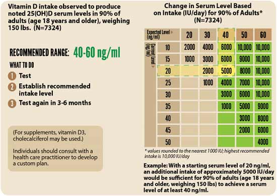
3. Retest in three to six months — Lastly, you’ll need to remeasure your vitamin D level in three to six months, to evaluate how your sun exposure and/or supplement dose is working for you.
- 1 Campbell Teaching
- 2, 3 French National Academy of Medicine May 22, 2020 (PDF)
- 4, 37, 38 Medrxiv May 18, 2020 DOI: 10.1101/2020.04.08.20058578
- 5 Clinicaltrials.gov Vitamin D Trials
- 6 Covidence UK Study
- 7, 10 The Guardian June 17, 2020
- 8, 9 My Green Pod Frontline Immune Support May 12, 2020
- 11 Scotland Herald June 17, 2020
- 12 MedRxiv DOI 10.1101/2020.05.08.20095893
- 13 Pharmacol Ther. 2020 May 13. doi: 10.1111/apt.15820
- 14 NutraIngredients.com April 28, 2020
- 15, 33 Emerginnova June 4, 2020
- 16 TILDA.tcd.ie Vitamin D deficiency in Ireland — Implications for COVID-19. Results from the Irish Longitudinal Study on Ageing (TILDA)
- 17 Medical Xpress April 6, 2020
- 18 Irish Medical Journal 2020; 113(5): 81
- 19, 21 Top Clin Nutr. 2022 Jul-Sep; 37(3): 203–217
- 20 J Clin Virol. 2011 Mar; 50(3): 194–200
- 22 Nutrients, 2020;12:988
- 23 Can Fam Physician. 2015 Jun; 61(6): 50
- 24 Endocr Pract. 2009 Jul–Aug; 15(5): 438–449
- 25 Clinical Therapeutics 2017; 39(5): 930-945 (PDF)
- 26, 27 Derma Endocrinology 2009 Jul-Aug; 1(4): 215–219
- 28, 30 The BMJ 2017;356:i6583
- 29 YouTube, Coronavirus Epidemic Update 30, 7:58 minutes
- 31 GrassrootsHealth.net, Vitamin D Reduces Colds and Flu
- 32 Nutrients April 2, 2020; 12(4): 988
- 34 MedRxiv June 10, 2020 DOI: 10.1101/2020.06.01.20112334
- 35 News Medical Life Sciences June 4, 2020
- 36 Infectious Diseases April 8, 2020 DOI: 10.21203/rs.3.rs-21211/v1
- 39, 40 Northwestern McCormick School of Engineering May 7, 2020
- 41, 43 DMinder app
- 42 GrassrootsHealth Magnesium and Vitamin K2 Combined Important for Vitamin D Levels
‘Anecdotals’ Documentary
Reproduced from original article:
https://articles.mercola.com/sites/articles/archive/2023/01/14/anecdotals-documentary.aspx
The original Mercola article may not remain on the original site, but I will endeavor to keep it on this site as long as I deem it to be appropriate.
Analysis by Dr. Joseph Mercola Fact Checked January 14, 2023
STORY AT-A-GLANCE
- The film “Anecdotals” provides a glimpse into the lives of people who have suffered significant adverse reactions from COVID-19 shots
- Those who spoke out about their shot-related injuries have been shamed, ridiculed and labeled unethical
- Those injured by the shots have been largely abandoned by the mainstream medical community; their medical issues have been politicized, while society provides no empathy
- With no programs in place to help those injured by COVID-19 shots, and many doctors afraid to even acknowledge the shot’s connection to patients’ symptoms, many of those harmed have nowhere to turn for help
- The film calls for an open dialogue and a movement from humanity to acknowledge the risks of COVID-19 shots, as well as those who are suffering due to them
People who have been harmed by COVID-19 shots have suffered a range of medical issues — everything from death and permanent disability to pericarditis, nerve damage and overwhelming fatigue. While their symptoms vary, they share several common themes:
- Abandonment — Those injured by COVID-19 jabs have been largely abandoned by the mainstream medical community and government.
- Shame — Those who spoke out about their injuries have been shamed, ridiculed and labeled unethical; their medical issues have been politicized, while society provides no empathy.
- Hopelessness — With no programs in place to help those injured by COVID-19 shots, and many doctors afraid to even acknowledge the shot’s connection to patients’ symptoms, many of those harmed feel lost and don’t know where to turn for help.
Bringing attention to the issue — and to the people whose lives have changed drastically since receiving a COVID-19 shot — is the first step to recovery. The film “Anecdotals” does just that, providing a glimpse into the lives of people who have suffered significant adverse reactions from COVID-19 shots.1
Many of them have been told their stories don’t matter. After all, they’re just anecdotes. But as you’ll see in the film, their journeys need to be heard, not only so they can access much-needed medical care but also so society becomes aware of the real risks of COVID-19 shots that have been covered up and censored.
Secrets From the Trials
One case involves Maddie de Garay, who was a healthy 12-year-old when she signed up for Pfizer’s COVID-19 trial for 12- to 15-year-olds. She suffered a severe systemic adverse reaction to her second dose of the shot, however, and struggled through 11 ER visits and four hospital admissions in the year and a half that followed.
Injuries from the shot have left her unable to walk or eat — she receives her nutrition via a feeding tube — and suffering from constant pain, vision problems, tinnitus, allergic reactions and lack of neck control.2
As though the physical trauma weren’t enough, Maddie and her family were continually dismissed by the medical professionals put in place to help, ignored by the U.S. Food and Drug Administration and denied the care needed to help Maddie. But the first red flag, Maddie’s mother Stephanie said at a hearing, was the way the trial was set up in the first place.3
Participants were given access to the TrialMax app to record side effects, like a swollen arm, but de Garay was surprised at the format it used. There wasn’t space for open-ended comments, only direct questions with “yes” or “no” options for answers, or check boxes to signify a set of predetermined potential effects.4 She explained:5
“I just want to give everybody a little better idea of what happened in our trial, because I did not know when you enter the trial, everybody uses a trial app. The app only allows you to record solicited adverse events — fever, redness, mild, moderate.
There’s no free form to fill in any other reaction that you have. What you have to do, if you have any other type of adverse event, is you have to call this study doctor. This leaves a lot of room for human error and concern of reporting bias coming from the principal investigator.”
In Pfizer’s April 2021 disclosure of Maddie’s case to the FDA, it’s stated only that she had abdominal pain:6
“One participant experienced an SAE [serious adverse event] reported as generalized neuralgia, and also reported 3 concurrent non-serious AEs (abdominal pain, abscess, gastritis) and 1 concurrent SAE (constipation) within the same week. The participant was eventually diagnosed with functional abdominal pain. The event was reported as ongoing at the time of the cutoff date.”
Then, a day before Pfizer submitted their request for emergency approval of the COVID-19 shot for 12- to 15-year-olds to the FDA, they added functional neurological disorder as a diagnosis in Maddie’s chart.7 Her mother noted in the film:8
“By the data cut off for the trial, Maddie experienced over 35 adverse events. None of these were mentioned … Maddie was in the hospital when the EUA [emergency use authorization] was approved. I thought that Maddie would be in the best hands possible in the rare chance she has a severe reaction. That was not the case. They did everything in their power to hide everything. Neither Pfizer, the FDA or the CDC has ever talked to us.”
Pfizer Trial ‘Like Nothing I’ve Ever Seen’
While health agencies continue to assure the public that COVID-19 shots are safe, those working closely on the trials had a different take. “I was working on Pfizer’s trial,” Brooke Jackson, a regional director formerly employed by Pfizer subcontractor Ventavia Research Group, which was testing Pfizer’s COVID-19 vaccine, said in the film.9 “What I saw was like nothing I’ve ever seen before.”
She witnessed falsified data, unblinded patients, inadequately trained vaccinators and lack of proper follow-up on adverse events that were reported. After notifying Ventavia about her concerns repeatedly, she made a complaint to the FDA directly — and was fired the same day.10 In her words:11
“The speed in which they were enrolling in the study — four to five coordinators pushing through 40, 50, 60 patients a day. We were not storing the vaccine at its appropriate temperature, the failures in reporting serious adverse events. We had so many reports of adverse events … we just could not keep up. The study doctor signed a physical exam when he wasn’t even in clinic.
Then Ventavia had unblinded every patient that was randomized in the trial. When we brought it to their attention, that’s what we were instructed to do — remove the evidence and destroy it. Emails about mislabeled blood specimens per Pfizer’s protocol, we should have immediately stopped enrolling, but they never told Pfizer.
I would bring the concerns to my managers and it was, ‘We’re understaffed.’ The FDA, they only see what Pfizer gives them. So I was documenting all of this. And on the 25th of September, I went directly to the FDA, and about six and a half hours later, I lost my job. I was fired.”
The FDA and Pfizer attempted to hide the COVID-19 shot clinical trial data for 75 years, but the FDA was ordered by the U.S. District Court for the Northern District of Texas to release redacted versions of trial documents on a much faster schedule. As part of the court order, 80,000 pages of documents related to the FDA’s approval of Pfizer’s COVID-19 shots were released June 1, 2022.12
Among those documents were case report forms (CRFs) revealing that deaths and severe adverse events took place during Phase 3 trials, but, as reported by Children’s Health Defense, Pfizer had “a trend of classifying almost all adverse events — and in particular severe adverse events (SAEs) — as being ‘not related’ to the vaccine.”13 Journalist Naomi Wolf explained:14
“We’ve got these amazing 2,500 volunteers — highly credentialed medical researchers, doctors and nurses — pouring over these 55,000 documents that a court order forced Pfizer and the FDA to release.
Well, they’re finding that there were horrible harms — deaths, spontaneous abortions, neurological problems, fainting, heart damage, debilitating muscle pain, debilitating joint pain — that were concealed by Pfizer and the FDA from the American people.”
Adverse Reactions — Real, Not Rare
The film details adverse reactions that have stolen careers, independence and the ability to function normally in daily life from countless people. Dr. Joel Wallskog, a former orthopedic surgeon, shared his story after getting the shot:15
“My life has dramatically changed after this adverse reaction. My career of 19 years, that I took almost 14 years to train for, is likely over. I’m just not safe to work as an orthopedic surgeon. Assuming the FDA and the CDC would be alarmed at my diagnosis, I expected to be contacted soon after my VAERS [Vaccine Adverse Event Reporting System] submission. No phone call, no contact.”
Kellai Rodriguez also detailed her struggles since receiving a COVID-19 shot:16
“I lost my ability to speak naturally. I have become unable to walk without a walker, and never know if or when the tremors will come or go. I can no longer cook, clean or even pick up and hold my baby for too long, before my body begins to shake uncontrollably or is thrown into excruciating amounts of pain.
I’ve seen countless ER doctors as well as two neurologists who have given me no diagnosis, no further testing besides regular bloodwork, CT scans, ECGs, EKGs and an MRI, all of which the doctors told me came back normal.”
At a rally for those injured by the shots, hundreds came together to share their experiences, with striking similarities. Many suffered from tremors that left them unable to walk, with onsets within days of receiving the shots. In the hospital, nurses shared that other patients were experiencing similar symptoms, but doctors refused to label the conditions shot-related. Jennifer Bridges, a former nurse with Houston Methodist Hospital, who was fired for not getting the shot, explained:17
“I’ve seen emails, where hospitals threatened their doctors — you cannot sign medical exemptions, you cannot talk about, you cannot report adverse reactions to these vaccines. And if somebody was actually brave enough to do that in writing, there were other people higher up to erase those. I have the proof, and I have the people that have shown me these things.”
Stories Censored and Silenced
Those injured by the shots were left abandoned during shot mandates. The film’s director, Jennifer Sharp, is among those who suffered from debilitating symptoms after the shot, including facial numbness, electric shock-like feelings and muscle weakness. She opted to not get a second dose of the shot after experiencing the serious adverse events after the first dose, and lost her job as a result:18
“In January 2022, I lost a job because I wasn’t vaccinated. I had a VAX card showing one shot, I had a blood test showing that I still had antibodies and a doctor’s exemption. And I was willing to get tested every day. They didn’t care. I couldn’t go to restaurants, gyms, malls, events.
So when the anti-mandate rally came to Los Angeles, I attended it to represent those of us who were suddenly societal outcasts just for doing what the government asked us to do. Even if you fundamentally disagree with someone else’s stance, does that justify the lack of compassion for them losing their livelihoods?”
Yet, when those affected tried to speak out about their experiences, they were silenced and shunned. One woman who was injured by the shots shared:19
“We are being so censored that we can’t get the message out that we’re even being censored, because if it’s through social media, they are one of the platforms that is censoring us. And even if it’s not outwardly, we’re being shadow banned …
So you could share something, but nobody acknowledges it. And you’re thinking, ‘Oh, I’m isolated, I’m alone,’ but they’re probably not seeing it. It’s been moved to the bottom of the timeline or it’s not in existence. You literally cannot post on social media about having a vaccine reaction without it being censored.”
When Sharp decided to film “Anecdotals,” she made a pitch video that she shared privately on the platform Vimeo. It described her reaction to the shot and the need for compassion. “It was removed for misinformation. They said they don’t allow content that goes against the CDC recommendations. I am not allowed to tell my own story,” she said.20
Suicides Due to COVID-19 Shot Reactions
Brianne Dressen, cofounder of React10, a nonprofit offering financial and other support to those suffering from long-term adverse events from COVID-19 shots, detailed several suicides among victims suffering from electric shocks, neuropathy, tinnitus, tremors and other effects from the shots. She also considered suicide due to adverse effects she suffered after participating in the AstraZeneca trial:21
“I don’t think people realize how debilitating the symptoms are. My husband couldn’t leave me alone for months. He’d leave the house and he didn’t know if he was going to come home to a wife that was alive. He was afraid, every moment of every day, and it seeps into our kids’ lives.
Six months, I was not mom, I was not a human. I was just going to drive down to the lake. And I was going to carbon monoxide my car. And I was gonna put AstraZeneca did this on a sign in the window. And I was too sick to do it. So only reason I’m alive is because I was too sick to do it. And I would like to finish with a letter from a friend, Bree:
‘I cannot take this any longer. This has taken everything away from me, my career, my family, my life, my body will not stop attacking itself. And this is beyond the worst amount of torture. Please accept my apologies. I must bid farewell to this world. Please make sure the world knows the cruelty that has been imposed upon us. Goodbye, my dear friend, I will see you on the flip side.’
Rochelle Walensky. Janet Woodcock, Peter Marks, Anthony Fauci, you erased her and the many others like her, their blood is on your hands. You cannot bring my friends back. But you can save others from their fate. If you finally just tell the truth.”
The film calls for an open dialogue and a movement from humanity to ask the difficult questions and acknowledge those who are suffering due to COVID-19 shots. “We must be seen, believed and helped,” Sharp said. “Our stories are anecdotal, but in a situation where the science is changing, the studies are flawed and political agendas regulate, anecdotes could quite possibly be the most reliable data that we have. Yes, we are anecdotal. And these are our stories.”22
- 1 Anecdotals Movie
- 2, 7 Life Funder, Help Maddie de Garay get essential medical care
- 3, 5 YouTube, Anecdotals Movie December 11, 2022, 6:46
- 4 The Highwire, Rigged: Maddie’s Story August 13, 2022
- 6 The Highwire, Rigged: Maddie’s Story August 13, 2022, 1:04
- 8 YouTube, Anecdotals Movie December 11, 2022, 8:00
- 9, 11 YouTube, Anecdotals Movie December 11, 2022, 12:01
- 10 BMJ 2021;375:n2635
- 12 Children’s Health Defense, The Defender, February 7, 2022
- 13 Children’s Health Defense, The Defender, June 21, 2022
- 14 YouTube, Anecdotals Movie December 11, 2022, 14:24
- 15 YouTube, Anecdotals Movie December 11, 2022, 19:00
- 16 YouTube, Anecdotals Movie December 11, 2022, 16:17
- 17 YouTube, Anecdotals Movie December 11, 2022, 26:00
- 18 YouTube, Anecdotals Movie December 11, 2022, 43:11
- 19 YouTube, Anecdotals Movie December 11, 2022, 55:21
- 20 YouTube, Anecdotals Movie December 11, 2022, 56:37
- 21 YouTube, Anecdotals Movie December 11, 2022, 1:15
- 22 YouTube, Anecdotals Movie December 11, 2022, 1:20
Top Foods to Improve Your Vision
Reproduced from original article:
https://articles.mercola.com/sites/articles/archive/2023/01/13/top-foods-to-improve-vision.aspx
The original Mercola article may not remain on the original site, but I will endeavor to keep it on this site as long as I deem it to be appropriate.
Analysis by Dr. Joseph Mercola Fact Checked January 13, 2023

STORY AT-A-GLANCE
- Nourishing your eyes with appropriate nutrients throughout the years can go a long way toward maintaining good eyesight well into your senior years
- Some of the most important nutrients for eye health include lutein, zeaxanthin, meso-zeaxanthin, astaxanthin, anthocyanins, animal-based omega-3 fats and vitamin C
- Foods rich in beneficial lutein include egg yolks, spinach, avocado and broccoli; black currants and bilberries also contain important antioxidants for vision health
- Avoiding blue light, especially from cool white LED lights, may help prevent vision deterioration
Aging is inevitable, but are age-related problems such as poor vision absolute givens? Contrary to popular belief, your vision is largely dependent on your lifestyle. Nourishing your eyes with appropriate nutrients throughout the years can go a long way toward maintaining good eyesight well into your senior years.
Even if your eyesight has started to deteriorate, evidence suggests you can stop the deterioration. You may even be able to turn back the clock, as it were, and improve your vision.
Foods Can Protect and Improve Your Eyesight
Medical journalist Michael Mosley discussed his vision problems and experiences with nutritional intervention in an episode of the BBC program “Trust Me, I’m a Doctor.”1 The shape and length of your eyeballs, and the thickness of your eye lens, affect your ability to see things close-up and at a distance. Your retina, located at the back of your eye, also contains light-sensitive cells that are critical for good vision.
Your macula — the part of your retina responsible for central vision — is protected by a yellow pigment made up of lutein, zeaxanthin and meso-zeaxanthin. These compounds absorb light and protect your macula from blue light and ultraviolet (UV) light from the sun and other light sources.2
Lutein, zeaxanthin and meso-zeaxanthin are plant compounds with potent antioxidant capacities. Your body cannot make them, so you must get them from your diet. As noted by the BBC:3
“Lutein and zeaxanthin are found commonly in dark green leafy veg such as kale and spinach, and also bell peppers … and saffron. Meso-zeaxanthin is generally not found in plants — it is thought to be made in our bodies from lutein (although it is also present in some fish …) These pigments, once we eat them, appear to be important in our vision and in helping keep the macula healthy.”
Lutein and Zeaxanthin Supplements Can Make a Big Difference
Mosley describes undergoing a number of extensive vision tests designed to evaluate the health of his retina, his ability to see colors, night vision and the level of protection his macula had against UV and blue light. He explained:4
“The results of my tests were both fascinating and depressing. My detection of yellow and blue colors was extremely poor — something that [Professor John] Barbur said was likely the result of my brush with diabetes many years ago.
My night vision and perception of details were also poor compared with younger people — but consistent with my age. The Trust Me team then handed me a 90-day supply of supplement pills that were supposed to help.”
The supplements in question contained lutein and zeaxanthin. Three months later, follow-up tests revealed remarkable improvement. Not only did his night vision and protective macular pigments improve, but his blue and yellow color perception moved within the normal range.
Some studies suggest these nutrients may also slow down or prevent age-related macular degeneration (ARMD),5 which is the leading cause of vision loss and blindness for Americans aged 65 and older.6
How Much Lutein and Zeaxanthin Do You Need?
While there’s no recommended daily intake for lutein and zeaxanthin, studies have found health benefits for lutein at a dose of 10 milligrams (mg) per day7 and at 2 mg per day for zeaxanthin.8 Meanwhile, studies suggest American adults get, on average, only 1 mg to 2 mg of lutein from their diet each day.9
Research evaluating the effect of lutein, zeaxanthin and meso-zeaxanthin in combination, using a dose of 10 mg of lutein, 10 mg of meso-zeaxanthin and 2 mg of zeaxanthin per day for one year, found it helped improve vision in those who had normal vision at the outset.10,11
“Should we all, then, be taking supplements to protect and even improve our eyesight? Well, the research certainly shows that supplements work,” Mosley wrote.12
“Even for someone like me, who has a relatively healthy diet with plenty of fruit and vegetables, and whose blood levels of the compounds that weren’t particularly low, the supplements helped. However, some researchers believe that diet can simply be enough, if we eat the right things.”
What Should You Eat to Protect or Improve Your Vision?
Lutein and zeaxanthin are primarily found in green leafy vegetables, with kale and spinach topping the list of lutein-rich foods. You’ll also find these nutrients in orange- and yellow-colored fruits and vegetables.
According to a study published in the British Journal of Ophthalmology, orange pepper had the highest amount of zeaxanthin of the 33 fruits and vegetables tested.13 According to the authors:
“Most of the dark green leafy vegetables, previously recommended for a higher intake of lutein and zeaxanthin, have 15 [to] 47 percent of lutein, but a very low content (0 to 3 percent) of zeaxanthin. Our study shows that fruits and vegetables of various colors can be consumed to increase dietary intake of lutein and zeaxanthin.”
Egg yolk is another good source of both lutein and zeaxanthin, along with healthy fat and protein, and while the total amount of carotenoids is lower than in many vegetables, they’re in a highly absorbable, nearly ideal form. For instance, one study found that adding a couple of eggs to your salad can also increase the carotenoid absorption from the whole meal as much as ninefold.14
Eggs from free-range, pastured hens have bright orange yolks, which is an indication of their elevated lutein and zeaxanthin content. Dull, pale yellow yolks are a sure sign you’re getting eggs form caged hens fed an unnatural grain diet, and hence will have low amounts of these valuable nutrients.
For a beverage, one of your best choices is chrysanthemum tea, as it contains vitamin A, a nutrient that plays a critical role in promoting healthy vision. This vitamin is an essential component of rhodopsin, a protein that absorbs light in the retinal receptors.
It also helps promote normal function of the conjunctival membranes and cornea, and helps reduce the risk for eye disorders like age-related macular degeneration.15
What Are the Best Food Sources of Lutein?
Following is a list of foods that are particularly rich in lutein.16,17,18 Most of these also contain zeaxanthin, albeit in lesser quantities than lutein.
- Egg yolks
- Kale and spinach
- Avocado
- Broccoli
- Green, red and yellow peppers
Ideally, you’ll want to buy the whole food and consume it as close to raw as possible, as the lutein and other carotenoids such as zeaxanthin are easily damaged by heat. Accessory micronutrients in the foods that enhance their action also tend to get easily damaged. Lutein and other carotenoids are fat-soluble, so to optimize absorption, be sure to add a little bit of healthy fat to your meal.
Vitamin C Combats Cataracts
Other nutrients are also beneficial for vision health. For instance, vitamin C is associated with a lower risk of cataracts. According to the National Eye Institute, more than half of all Americans end up getting cataracts by the time they’re 80.19
One study compared vitamin C intake and the progression of cataracts in more than 320 pairs of female twins over the course of a decade.20 It found those who ate more vitamin C-rich foods lowered their risk of cataracts by one-third. Interestingly, vitamin C supplements were not associated with a reduction in risk.
Citrus fruits such as kiwis, oranges, lemons, limes and grapefruits are well-known for being high in vitamin C, but the fruit with the highest concentration of all is actually acerola cherries (also known as Barbados cherries). Each cherry has only 1 calorie, but 80 mg of vitamin C and the associated micronutrients. I have two of these trees in my front yard and for months I am able to gather 50 to 70 cherries a day. It is by far my favorite and healthiest fruit.
Animal-Based Omega-3 Lowers Risk of Blindness in Diabetics
Another study21 found that diabetics who routinely ate 500 mg of omega-3-rich fish (two servings per week) reduced their risk of diabetic retinopathy by an impressive 48%. Diabetic retinopathy is a serious complication of Type 2 diabetes that occurs when blood flow to your retina is reduced. It’s the most common cause of blindness in diabetics.
This significant risk reduction was primarily attributed to lower inflammation levels. Animal-based omega-3 fats also provide structural support to cell membranes that boost eye health and protect retinal function. Earlier research14 has shown that those with the highest intake of animal-based omega-3 fats have a significantly lower risk of advanced macular degeneration compared to those who consume the least.22
In a survey of optometrists in Australia and New Zealand, 79% said they recommended their patients consume omega-3 fats to improve eye health.23 Specifically, 68% recommended omega-3-rich foods for ARMD while 62% recommended omega-3 supplements for this purpose.
Seventy-eight percent also recommended omega-3-rich foods or supplements for dry eye disease. Marine sources that are high in omega-3 and low in environmental pollutants include:
- Wild Alaskan salmon (which also contains astaxanthin — one of the most potent promoters of eye health; see below)
- Small fatty, cold-water fish such as herring, sardines and anchovies
- Fish roe
- Krill oil
While fish oil is a well-known source of omega-3 fats, it has several drawbacks, the lack of phospholipids being one of them. The omega-3 fats DHA and EPA are water insoluble and therefore cannot be transported in their free form in your blood. They must be packaged into lipoprotein vehicles such as phospholipids. This is primarily why the bioavailability of krill oil is so much higher than fish oil, because in fish oil, the DHA and EPA are bound to triglycerides.
Astaxanthin — The Most Powerful Promoter of Eye Health
Astaxanthin is produced by the microalgae Haematococcus pluvialis when its water supply dries up, forcing it to protect itself from UV radiation. Besides the microalgae that produce it, the only other source are the sea creatures that consume the algae, such as wild salmon, shellfish and krill.
Astaxanthin is far more powerful an antioxidant than both lutein and zeaxanthin, and many researchers believe it to be the most powerful antioxidant ever discovered for eye health.24,25 It’s been found to have protective benefits against a number of eye-related problems, including ARMD and cataracts, as well as:
- Cystoid macular edema
- Diabetic retinopathy26
- Retinal arterial occlusion and venous occlusion
- Glaucoma27
- Inflammatory eye diseases (retinitis, iritis, keratitis and scleritis)
Astaxanthin easily crosses into the tissues of your eye and exerts its effects safely and with more potency than any of the other carotenoids, without adverse reactions. Specifically, astaxanthin has been shown to ameliorate or prevent light-induced damage, photoreceptor cell damage,28 ganglion cell damage and damage to the neurons of the inner retinal layers.
Astaxanthin also helps maintain appropriate eye pressure levels that are already within the normal range, and supports your eyes’ energy levels and visual acuity. Depending on your individual situation, you may want to take an astaxanthin supplement.
I recommend starting with 4 mg per day and working your way up to about 8 mg per day — or more if you’re suffering from chronic inflammation. Taking your astaxanthin supplement with a small amount of healthy fat, such as grass fed butter, coconut oil, MCT oil or eggs, will optimize its absorption.
Krill oil also contains high quality animal-based omega-3 fat in combination with naturally occurring astaxanthin, albeit at lower levels than what you’ll get from an astaxanthin supplement.
How Anthocyanins in Berries Benefit Your Eyes
Dark blue or purplish, almost black-colored, berries like black currants and bilberries contain high amounts of antioxidant anthocyanins that are beneficial for vision and eye health.29 Black currants contain some of the highest levels. They’re also rich in essential fatty acids, lending added support to their anti-inflammatory properties.
For medicinal purposes, many opt for using black currant seed oil, but eating the whole food is always an option, especially when they’re in season. Bilberry, a close relative of the blueberry, also contain high amounts of anthocyanins, just like black currant. Research suggests bilberry may be particularly useful for inhibiting or reversing macular degeneration.
A 2005 study found that rats with early senile cataract and macular degeneration that received 20 mg of bilberry extract per kilogram of body weight suffered no impairment of their lens and retina, while more than 70% of the control group suffered degeneration over the three-month-long study.30 According to the authors, “The results suggest that … long-term supplementation with bilberry extract is effective in prevention of macular degeneration and cataract.”
Even More Important: Avoid Artificial Blue Light
The full extent of the health benefits associated with blue light avoidance are only beginning to be understood. Glasses that block blue light have the benefit of making objects appear sharper, but mounting research suggests blocking blue light also serves an important biological purpose by regulating your internal body clock, which controls your sleep patterns and other body functions.
Indeed, exposure to artificial light is among the most often-overlooked health risks of living in the 21st century.
One of the simplest and least expensive ways to protect your body’s internal rhythm, and thereby support healthy sleep and a lowered risk of many chronic diseases, is to wear amber-colored glasses that block blue light — not just at night but anytime you are exposed to fluorescent or LED lights.
This is because LEDs and fluorescents, although far more energy efficient, are not analog thermal light sources but digital ones. They have unbalanced wavelengths that are very different from the sun. They have a predominance of blue frequencies that contributes to free radical production, and lack the red, infrared and near infrared healing frequencies that stimulate repair and regeneration that are present in thermal light sources like clear incandescent bulbs.
LEDs found in many screens have virtually no beneficial infrared light and an excess of blue light that generates reactive oxygen species (ROS), harming your vision and possibly leading to age-related macular degeneration.31 LED lights may also exacerbate mitochondrial dysfunction leading to chronic conditions ranging from metabolic disorders to cancer.
If you view screens at night, it’s therefore essential to block your exposure to blue light while doing so. In the case of your computer, you can install a program to automatically lower the color temperature of your screen. In addition, when watching TV or other screens, be sure to wear blue-blocking glasses after sundown. Better yet, eliminate the use of screens entirely after sunset.
Additionally, one of the best strategies to establish your circadian rhythm is to spend some time outdoors shortly after sunrise. The red and infrared frequencies will help prepare your retina for the blue light emitted a bit after sunrise, which will produce a reactive oxygen signal to produce melatonin later that night — assuming you sleep in complete darkness.
Establishing a healthy circadian rhythm is essential for good health, so please consider the healthy habit of getting outside every day shortly after sunrise without glasses to expose your retina to the full spectrum of sunlight.
It is likely that chronic exposure to unopposed digital blue light from LED and fluorescents will likely contribute to an epidemic of blindness in the next two decades, as it will increase the incidence of ARMD. The benefits of blue-blocking glasses and avoidance of blue light include but are not limited to:
- Preventing damage to the DHA in your retinal pigmented epithelium (RPE), which is responsible for converting the photons from sunlight into electrons via the photoelectric effect that Einstein received a Nobel prize for in 1921. These electrons provide a vital DC electric current that your body needs for optimal functioning
- Increasing mitochondrial health and efficiency, as blue light has the side effect of increasing the distance of the proteins in the respiratory electron transport chain in your mitochondria, making them far less efficient in producing ATP. Blocking blue light prevents this decline in efficiency from occurring
- Significantly improving symptoms of bipolar disorder. Those who wore blue-blocking orange-tinted glasses from 6 p.m. to 8 a.m. for seven days had significant improvements in symptoms of mania compared to those who wore clear glasses.32 What’s more, the improvements began after just three nights of use
The dramatic results make sense in light of the relatively recent discovery of intrinsically photo-responsive retinal ganglion cells, which are receptors in your eyes that detect only blue light.
These receptors communicate with areas of your brain linked to control of your biological clock (hypothalamus) as well as mood and emotions (the limbic system).33 Earlier research also found dramatic improvements in insomnia and mood in about half of bipolar patients who wore blue-blocking glasses.34
Optimize Your Eyesight by Eating Right and Living Healthy
Besides eating plenty of carotenoid-rich vegetables, organic pastured egg yolks and omega-3 and astaxanthin-rich wild Alaskan salmon, another important dietary aspect is to normalize your blood sugar. Excessive sugar in your blood can pull fluid from the lens of your eye, affecting your ability to focus. It can also damage the blood vessels in your retina, thereby obstructing blood flow.
It’s important to ignore the mainstream advice to eat vegetable oils, which are more accurately defined as seed oils. Seed oils, such as soybean, cottonseed, sunflower, rapeseed (canola), corn and safflower, are hidden in virtually every processed food, including restaurant foods, and there’s virtually nothing more destructive to your body in producing age-related macular degeneration35 and other chronic health problems.
These seed oils contain the omega-6 fat linoleic acid, which is toxic at the high doses many Americans consume daily. In your own cooking, examples of healthy fats to use instead of seed oils are beef tallow, butter or coconut oil. As mentioned, it’s also important to avoid ultraprocessed foods.
Besides addressing your diet, you’ll also want to exercise regularly, and be sure to optimize your vitamin D level. Vitamin D is particularly important for those with genetic risk factors for ARMD. In one study, middle-aged women who have a high-risk genotype and are vitamin D deficient were found to be 6.7 times more likely to develop ARMD than those without this genetic risk factor who also have sufficient vitamin D.36,37
- 1, 4, 12 BBC News September 6, 2016
- 2 Annual Review of Nutrition 2003;23:171-201
- 3 BBC, Trust Me, I’m a Doctor, Can I Improve My Eyesight?
- 5 Med Hypotheses. 2003 Oct;61(4):465-72
- 6 U.S. CDC, Learn About Age-Related Macular Degeneration
- 7 Investigative Ophthalmology & Visual Science December 2006, Vol.47, 5227-5233
- 8 Verywell Health December 9, 2022
- 9 The Journal of Nutrition March 1, 2002; 132(3): 518S-542S
- 10 Investigative Ophthalmology and Visual Sciences June 1, 2016;57(7):3429-39
- 11 Nutraingredients-USA.com July 14, 2016
- 13 British Journal of Ophthalmology Aug 1998; 82(8): 907–910
- 14 Science Daily March 9, 2015
- 15 NIH Office of Dietary Supplements, Vitamin A
- 16 Self Nutrition Data Lutein + Zeaxanthin
- 17 Nutrients 2013 Apr; 5(4): 1169–1185
- 18 USDA, Healthy Colors of Your Diet
- 19 National Eye Institute, Cataracts
- 20 Opthalmology June 2016; 123(6): 1237-1244
- 21 JAMA Ophthalmol. 2016;134(10):1142-1149
- 22 Ophthalmology 117 (12): 2395-2401, December 2010
- 23 Nutrients. 2020 Apr 22;12(4):1179. doi: 10.3390/nu12041179
- 24 Journal of the Science of Food and Agriculture May 1, 2001: 81(6); 559-568
- 25 Oxid Med Cell Longev. 2019; 2019: 3849692
- 26 J Ophthalmol. 2022; 2022: 8071406
- 27 Mar Drugs. 2020 May; 18(5): 239
- 28 Antioxidants (Basel). 2020 Aug; 9(8): 729
- 29 Molecules. 2019 Sep; 24(18): 3311
- 30 Advances in Gerontology 2005;16:76-9
- 31 Cleveland Clinic August 9, 2019
- 32 Bipolar Disorders May 26, 2016
- 33 Newsweek July 26, 2016
- 34 Chronobiol Int. December 2009;26(8):1602-12
- 35 Free Radic Biol Med. 2019 Dec;145:349-356. doi: 10.1016/j.freeradbiomed.2019.10.007. Epub 2019 Oct 9
- 36 Science Daily August 31, 2015
- 37 JAMA Ophthalmol. 2015;133(10):1171-1179
Bradykinin Hypothesis Explains COVID-19 Complexities
Reproduced from original article:
https://articles.mercola.com/sites/articles/archive/2023/01/13/bradykinin-hypothesis.aspx
The original Mercola article may not remain on the original site, but I will endeavor to keep it on this site as long as I deem it to be appropriate.
Analysis by Dr. Joseph Mercola Fact Checked January 13, 2023
STORY AT-A-GLANCE
- Genetic analysis using the Oak Ridge National Lab supercomputer has revealed an interesting new hypothesis — the bradykinin hypothesis — that helps explain the disease progression of COVID-19
- The bradykinin hypothesis also strengthens the hypothesis that vitamin D plays a really important role in the disease
- SARS-CoV-2 not only infects cells that naturally have high numbers of ACE2 receptors but also tricks your body into upregulating ACE2 receptors in places where they’re usually expressed at lower levels, such as your lungs
- SARS-CoV-2 also downregulates your body’s ability to degrade or break down bradykinin, a chemical that helps regulate your blood pressure and is controlled by your renin-angiotensin system
- The end result is a bradykinin storm, which appears to be the primary cause behind many of COVID-19’s lethal effects, even more so than the cytokine storms associated with the disease
- The virus also increases production of hyaluronic acid (HLA) in your lungs. HLA has the ability to absorb more than 1,000 times its own weight in fluid, and when it combines with the built-up fluid in the lungs, it forms a thick hydrogel that makes breathing very difficult
Genetic analysis using the Oak Ridge National Lab supercomputer called the Summit has revealed an interesting new hypothesis that helps explain the disease progression of COVID-19. A September 1, 2020, Medium article1 by Thomas Smith reviewed the findings of what is now referred to as the bradykinin hypothesis.
As reported by Smith, the computer crunched data on more than 40,000 genes obtained from 17,000 genetic samples.
“Summit is the second-fastest computer in the world, but the process — which involved analyzing 2.5 billion genetic combinations — still took more than a week. When Summit was done, researchers analyzed the results. It was, in the words of Dr. Daniel Jacobson, lead researcher and chief scientist for computational systems biology at Oak Ridge, a ‘eureka moment.’”
The Bradykinin Hypothesis
Bradykinin is a chemical that helps regulate your blood pressure and is controlled by your renin-angiotensin system (RAS). As explained in the Academic Press’ book on vitamin D (which has a significant impact on the RAS):2
“The renin-angiotensin system (RAS) is a central regulator of renal and cardiovascular functions. Over-activation of the RAS leads to renal and cardiovascular disorders, such as hypertension and chronic kidney disease, the major risk factors for stroke, myocardial infarction, congestive heart failure, progressive atherosclerosis, and renal failure.”
The bradykinin hypothesis provides a model that helps explain some of the more unusual symptoms of COVID-19, including its bizarre effects on the cardiovascular system. It also strengthens the hypothesis that vitamin D plays a really important role in the disease.
The findings3 were published in the journal eLife July 7, 2020. Based on this new hypothesis, the researchers also suggest more than 10 potential treatments, most of which are readily available drugs already approved by the U.S. Food and Drug Administration. I’ll review those later on.
As detailed in previous articles, your ACE2 receptors are the primary gateways of the virus, as the virus’ spike protein binds to the ACE2 receptor. As explained by Smith:4
“… COVID-19 infection generally begins when the virus enters the body through ACE2 receptors in the nose … The virus then proceeds through the body, entering cells in other places where ACE2 is also present: the intestines, kidneys, and heart. This likely accounts for at least some of the disease’s cardiac and GI symptoms.
But once Covid-19 has established itself in the body, things start to get really interesting … The data Summit analyzed shows that COVID-19 isn’t content to simply infect cells that already express lots of ACE2 receptors. Instead, it actively hijacks the body’s own systems, tricking it into upregulating ACE2 receptors in places where they’re usually expressed at low or medium levels, including the lungs.
In this sense, COVID-19 is like a burglar who slips in your unlocked second-floor window and starts to ransack your house. Once inside, though, they don’t just take your stuff — they also throw open all your doors and windows so their accomplices can rush in and help pillage more efficiently.”
Bradykinin Storm Likely Responsible for Lethal Effects
In addition to upregulating ACE2 receptors throughout your body, the SARS-CoV-2 virus also downregulates your body’s ability to degrade or break down bradykinin.
The end result is a bradykinin storm, and according to the researchers, this appears to be an important factor in many of COVID-19’s lethal effects, even more so than the cytokine storms associated with the disease. As bradykinin accumulates, the more serious COVID-19 symptoms appear.
Mounting clinical data suggest COVID-19 is actually primarily a vascular disease rather than a respiratory one, and runaway bradykinin buildup can help explain this.
How SARS-CoV-2 Attacks the Lungs
That said, COVID-19 certainly has a respiratory component, and it appears the virus attacks the lungs in more ways than one. For starters, bradykinin increases vascular permeability, essentially causing your blood vessels to leak fluid. In the lungs, this leads to fluid buildup that can trigger inflammation when immune cells also leak out into the lungs.
But the Summit data also show the virus uses yet another pathway, which raises production of hyaluronic acid (HLA) in your lungs. HLA has the ability to absorb more than 1,000 times its own weight in fluid, and when it combines with the built-up fluid in the lungs, the effect is devastating, as it ends up forming a thick hydrogel that makes breathing near-impossible.
When this happens — in severe cases — even mechanical ventilation becomes ineffective, as the alveoli in the lungs are simply too clogged with this gel-like substance that prevents oxygen uptake.
Note that the HLA produced in your lungs does not mean that using supplemental HLA is a bad strategy. It is only when HLA is produced locally in high concentrations in pathologic conditions like COVID-19 that it becomes problematic. Otherwise it has important physiologic benefits.
How SARS-CoV-2 Attacks Your Heart and Brain
SARS-CoV-2 can also affect heart function, causing arrhythmias and low blood pressure. About 1 in 5 COVID-19 patients requiring hospitalizations have experienced damage to their heart. Your heart has ACE2 receptors, so SARS-CoV-2 has the ability to infect your heart directly. Arrhythmias and low blood pressure can also be the result of a bradykinin storm.
In some cases, COVID-19 has also been known to trigger neurological symptoms such as dizziness, seizures, delirium and stroke, and this too can be explained by bradykinin buildup.
At high levels, bradykinin can lead to a breakdown of your blood-brain barrier, thereby allowing harmful compounds to flood your brain. Bradykinin itself also causes blood vessel leakage. Together, these effects can trigger inflammation, brain damage and a variety of neurological symptoms.
SARS-CoV-2 Mimics ACE Inhibiting Drugs
Interestingly, as reported by Smith:5
“Increased bradykinin levels could also account for other common COVID-19 symptoms. ACE inhibitors — a class of drugs used to treat high blood pressure — have a similar effect on the RAS system as COVID-19, increasing bradykinin levels.
In fact, Jacobson and his team note in their paper that ‘the virus … acts pharmacologically as an ACE inhibitor’ — almost directly mirroring the actions of these drugs.
By acting like a natural ACE inhibitor, COVID-19 may be causing the same effects that hypertensive patients sometimes get when they take blood pressure-lowering drugs. ACE inhibitors are known to cause a dry cough and fatigue, two textbook symptoms of COVID-19.
And they can potentially increase blood potassium levels, which has also been observed in COVID-19 patients. The similarities between ACE inhibitor side effects and COVID-19 symptoms strengthen the bradykinin hypothesis, the researchers say.”
Another side effect associated with ACE inhibiting drugs is the loss of smell and taste. This is also an early sign associated with SARS-CoV-2 infection, and it’s a primary symptom of zinc deficiency too.
Zinc, as explained in “Swiss Protocol for COVID — Quercetin and Zinc,” plays a vital role in immunity as well as in blood clotting, cell division, thyroid health, smell and taste, vision and wound healing, and can effectively inhibit viral replication.
Your body does not store zinc, and it’s poorly absorbed, which appears to be why the combination of zinc and zinc ionophores such as quercetin and hydroxychloroquine are so effective when taken at first symptoms.
Bradykinin Hypothesis Explains Other COVID-19 Symptoms Too
The bradykinin storm also helps explain other odd COVID-19 symptoms such as “COVID toes,” a condition in which your toes become swollen and bruised. This may be due to leaky vasculature in your toes.
As explained by Smith, it can also shed helpful light on the gender differences seen in COVID-19. Women tend to have a lower mortality rate than men, and this may be due to the fact that women have twice the level of certain proteins involved in the RAS system.
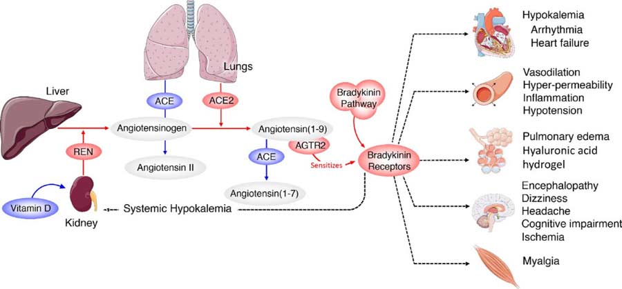 Image source: Daniel Jacobson et al. via eLife Sciences
Potential Treatments
The good news is that if bradykinin storms are to blame, there are a number of already existing drugs that can help prevent bradykinin storms, either reducing bradykinin or blocking its receptors. As noted in the study:6
“Several interventional points (most of them already FDA-approved pharmaceuticals) could be explored with the goal of increasing ACE, decreasing BK [bradykinin], or blocking BK2 receptors.
Icatibant is a BKB2R antagonist whereas Ecallantide acts to inhibit KLKB1, reducing levels of BK production. Androgens (danazol and stanasolol) increase SERPING1, although the side effects likely make these undesirable, but recombinant forms of SERPING1 could be administered to reduce BK levels.
It should be noted that any intervention may need to be timed correctly given that REN levels rise on a diurnal cycle, peaking at 4AM which corresponds with the commonly reported worsening of COVID-19 symptoms at night …
4-methylumbelliferone (Hymecromone) is a potent inhibitor of HAS1, HAS2, and HAS3 gene expression and results in the suppression of the production of hyaluronan in an ARDS model.
Hymecromone (4-methylumbelliferone) is approved for use in Asia and Europe for the treatment of biliary spasm. However, it can cause diarrhea with subsequent hypokalemia, so considerable caution should be used if this were to be tried with COVID-19 patients … Timbetasin may reduce COVID-19 related coagulopathies by increasing fibrinolysis.”
However, please understand that taking these drugs is absolutely not my recommendation. These are simply the conventional strategies that can be used if this hypothesis is correct.
Why on earth would we use dangerous drugs like these for a disease that they have not been tested on when we have so many other safe, inexpensive and highly effective interventions for COVID-19? Strategies like nebulized peroxide, ozone, molecular hydrogen, exogenous ketones, and quercetin with zinc.
Evidence for Vitamin D Strengthens
The researchers also highlight the usefulness of vitamin D, noting that “Another approach would be the modulation of REN levels via Vitamin D supplementation …” Vitamin D is involved in the RAS system,7,8,9 and can reduce a compound called renin (REN), thereby preventing a deadly bradykinin storm. Renin is an endopeptidase, the function of which is to generate angiotensin 1 from angiotensinogen in your plasma.
If you are vitamin D deficient, your renin expression is stimulated, and based on the latest data, that may render you more prone to bradykinin storm.
Several investigations have highlighted the apparent influence of vitamin D in COVID-19 incidence, severity and mortality, and its effects on RAS further strengthens the idea that vitamin D may be a crucial component in your COVID-19 defense arsenal.
As explained in the 2004 paper,10 “Vitamin D: A Negative Endocrine Regulator of the Renin-Angiotensin System and Blood Pressure,” when the RAS system is inappropriately activated, high blood pressure can result.
One factor that influences your RAS is your vitamin D level, as it suppresses renin biosynthesis. If you are vitamin D deficient, your renin expression is stimulated, and based on the latest data, that may render you more prone to bradykinin storm.
The Evidence for Vitamin D
In a November 1, 2020 commentary11 in the journal Metabolism Clinical and Experimental, JoAnn Manson and Shari Bassuk call for the elimination of vitamin D deficiency to effectively squelch the COVID-19 pandemic, noting that 23.3% of the total U.S. population have insufficient or deficient vitamin D levels, with people of color having disproportionately lower levels than non-Hispanic whites.
They list several types of studies showing vitamin D deficiency is “an important modifiable risk factor for COVID-19,” including:12
• Laboratory studies that demonstrate how vitamin D helps regulate immune function and the RAS, and modulate inflammatory responses to infection.
• Ecologic studies showing populations with lower vitamin D levels or lower UVB radiation exposure have higher COVID-19 mortality,13,14,15 and the fact that people identified as being at greatest risk for COVID-19 hospitalization and death (people of color, the elderly, nursing home residents and those with comorbidities such as obesity, vascular conditions and chronic kidney disease) also have a higher risk of vitamin D deficiency.
A pilot randomized clinical study16,17 published online August 29, 2020, found hospitalized COVID-19 patients in Spain who were given supplemental vitamin D (calcifediol) in addition to standard of care — which included the use of hydroxychloroquine and azithromycin — had significantly lower intensive care unit admissions.
Patients in the vitamin D arm received 532 micrograms of calcifediol on the day of admission (equivalent to 106,400 IUs of vitamin D18) followed by 266 mcg on Days 3 and 7 (equivalent to 53,200 IUs19). After that, they received 266 mcg once a week until discharge, ICU admission or death.
Of those receiving calcifediol, only 2% required ICU admission, compared to 50% of those who did not get calcifediol. None of those given vitamin D supplementation died, and all were discharged without complications.
• Observational studies showing low vitamin D levels are associated with a greater risk of testing positive for SARS-CoV-2 and contracting acute respiratory infections.
Most recently, a September 3, 2020 JAMA study20 reported that people who tested positive for SARS-CoV-2 were 1.77 times more likely to be deficient in vitamin D than those who tested negative for the virus.
• Randomized clinical trials showing vitamin D inhibits respiratory tract infections, especially in those with lower vitamin D levels at baseline.
REALLY IMPORTANT: Optimize Your Vitamin D Level Now
For years, I’ve stressed the importance of optimizing your vitamin D level, especially in anticipation of flu season, and it seems clear it can go a long way toward protecting yourself against COVID-19 as well.
Aside from what’s already been mentioned, vitamin D also helps Type II cells in your lungs produce surfactant that aids in fluid clearance. When you’re vitamin D deficient, your entire RAS is deranged or dysfunctional, thereby raising your risk of both bradykinin storm and cytokine storm.
In closing, experts have been warning that SARS-CoV-2 may reemerge in the fall when temperatures and humidity levels drop, thereby increasing the virus’ transmissibility.
Now is the time to check your vitamin D level and start taking action to raise it if you’re below 60 ng/mL. An easy and cost-effective way of measuring your vitamin D level is to order GrassrootsHealth’s vitamin D testing kit and learn more about vitamin D and its impact on your health.
Knowledge is empowerment, and that is particularly true during this pandemic. To learn more about the influence of vitamin D on your health in general and COVID-19 in particular, see my vitamin D report.
- 1, 4, 5 Medium September 1, 2020
- 2, 9 Vitamin D (Third Edition) 2011: 707-723
- 3, 6 eLife July 7, 2020; 9:e59177
- 7 Journal of Cellular Biochemistry February 1, 2003; 88(2):327-31
- 8 Journal of Nephropathology 2014; 3(2): 41–43
- 10 The Journal of Steroid Biochemistry and Molecular Biology May 2004; 89-90: 387-392
- 11 Metabolism Clinical and Experimental November 1, 2020; 112: 154322
- 12 Metabolism Clinical and Experimental November 1, 2020; 112: 154322, Table 1
- 13 MedRxiv DOI: 10.1101/2020.04.08.20058578
- 14 Northwestern.edu May 7, 2020
- 15 Fox News May 7, 2020
- 16 The Journal of Steroid Biochemistry and Molecular Biology August 29, 2020 [Epub ahead of print]
- 17, 18, 19 Chrismasterjohnphd.com September 3, 2020
- 20 JAMA Network Open 2020;3(9):e2019722
Key Food and Lifestyle Strategies for a Healthier Life
Reproduced from original article:
https://articles.mercola.com/sites/articles/archive/2023/01/08/lifestyle-strategies-healthier-life.aspx
The original Mercola article may not remain on the original site, but I will endeavor to keep it on this site as long as I deem it to be appropriate.
Analysis by Dr. Joseph Mercola Fact Checked January 08, 2023
STORY AT-A-GLANCE
- Avoiding industrial seed oils, due to their high linoleic acid (LA) content, can go a long way toward safeguarding your health. Examples of cooking oils to avoid include canola, corn, rapeseed, cottonseed and soybean oil. As a general rule, anything over 10 grams of LA a day is likely to cause problems
- For cooking, excellent substitutions include butter, tallow, ghee and organic coconut oil. Olive oil and avocado oil are problematic, as most are adulterated with cheap seed oils
- If you’re older, or eat a vegan or vegetarian diet, you’re at high-risk for vitamin B12 deficiency, which can have serious ramifications for your neurological function. Low B12 can also lead to elevated homocysteine, which raises your risk for dementia and cardiovascular problems
- Carnosine, found in meat, helps reduce oxidative stress, especially as it relates to LA. Carnosine binds to harmful LA byproducts and helps your body excrete them. It’s also important for building muscle, and acetyl carnitine is very beneficial for your memory
- Oxalates, found in a wide variety of fruits, vegetables and nuts, can cause problems when they overaccumulate. Harmful effects include fibromyalgia, chronic inflammation and kidney stones
In the “Inspired” interview above, I discuss what I believe are some of the most important dietary and lifestyle strategies to safeguard your health, starting with avoiding seed oils (also commonly referred to as vegetable oils). Examples of cooking oils to avoid include canola, corn, rapeseed, cottonseed and soybean oil.
The reason for this recommendation is these kinds of industrial seed oils contain high amounts of a harmful omega-6 fat called linoleic acid (LA). LA is not dangerous in and of itself. The danger is really in the dosing. Most people consume up to 10 times more LA than they need, and the excessive amount is what causes the problem.
LA is referred to as an essential fat, which is true. You do need some. But it’s present in nearly all foods — including whole foods but processed foods and condiments in particular — so there’s virtually no way for you to become deficient.
In all likelihood, you’re getting far more than your body needs, and in excess, LA creates free radicals that cause oxidative stress and damage your DNA, cell membranes and mitochondria. In this way, they contribute to chronic degenerative diseases of all kinds and premature aging.
How to Calculate Your LA Intake
The best way to ensure your LA intake is within the safe range is to use a nutritional calculator such as Cronometer. Ideally, it is best to enter your food for the day before you actually eat it. The reason for this is quite simple: It’s impossible to delete the food once you have already eaten it, but you can easily delete it from your menu if you find something pushes you over the ideal limit.
Once you’ve entered the food for the day, go to the “Lipid” section on the lower left side of the Cronometer app. To find out how much LA is in your diet for that day, just note how many grams of omega-6 is present. About 90% of the omega-6 you eat is LA. You can also move your cursor over the omega-6 field and the program will rank the order your largest contributors of LA, and tell you how much is in each food.
How to Reduce LA in Your Diet
As a general rule, anything over 10 grams of LA a day is likely to cause problems. The lower the better, but a reasonable goal for most people is to get your level below 5 grams per day. So, how do you cut seed oils out of your diet? Top culprits to minimize or eliminate include:
| Vegetable oils or seed oils used in cooking | Processed foods, especially sauces, dressings and other condiments |
| All restaurant foods (not just fast food), as most will cook the food in seed oil, not butter or lard | Conventionally raised chicken and pork (both are high in LA due to being fed omega-6 grains1) |
| Most seeds and nuts (most, with the exception of macadamia nuts are loaded with LA) | Bread and other grain products |
Safer Alternatives
The next question then becomes, what do you replace these oils with? While I’m not a fan of avocado oil, fresh avocados are a great source of healthy fat as long as you limit them to one per day or less. Both olive oil and avocado oil, however, are problematic in that most of these oils are adulterated with cheap seed oils and/or have gone rancid.
It’s very difficult to find really high-quality, fresh, unadulterated olive and avocado oil. For example, a 2020 Food Control report2 found a vast majority of commercially available avocado oils labeled as “extra virgin” and “refined” were in fact adulterated and of poor quality; 82% were found to have gone rancid before their expiration date.3 The same goes for olive oil.
For cooking, excellent substitutions include butter, tallow, ghee and organic coconut oil. Lard used to be fine, but virtually all commercial hogs for the past 50 years have been fed grains, which makes lard relatively high in LA.
Still, even if you do find a good brand, I would limit the amount of olive oil or avocado oil to 1 tablespoon a day, as they can be high in LA. The total amount varies from brand to brand. In the case of olive oil, it could be as low as 3% or as high as 25%, depending on the type of olives used. Your best bet is to simply assume there’s LA in there and limit the amount you use each day.
For cooking, excellent substitutions include butter, tallow, ghee and organic coconut oil. Lard used to be fine, but virtually all commercial hogs for the past 50 years have been fed grains, which makes lard relatively high in LA.
Butter contains retinol (the active form of vitamin A), vitamin D, E and K2, antioxidants, minerals and iodine, all of which are important for health.4 About 20% of butterfat also consists of short- and medium-chain fatty acids, which are used right away for quick energy and therefore don’t contribute to fat levels in your blood. Therefore, a significant portion of the butter you consume is used immediately for energy — similar to a carbohydrate.
Chicken fat is lower on my list of cooking oils primarily because of the unhealthy diet chickens are fed. Conventional chickens are routinely fed genetically engineered grains loaded with glyphosate, so their fat will be negatively affected. The grains and corn are also loaded with LA, which will make the fat higher in LA. The same is true for pork, so when buying lard, make sure it’s from organically raised animals.
The Importance of B12
Chemical-dependent farming has depleted soils of many important minerals. While many are frantically concerned about the looming fertilizer shortage, there are very effective and efficient ways to grow food without synthetic fertilizers. The switch-over, however, does take time, so it’s not an immediate solution. But, in the long-term, if farmers do the right thing, we could actually improve our food production.
Soil depletion is the reason why many of us need to take nutritional supplements. This is particularly true if you’re eating a primarily plant-based diet. If you’re a vegan or vegetarian, you’re at high-risk for vitamin B12 deficiency, which can have serious ramifications for your health, especially your neurological function. Low B12 can also lead to elevated homocysteine, which raises your risk for dementia and cardiovascular problems.
B12 is best taken either by injection or sublingually (under the tongue). You also want to make sure you’re taking methylcobalamin, not cyanocobalamin (which is the most commonly found B12). When taken sublingually (either by tablet or spray), it goes straight into your bloodstream.
If you take it as an oral supplement, you have to rely on a glycoprotein produced in your stomach called intrinsic factor, which binds to the B12 and shuttles it into the intestine to the end of the small intestine where it’s absorbed. As you get older, you lose the ability to produce intrinsic factor, making you more likely to suffer from B12 deficiency.
Carnosine, Carnitine, Magnesium and Copper
Another important nutrient is carnosine, a dipeptide (meaning it has two amino acids — beta alanine and histidine — and carnosine is found in meat. It’s really important for reducing oxidative stress, especially as it relates to LA.
LA is highly susceptible to oxidation, and as the fat oxidizes, it breaks down into harmful subcomponents such as advanced lipid oxidation end products (ALEs) and oxidized LA metabolites (OXLAMs). These ALEs and OXLAMs are what cause the damage.
Carnosine binds to ALEs like a magnet and acts as a sort of sacrificial sink. It’s a substitute target for this really profoundly damaging molecule. In this way, carnosine allows your body to excrete the ALEs from your body before they damage you.
However, taking a carnosine supplement is not a good idea. Not only is it expensive, but your body doesn’t break it down well. If you do opt for a supplement, I recommend taking beta alanine, as this precursor will help raise your carnosine level. The best way is to eat animal products. You typically won’t need a supplement if you eat sufficient amounts of animal proteins.
Carnitine is another important nutrient found in animal foods. It is especially useful for building muscle and facilitating fats into your mitochondria for energy production. A special type of carnitine called acetyl carnitine, taken in large amounts (1 to 2 grams a day), is very beneficial for your memory, and when used topically in your eyes, as an eye drop formulation, can help prevent cataracts.
Magnesium and copper deficiencies are also very common. Copper deficiency is particularly problematic as it contributes to high iron levels in your tissues, which is extremely potent oxidative stressor.
One of the best way to raise your copper level is to eat whole foods high in copper, acerola cherries being the classic example. The acerola cherry is high in tyrosinase, which is high in copper. Magnesium is also something that most people need these days.
The Danger of Oxalates
Vegans and vegetarians can also run into problems through oxalates. Oxalate, also known as oxalic acid, is found in a wide variety of fruits and vegetables. While not harmful in and of itself — your liver actually produces oxalate naturally5 — oxalates can cause problems when they overaccumulate.
Overaccumulation can occur either from overconsumption, or because your body either absorbs exceptionally high amounts of soluble oxalates or overproduces oxalate (primary hyperoxaluria6), or if you excrete excessive amounts of urinary calcium (hypercalciuria7).8
An overaccumulation of oxalate in your kidneys can lead to the development of calcium oxalate kidney stones, the most common type of kidney stones.9 If you are predisposed to kidney stones or have calcium oxalate stones already, your doctor may recommend avoiding foods rich in oxalates, such as dark green vegetables (especially spinach and Swiss chard), bran, rhubarb, beets and beet greens, chocolate, nuts (especially almonds, cashews and peanuts), nut milks and nut butters.10,11
It can also be helpful to increase your calcium intake. While this may seem counterintuitive, seeing how calcium is the largest component of these stones, the answer to this paradox is that high dietary calcium actually blocks a chemical action that causes the formation of the stones in the first place.12,13
Oxalates can affect other organs and conditions as well. Examples include fibromyalgia, vulvodynia (vulvar pain)14 and chronic inflammation.15 Oxalate also impairs the absorption of nonheme iron and can lower your iron stores,16 needed for red blood cell formation.
What’s more, oxalates have the ability to chelate a number of toxic metals, including mercury and lead. Unfortunately, they do this by trapping the heavy metals in your tissues, which makes it difficult to eliminate them.17
The Importance of Strength Training
Exercise is, of course, a foundational aspect of good health, but the type of exercise you do can make a big difference. I’ve been an avid exerciser since 1968, and for the first 40 years of my life I focused almost exclusively on cardiovascular exercise such as running.
I completely ignored resistance training. Now, as I’ve gotten older, reality has driven home the importance of building and maintaining muscle. An estimated 9 out of 10 Americans are metabolically inflexible, insulin resistant or both, which contributes to obesity, diabetes, heart disease, cancer, inflammation and arthritis, just to name a few.
One of the ways you can improve your metabolic flexibility is to have more muscle mass, because muscle is the biggest sink in your body for glucose (sugar). So, when you eat a meal, the receptors in the cells of your muscles, called GLUT4 receptors, absorb blood sugar, thereby lowering the level of sugar in your blood. Once inside your muscle, the blood sugar is converted to glycogen and stored for when your muscle needs it.
So, if you have enough muscle mass, you have built-in protection against high blood sugar and insulin spikes that cause insulin resistance and diabetes. You also need to be mindful of your LA intake, because high levels of LA are more damaging to metabolic flexibility than high levels of carbs.
What’s more, glutamine, a nonessential amino acid (meaning your body can generate it) is an important nutrient source for your immune cells, and the way your body generates glutamine is through muscle contraction. So, when you’re contracting skeletal muscle, you’re quite literally nourishing your immune system.
Are You Getting Enough Protein?
To build muscle, it’s really important to get enough protein in your diet, ideally animal protein. There are 20 amino acids and nine of them are essential, which means you have to get them from your diet, as your body cannot make them from other substrates. In particular, skeletal muscle requires branched-chain amino acids — leucine, isoleucine and valine.
Must you eat animal protein? No, but one of the problems with avoiding animal protein is that it is quite difficult to obtain enough complete essential amino acids, especially branched chained amino acids to stimulate the mammalian target of rapamycin (mTOR), an important anabolic switch that is activated by about 3 grams of branched chain amino acids.
While there are plants that are high in protein, they’re not identical or even equivalent to animal protein in most cases. Certain micronutrients found in animal foods you simply cannot get from plants. This includes vitamin B12, vitamin A (retinol not beta-carotene), creatine, bioavailable iron, carnitine and carnosine, all of which are important for muscle growth and health in general.
To determine your personal protein requirement, Lyon recommends 1 gram of protein per pound of ideal body weight (the weight you would ideally be, not necessarily the weight you are now). Once you have that number, you can divide it by the number of meals you eat to get your per-meal quota which, for older adults should be around 30 to 50 grams per meal.
For reference, there’s approximately 7 grams of protein in each ounce of steak, so a 5-ounce steak would give you 35 grams of high-quality protein. For children, the average amount per meal is around 5 to 10 grams, while young adults typically can get away with 20 grams per meal.
For most normal-weight adults, 30 grams per meal is really the minimum you need to stimulate muscle protein synthesis. If you have a robust strength training program, you may need to go even higher. To make sure you’re getting enough protein in your meals, consider using Cronometer. That way, you’re not guessing.
Time-Restricted Eating Helps Optimize Your Health
Another core lifestyle strategy that will help optimize your health by making you metabolically flexible is time-restricted eating (TRE), a form of intermittent fasting. I believe TRE is best done 16 to 18 hours a day, every day, which is probably the sweet spot to optimize metabolic health if you are insulin resistant or metabolically inflexible.
This schedule will give you virtually all the same benefits as calorie restriction with respect to longevity benefits, but without any of the downsides, the primary one being compliance. However, if you are one of the one in 20 that is not insulin resistant than you don’t want to decrease your eating time to less than 8 hours per day.
Most people eat more or less continuously throughout each day, which will keep mTOR continuously activated. When you’re doing TRE, mTOR gets stimulated only once or twice a day, which is not a problem. mTOR is best activated twice a day in a pulsatile fashion. When you activate it continuously as most people do, it can lead to an increase in risk in diseases like cancer.
I prefer to do my workout in a fasted state, followed by an infrared sauna and swim, and then break my fast afterward. This will reduce the carbohydrate load in the muscles as they’re using up glucose during the workout. This, in turn, gives you the additional benefits of autophagy.
In addition, when muscle contracts, it releases myokines, which play a role in both lipolysis (the breakdown of fat) and glucose utilization, and when you train in a low-glycogen fasted state, myokine release is increased.
- 1 YouTube, Omega-6 Apocalypse 2, Chris Knobbe August 25, 2021, 15:01
- 2 Food Control October 2020; 116: 107328
- 3 The Counter June 17, 2020
- 4 Weston A. Price Foundation January 1, 2000
- 5, 8, 11, 13 Asia-Pacific Journal of Clinical Nutrition. 1999
- 6 NIH.gov Primary Hyperoxaluria
- 7 Medscape, Hypercalciuria
- 9 Kidney Fund, Four Main Types of Kidney Stones
- 10, 12 The Cleveland Clinic, Oxalate-Controlled Diet
- 14 Restormedicine.com Oxalates and Their Role in Fibromyalgia Syndrome
- 15 BJUI International, Oxalate at physiological urine concentrations induces oxidative injury in renal epithelial cells: effect of α-tocopherol and […]
- 16 Iron Disorders Institute, Iron Balance With Diet
- 17 Great Plains Laboratory November 16, 2015