Vitamins
now browsing by category
What’s the Real Story Behind Vaccines?
Reproduced from original article:
https://articles.mercola.com/sites/articles/archive/2025/05/04/whats-the-real-story-behind-vaccines.aspx
Analysis by Dr. Joseph Mercola May 04, 2025
STORY AT-A-GLANCE
- Dr. Suzanne Humphries challenges conventional vaccine narratives, arguing that improvements in sanitation and nutrition, not vaccines, were primarily responsible for declining disease rates
- Humphries’ journey from kidney specialist to vaccine researcher began after noticing patterns of kidney failure in patients following flu vaccination
- The 1986 National Childhood Vaccine Injury Act shields vaccine manufacturers from lawsuits, prioritizing profit over rigorous safety testing
- Early vaccines contained contaminants like SV40, a cancer-causing monkey virus that entered polio vaccines through production methods using monkey kidneys
- Humphries emphasizes the importance of avoiding dogmatic thinking about vaccines, advocating for open-minded examination of medical practices
Joe Rogan recently sat down with Dr. Suzanne Humphries, co-author of “Dissolving Illusions: Disease, Vaccines, and the Forgotten History,” one of my favorite books on vaccines.1 I previously interviewed Humphries about how vaccine science has been misrepresented to portray them as safe and effective, when in reality they’re neither.
She absolutely crushed it in this Rogan interview! I honestly don’t think anyone in history has laid out such a clear, convincing, and downright compelling case about the vaccine downsides. After decades of grinding away, her hard work’s finally getting the spotlight it deserves, and I’m beyond thrilled to have written the forward to her fantastic book.
Rogan also asked Humphries questions about the history, science and real impact of vaccines, and she didn’t hold back with her answers. Their conversation challenges conventional narratives about vaccines, explores the efficacy of natural remedies and uncovers an important history of medicine that is often overlooked.
The Importance of an Open Mind
Rogan opens the episode by emphasizing a key principle: avoiding dogma. “You can’t be dogmatic when you’re talking about vaccines — or about anything,” he says, advocating for a flexible, 360-degree perspective rather than the tunnel vision often fostered by indoctrination. Humphries agrees, noting that intentional and profitable indoctrination has shaped public perception of medical practices.
• Beneficial practices are often unfairly dismissed — Rogan praises “Dissolving Illusions” for highlighting the use of natural remedies like cinnamon, which are often discredited as “hippie nonsense.” Humphries explains that cinnamon, a powerful herb, contains significant vitamin C, a nutrient she believes underpinned the effectiveness of many traditional remedies.
She recalls dismissing such ideas early in her career, only to later recognize their value. Garlic, too, emerges as a standout, effective against staph infections without fostering drug resistance — a stark contrast to engineered pharmaceuticals.
• This shift in perspective — from skepticism to appreciation — mirrors a broader theme — The conventional medical establishment has a tendency to reject natural solutions in favor of standardized, profitable interventions. Humphries argues that doctors should recommend these remedies alongside conventional treatments, citing, for example, vitamin D and vitamin A as important yet underutilized tools.
Vaccines and Vitamin A — A Hidden Connection
The conversation pivots to a striking revelation about the measles vaccine. Humphries explains that both natural measles infection and the vaccine deplete vitamin A levels in the body. “They don’t tell you that,” she says, noting that post-vaccination advice often only recommends Tylenol, which she notes impairs immune response and causes “immunological disturbances.”
• The medical system prioritizes standardized procedures over holistic care — This vitamin A depletion, Humphries argues, should prompt vitamin A supplementation to be recommended along with the measles shot, but this advice is absent from standard protocols. This point underscores a recurring critique of the medical system focusing on disease care instead of health care.
• Variability in vaccine production contributes to inconsistent outcomes — This problem is made worse by legal immunity granted to vaccine manufacturers. Rogan probes this further, asking if the 1986 National Childhood Vaccine Injury Act, which shields vaccine companies from lawsuits, fueled this variability.
Humphries traces the precedent to the 1976 swine flu vaccine fiasco, where injuries forced the government to absorb liability, setting the stage for 1986’s broader measure.
• Post-1986, vaccine makers prioritized profit over safety — They introduced vaccine enhancers, or adjuvants, like aluminum and, eventually, mRNA technology. The legal protection afforded under the 1986 Act, she contends, allowed companies to prioritize profit over rigorous safety testing, a theme that reverberates throughout their discussion.

Save This Article for Later – Get the PDF Now
The Polio Paradox — Vaccines or Sanitation?
Polio remains the poster child for vaccine success, but Humphries challenges this narrative with a detailed historical analysis. Rogan asks what caused polio’s decline, questioning the vaccine’s role. Humphries responds that the facts don’t align with popular belief. “Polio is still here,” she asserts, rebranded as conditions like Guillain-Barré syndrome — diagnostic criteria shifted post-vaccine to mask its persistence.
• Environmental factors — pesticides like DDT, arsenic and lead — are primary culprits — DDT production, she notes, mirrored polio outbreaks, with rural communities exposed through livestock dipping and crop spraying. “Arsenic causes the exact same spinal pathology,” she says, citing medical references that link these toxins to symptoms attributed to polio.
• Up to 95% to 99% of polio cases are asymptomatic — This suggests it’s a virus naturally present and benign in healthy individuals. Humphries cites a study of the Xavante Indians in South America, where nearly all tested had immunity to polio strains without paralysis, implying robust natural immunity negated its threat.
Rogan marvels at this, pointing out that viruses often weaken on their own and become less harmful over time — yet human interventions, like the 1916 Rockefeller lab’s engineered polio strain, made it more deadly.
• The threat of vaccine-derived polio — This is particularly related to oral vaccines still used in India and Israel. These live strains, she says, caused more paralytic cases than they prevented in early trials, a fact obscured by redefined diagnostics and propaganda. This complexity dismantles the simplistic “vaccine eradicated polio” story, pointing instead to sanitation, nutrition and reduced toxin exposure as key drivers of its decline.
Smallpox — A Tale of Sanitation and Suffering
Smallpox, another supposed vaccine triumph, faces similar scrutiny. Humphries describes its vaccine as a crude concoction of animal pus — often from cows, horses or cadavers — mixed with glycerin and dubbed “pure lymph.”
• Far from pure, these vaccines harbored bacteria and fungi — Contamination persisted into the 20th century. Rogan is incredulous: “Can you believe that fairy tale?” he asks, as Humphries details how these vaccines spread disease, including tuberculosis, a side effect she dubs “the white plague.”
• Public health conditions amplified smallpox’s toll — Rogan paints a vivid picture: streets awash with feces, no running water and rampant malnutrition. Humphries agrees, noting that in the late 1600s, smallpox was “one of the easiest diseases to treat” with supportive care. The Industrial Revolution worsened conditions, cramming people into filthy slums where disease thrived.
Death rates, she argues, plummeted not due to vaccines but alongside improvements in water, shelter and labor laws. Death rates from conditions like diarrhea, which had no vaccine, also declined during this time.
• Doctors of the era often worsened outcomes with toxic treatments — Mercury, arsenic and bloodletting were prescribed until vomiting or diarrhea ensued. These “purges,” meant to expel illness, instead debilitated patients. Yet, natural remedies like apple cider vinegar showed promise, with historical reports of doctors using it to prevent smallpox infection — a practice echoing its modern resurgence for gut health.
Natural Remedies Reclaimed
The dismissal of natural remedies frustrates both Rogan and Humphries. “The hippies seem to have got it right,” Rogan quips. Humphries recounts treating tetanus — a vaccine-targeted disease — with vitamin C and wound care, achieving better outcomes than in vaccinated cases. Studies, she says, show vitamin C prevents tetanus in rabbits if administered early, challenging the vaccine’s necessity.
• Breast milk emerges as a nutritional powerhouse — This food is rich in stem cells, immune factors and memory T-cells that confer cellular immunity. Humphries laments its replacement by formula, a profitable industry that downplays these benefits. Rogan agrees, decrying the arrogance of assuming artificial substitutes match nature’s design.
• Vitamins A, D and C recur as unsung heroes — Humphries ties vitamin A deficiency to vaccine side effects, vitamin D to immune resilience and vitamin C deficiency to hospital-acquired scurvy and more. “Most people are walking around with subclinical scurvy,” she warns, worsened by stress, smoking and poor diets — conditions vaccines can’t fix.
The Dark Side of Vaccine Production
How do contaminants like SV40, a cancer-causing monkey virus, end up in vaccines? Humphries explains the process: vaccines require living tissue — rotten meat for tetanus, monkey kidneys for polio, E. coli for COVID-19 mRNA shots.
SV40, harmless in monkeys, infiltrated polio vaccines via African green monkey kidneys, and remained undetected until Dr. Bernice Eddy flagged it in the 1950s. Eddy’s warnings were ignored and suppressed, and contaminated stocks persisted into the 1990s.
• Now transmissible among humans, SV40 enhances cancer-promoting genes — It also inhibits cancer-suppressing genes, driving kidney, brain and lung tumors. Humphries links its introduction to rising cancer rates.
Rogan is stunned: “How could they keep injecting that into people?” Humphries cites suppression — “any doubts … must not be allowed to exist” — and profit motives, noting research into SV40’s long-term effects was axed despite clear correlations.
• COVID shots contain compounds that amplify side effects — These effects include blood clots and stem cell loss in placentas. These issues went unreported amid media silence. This opacity, Humphries argues, reflects a system that prioritizes industry over inquiry.
A Doctor’s Awakening
Humphries’ journey from kidney doctor to advocate began with the 2008-2009 flu vaccine, which she linked to kidney failure in her patients. “We’re not told to take a vaccine history,” she says, yet patterns emerged. Many patients experienced high blood pressure and dialysis post-vaccination. Her requests to delay shots for chemotherapy patients were rebuffed, sparking her research into polio, smallpox and beyond.
• Humphries went on to co-author “Dissolving Illusions” — The book was self-published after multiple rejections. The book, now in eight languages, challenges vaccine efficacy with statistics showing death rates dropped before widespread vaccination, driven by improved sanitation and nutrition. Threats followed, but she remains undeterred, determined to spread the word.
• Rogan, once a vaccine advocate, credits Humphries’ book with shattering his illusions — “I would have told you vaccines saved us from polio,” he admits, now seeing propaganda’s power. Humphries, meanwhile, urges a return to healing’s roots — nutrition, natural remedies and patient-centered care.
• This conversation isn’t anti-science; it’s a call for true science — It’s time for open, unbiased debate, unshackled from profit and dogma. Humphries invites us to question, explore and reclaim health through knowledge, not blind trust. For more, visit dissolvingillusions.com, where Humphries’ work continues to challenge and enlighten.
FAQs About Vaccines
Q: What is the main argument against the conventional narrative of vaccines?
A: The widely accepted belief that vaccines are solely responsible for the decline of diseases like polio and smallpox is oversimplified. Improved sanitation, better nutrition and natural remedies played significant roles in reducing disease rates.
Q: How does vitamin A relate to vaccines, particularly the measles vaccine?
A: Both natural measles infection and the measles vaccine deplete vitamin A levels in the body, which may lead to negative health effects. Humphries highlights that this depletion is rarely mentioned in standard medical advice, which often limits post-vaccination recommendations to Tylenol. She argues that vitamin A supplementation should be advised alongside the measles vaccine to support immune health, a practice currently overlooked.
Q: What are the concerns about vaccine production and legal immunity?
A: Variability in vaccine production results in inconsistent safety and efficacy outcomes. The legal immunity granted to vaccine manufacturers through the 1986 National Childhood Vaccine Injury Act, which protects them from lawsuits, is also problematic. This legal shield has allowed vaccine manufacturers to prioritize innovation and profit over thorough safety testing, putting public health at risk.
Q: What are some of the issues with smallpox vaccines?
A: Early smallpox vaccines were crude, made from animal pus and often contaminated with bacteria and fungi, spreading diseases like tuberculosis. Smallpox declined primarily due to improvements in sanitation, living conditions and nutrition — not the vaccine.
Q: What is SV40 and how did it end up in vaccines?
A: SV40 is a cancer-causing monkey virus that contaminated polio vaccines in the 1950s and 1960s. It entered the vaccines because they were produced using monkey kidneys, and the virus went undetected until later flagged by Dr. Bernice Eddy. Despite warnings, contaminated vaccines were used for years, contributing to increased cancer rates. This is as example of flaws in vaccine production and oversight.
The Crucial Connection Between Vitamin K2, Calcium Metabolism and Disease Prevention
Reproduced from original article:
https://articles.mercola.com/sites/articles/archive/2025/04/28/vitamin-k2-calcium-metabolism-disease-prevention.aspx
Analysis by Dr. Joseph Mercola April 28, 2025
STORY AT-A-GLANCE
- Vitamin K2 directs calcium into your bones instead of arteries, which helps keep them strong and reduces calcification that leads to heart disease
- A recent Nutrients review revealed that higher K2 intake improved bone mineral density (BMD) and slashed fracture risk, particularly among older adults and postmenopausal women
- Your apolipoprotein E (ApoE) genotype influences how your body processes vitamin K2. ApoE 2/2 individuals clear it slowly, risking buildup, while ApoE 3/4 or 4/4 clear it fast, requiring higher doses
- MK-4, found in animal foods, has a short half-life; MK-7, from natto or supplements, lasts longer. Matching the correct form to your genetic profile helps ensure optimal calcium transport
- To direct calcium where it belongs, balance vitamin K2 with D3, avoid vegetable oils, and get enough magnesium. This will help prevent arterial calcification while maintaining strong bones and reducing your fracture risk
For decades, the conventional advice for optimal bone health has been to take calcium supplements — but the fact is that without the right co-factors, like vitamin K2, this nutrient doesn’t end up strengthening bones, and could cause damage instead.
Vitamin K2 ensures that calcium binds to bone where it belongs. Without sufficient amounts of K2, calcium builds up in places where it shouldn’t — like your arteries — while leaving your bones weak and brittle. This is why people with low vitamin K2 levels often develop both osteoporosis and hardened arteries, a paradox that drastically increases their risk of fractures and heart disease.
Vitamin K2 Strengthens Bones and Protects Against Fractures
A recent review published in Nutrients examined the role of vitamin K2 in bone health, specifically its impact on bone mineral density (BMD) and fracture risk in adults. The researchers focused on how K2 influences calcium utilization in the body, ensuring that this essential mineral strengthens bones rather than accumulating in arteries.1
• Vitamin K2 strengthens skeletal function — The study analyzed a population of older adults, who are particularly vulnerable to bone loss and fractures. They found that participants who regularly consumed vitamin K2, whether through diet or supplementation, exhibited stronger bones and a lower risk of fractures compared to those with lower intake levels.
The researchers noted that K2 supplementation was especially beneficial for postmenopausal women, who are at the highest risk for osteoporosis-related fractures.
• Bone mineral density is higher — One of the most striking findings was the measurable increase in BMD among those supplementing with K2. Bone density improved significantly within months, reducing the likelihood of fractures over time. Individuals with low K2 intake had signs of weakened bone structure, making them more susceptible to breaks from minor falls or injuries.
• Risk of fractures is reduced — The study also looked at fracture rates over time. Participants with higher K2 intake had fewer fractures, particularly in the hip and spine, two of the most vulnerable areas in aging individuals.
The difference was significant — those with insufficient K2 intake experienced nearly twice as many fractures as those who maintained adequate levels. This suggests that K2 not only improves bone density but also enhances bone strength and resilience.
• Vitamin K2 directs calcium — The biological mechanism behind these findings is straightforward but powerful. Vitamin K2 activates key proteins — osteocalcin and matrix Gla protein (MGP) — which direct calcium where it is needed and prevent it from accumulating where it shouldn’t.
Osteocalcin ensures calcium binds to the bone matrix, while MGP prevents calcium deposits from forming in arteries. Without sufficient K2, these proteins remain inactive, leading to both weakened bones and arterial calcification.
• Vitamin D interplays with vitamin K2 — To summarize, the researchers said, “[W]e find that an adequate supply of vitamin K, on top of an optimal vitamin D status, seems to add to the benefit of maintaining bone health. More research related to synergism and the possible mechanisms of vitamins D3 and K interaction in bone health is needed.”2
Concerns and Challenges Regarding Vitamin K2 Absorption
However, there are significant challenges when it comes to optimizing vitamin K2. For example, testing isn’t widely available, as most doctors do not consider it part of routine health screenings. There are also individual factors that affect your ability to utilize K2, one of which is your genetics.
• The journey to testing vitamin K2 — In the video above, Nadir Shah, a licensed structural engineer and educational consultant who runs the YouTube channel “Knowledge for Quality of Life,” shares his personal health journey and how he discovered that your genetic makeup influences the amount of vitamin K2 you absorb.
His research began back in 2021, when he and his wife (who has osteopenia, or bone density loss) became curious if they are meeting the optimal levels of nutrients — particularly for vitamin K2.3
• Vitamin K2 testing is challenging — According to Shah, “There is no such test that which can test all the micronutrients and all the vitamins, all the minerals, because I’ve been hunting those tests, basically in the insurance literature, health insurance, which I’m covered with, and there’s none basically.
We’ve been taking vitamin K2 along with vitamin D, which I have mentioned in my previous vlogs. We’ve been taking 100 micrograms, myself and my wife, both. We’ve been taking the same dosage. But there is no test for vitamin K2,” Shah explained.4
After an extensive search, Shah was able to find two labs that offered testing for K2 — Genova Diagnostic and Vibrant Labs of America. However, there is no separate K2 test; it’s actually part of a micronutrient panel test that costs nearly $400. What’s more, the test needs to be prescribed by a physician.5
• Finding a solution — According to Shah, he found an integrative medicine doctor in Phoenix, Arizona who recommend Vibrant Labs of America for micronutrient panel testing. After this encounter, his family got their results after four to six weeks.
• The importance of getting tested — Shah noted that, “[T]he eye-opening thing for me in that test … was the vitamin K2 level. I came OK in vitamin K2, but my wife was a bit short. She was OK at the serum level, when I say serum level means it’s the blood level, but at the blood cells at the cellular level, she falls short. And that was an alarming point for me …”
As Shah and his wife were taking the same dose, he then sought answers as to why there was such a significant difference in their levels. Her cellular levels of this nutrient were not reaching the optimal amount, which means it’s not activating the vitamin K-dependent proteins, matrix GLA protein and osteocalcin. It took him months, but he eventually found the answer in a research paper.6
Save This Article for Later – Get the PDF Now
The Role of ApoE Genotype in Vitamin K2 Absorption
Your apolipoprotein E (ApoE) genotype is one of the key genetic markers involved in processing vitamin K2. This gene influences how your body processes fats and vitamins, including vitamin K2. There are different variations of the ApoE gene, and depending on which one you inherit, your body may either clear vitamin K2 quickly or retain it for a longer time. In Shah’s case, he and his wife have different ApoE genotypes.7
• The differences in genotypes — According to Shah, “There are genotypes which we have inherited from mom and dad. There are basically three copies: 2, 3 and 4. Then there is a combo 2/3, 3/3, 3/4 and 4/4. So, there’s a combination of six altogether. These are called ApoE genotype alleles.”
• Genotypes determine vitamin K2 clearing — Shah continues, “I found out in that paper that people who are ApoE 2/2, they clear vitamin K2 very slow; particularly, their clearance rate is very slow. Now, 3/3 are typically neither slow nor fast, but 3/4 and 4/4 people — people who carry ApoE one copy of 4 or double copy of 4 — they clear their vitamin K2 from the body pretty fast.
It means that the dosage which I’ve been taking, 100 micrograms … was okay because I’m 3/3. We got both tested after that and that was like a light bulb went on,” Shah says.8
Many people do not know their ApoE genotype because it is not a standard diagnostic test. However, getting tested provides valuable insights into how your body processes vitamin K2 and other nutrients. After learning this information, Shah increased his wife’s K2 dose, and since then they have seen noticeable results.
• The impact of increasing vitamin K2 intake — Shah recalls their story, “Since 2021, we did another DEXA scan. Her osteopenia stopped progressing at two locations. That’s a good sign. Very good sign. Because if … the T-score doesn’t go in the negative further, it means that it has stopped. It means that she is getting enough K2 …”
• Boosting your vitamin K2 intake has no side effects — According to Shah, his wife is “eating natto as well. She is in the range of probably 800, 900 micrograms — eight- or ninefold than what she was taking before. And I am the same way. I increased even though I was okay, but I increased because there is no, we don’t feel any side effect, nothing whatsoever.”9
He adds that understanding your ApoE genotype is also useful for other aspects of your health. Certain variations are linked to a higher risk of Alzheimer’s disease, cardiovascular issues, and how your body handles cholesterol. Knowing this information will help you make better choices about your diet, supplements, and overall lifestyle.
The Type of Vitamin K2 You Take Matters
Shah also mentions that the form of vitamin K2 affects how efficiently you absorb it. The most common forms are menaquinone-4 (MK-4) and menaquinone-7 (MK-7).
• The difference between MK-4 and MK-7 — Shah explains, “MK-4’s half-life is only four hours and MK-7’s half-life is 72 hours. It stays in the system in the body longer … So, it means that MK-7 is not good for people who are ApoE genotype 2/2, because I’ve been seeing a lot of people when they take K2 MK-7, they feel palpitation.
What happens is that, based on the literature, if they take [it] on daily basis, the body is not clearing because it’s clearing slow, it’s going to back up and then it’s going to give you a side effect.”
• Your genotype dictates which form of vitamin K is better — From Shah’s findings, he noted that “People who are ApoE genotype 2/2, based on the literature, they are better off with MK-4 because it clears fast. Now, if you are 2/2 — the only way we are going to know what type of ApoE genotype we carry is we need to get tested, and it’s a once in a lifetime test.”10
• Top sources of MK-4 — MK-4 is a short-chain form of vitamin K2 found in animal products such as meat, eggs, liver and dairy.11,12 However, it has a short biological half-life, which makes it a poor candidate as a dietary supplement. However, MK-4 from food is important for good health as it plays a role in gene expression. For example, research has found it helps lower your risk of liver cancer.13
• Top sources of MK-7 — Meanwhile, MK-7 is a longer-chained vitamin K2 found in fermented foods such as sauerkraut, certain cheeses and natto. It’s produced by specific bacteria during the fermentation process. However, not all strains of bacteria make it, so not all fermented foods will provide it.14
How to Make Sure Calcium Goes to the Right Places
If you want strong bones and flexible arteries, getting enough vitamin K2 is non-negotiable. Without it, calcium ends up in all the wrong places — clogging your arteries instead of strengthening your bones. The good news is, you have control over this. By making a few simple adjustments, the calcium you ingest will work for you, not against you.
1. Get enough vitamin K2 from the right sources — Your body needs K2 to activate the proteins that direct calcium into your bones and keep it out of your arteries. The best natural sources are natto, hard cheeses like Gouda, egg yolks and organ meats.
If you don’t regularly eat these foods, consider a high-quality vitamin K2 supplement. If you opt for an oral K2 supplement, it’s best taken with your evening meal, along with any vitamin D and/or calcium and magnesium you’re taking.
2. Balance your vitamins D3 and K2 intake — If you are supplementing with calcium and vitamin D3 but not vitamin K2, you’re making a huge mistake. Vitamin D3 increases calcium absorption, but, as discussed, you need K2 to prevent that extra calcium from going where it doesn’t belong.
Always make sure that for every 5,000 IUs of vitamin D3 you take, you’re also getting around 180 micrograms (mcg) of vitamin K2. This keeps calcium metabolism in balance and prevents calcification where you don’t want it.
3. Eliminate vegetable oils and processed foods — Too much linoleic acid (LA), found in vegetable oils like soybean, canola and corn oil, damages your arteries, making them more vulnerable to calcium buildup.
Processed foods are packed with these inflammatory oils, so eliminating them from your diet is one of the best strategies to support your heart and overall health. Instead, cook with saturated fats like grass fed butter, ghee or tallow, and avoid anything that lists vegetable oil as an ingredient.
4. Get more magnesium to keep calcium in check — Along with vitamin K2, magnesium is another key to proper calcium regulation. It helps prevent calcium from accumulating in soft tissues. If you’re not getting enough magnesium, your body struggles to properly use both vitamins K2 and D3.
The easiest way to find your ideal magnesium dose is by using magnesium citrate — slowly increasing the amount until you notice loose stools, then backing off slightly. Once you know your threshold, you can switch to other forms of magnesium, such as magnesium threonate (which doesn’t cause loose stools), for better brain and bone benefits.
If you’re eating a balanced diet with dairy, leafy greens and bone broth, you probably don’t need extra calcium. Instead, focus on getting the right co-factors mentioned above, and let your body handle the rest naturally.
Frequently Asked Questions on Vitamin K2 and Bone Health
Q: Why is vitamin K2 important for bone health?
A: Vitamin K2 plays a crucial role in directing calcium to the bones, where it strengthens bone mineral density (BMD) and reduces fracture risk. Without sufficient K2, calcium accumulates in arteries instead, increasing the risk of both osteoporosis and cardiovascular disease.
Q: How does vitamin K2 impact bone density and fracture risk?
A: Studies show that vitamin K2 supplementation improves BMD and significantly reduces fractures, especially in older adults and postmenopausal women. It activates osteocalcin and matrix Gla protein (MGP), which ensure calcium binds to bones rather than accumulating in arteries.
Q: What factors affect vitamin K2 absorption in the body?
A: Genetics, specifically the ApoE genotype, influence how quickly your body clears vitamin K2. Individuals with ApoE 3/4 or 4/4 genotypes clear K2 faster and will need higher doses, while those with ApoE 2/2 clear it slowly and are better suited to the MK-4 form of vitamin K2.
Q: What are the best sources of vitamin K2?
A: Natural sources of vitamin K2 include fermented foods like natto, cheese made from grass fed dairy, egg yolks and organ meats. For supplementation, MK-4 has a shorter half-life and is better suited for fast K2 metabolizers, while MK-7 remains in the body longer and is preferable for slower metabolizers.
Q: How can I ensure calcium is used properly in my body?
A: To direct calcium to bones and prevent arterial calcification, maintain a balance of vitamins K2, D3 and magnesium. Aim for 180 mcg of vitamin K2 for every 5,000 IUs of D3. Also, avoid processed foods and vegetable oils, which contribute to arterial damage and improper calcium distribution.
- 1, 2 Nutrients 2024, 16(15), 2420
- 3, 4, 5, 7, 8, 9, 10 YouTube, Nadir Shah, “Knowledge for Quality of Life”, September 22, 2024
- 6 Longevity Medicine Review, July 20, 2018 (Archived)
- 11 Journal of Agricultural and Food Chemistry 2006; 54: 483-468 (PDF)
- 12 Haemostasis, 2000; 30: 298
- 13 Journal of Hepatology, 2007; 47(1): 83
- 14 J Food Sci Technol. 2014 Oct 19;52(8):5212–5219
Understanding the Effects of Vitamin D Toxicity
Reproduced from original article:
https://articles.mercola.com/sites/articles/archive/2025/04/06/understanding-effects-of-vitamin-d-toxicity.aspx
Analysis by Dr. Joseph Mercola April 06, 2025
STORY AT-A-GLANCE
- Vitamin D metabolism involves a three-step process — initial production in skin, conversion to 25-hydroxyvitamin D in the liver, and final activation to 1,25-dihydroxyvitamin D in the kidneys
- While vitamin D deficiency causes health problems, excessive intake results in hypercalcemia, causing symptoms like increased thirst, fatigue, and digestive issues
- Natural sunlight exposure is recommended for vitamin D production, but you should consider your skin color and linoleic acid intake when determining appropriate sun exposure times
- Regular monitoring through blood tests twice a year and maintaining levels between 60 and 80 ng/mL is recommended for optimal health benefits
- Approaches to optimize your vitamin D level includes proper sunlight exposure at the right time, consuming vitamin D-rich foods like pastured eggs and wild-caught fish, and taking supplements at the right dose
I’m a big believer in the role of vitamin D in optimizing your health. As noted in previous articles, it’s been shown to help promote a diverse range of functions, including mental health, skeletal health, and immune function.
However, there’s one topic about vitamin D that deserves more attention — toxicity. Just as a deficiency will lead to various health problems, it’s important to maintain a proper balance because too much vitamin D will also cause problems. This issue, as well as other topics, was explored by Reinhold Vieth, Ph.D., during a presentation at the University of Minnesota, which I will summarize below.
Vitamin D Metabolism Explained
Vieth begins his presentation, featured in the video above, by providing an overview of how vitamin D is produced and metabolized in your body.
• Your skin creates an early version of vitamin D — This precursor substance travels through your bloodstream to your liver, where it is converted into 25-hydroxyvitamin D. When your doctor asks for a vitamin D blood test, this is the vitamin D measurement they’re looking for.1
• The kidneys help synthesize vitamin D — To become fully active, 25-hydroxyvitamin D then travels to your kidneys, where it is converted into 1,25-dihydroxyvitamin D, which works more like a hormone.
This active form helps your intestines absorb calcium from the food you eat. It also supports a stable environment in your body by making sure cells get what they need to function correctly. Your body stores this nutrient in places such as your muscles and fatty tissue.2
• An easy way to understand how vitamin D is created — To paint a picture explaining this process, visualize the flow of vitamin D as if it moves through several buckets, each one connected by holes that let the vitamin drop to the next step. The first bucket is the form made in your skin. The second is the 25-hydroxyvitamin D in your liver. The third is the active 1,25-dihydroxyvitamin D produced in your kidneys.
The reason proper vitamin D levels matter is that this nutrient does not act alone. It works alongside calcium and other substances to keep many parts of your body healthy. Two common symptoms of vitamin D deficiency include fatigue and brittle bones. However, too much vitamin D also has its risks.
Balancing Calcium with Vitamin D — Your Body’s Tightrope Act
Another crucial topic that Vieth discusses is the interaction between vitamin D and calcium. Essentially, vitamin D boosts your ability to absorb calcium from meals.
• Vitamin D increases the bioavailability of calcium — When you eat foods rich in calcium, such as dairy products, your intestines take in more of that mineral if there is enough vitamin D available. In addition to supporting your bones, calcium is important for muscle and nerve health.
• Parathyroid hormone (PTH) also supports calcium management — It acts as a messenger in your body’s calcium system. When calcium levels are low, PTH sends signals to your bones to release some calcium into your bloodstream. Vitamin D works alongside PTH by helping your body absorb calcium from food through your digestive system, which helps replenish your bones.3
If your vitamin D level is too high, your blood calcium levels become excessive, leading to hypercalcemia. You’ll feel thirstier, develop fatigue faster, or sense that your digestive habits have changed. Although hypercalcemia is rare, it does occur if you take large doses of supplemental vitamin D on a regular basis.4
Save This Article for Later – Get the PDF Now
Effects of Vitamin D Overload
While low levels of vitamin D are not good, mega-doses put extra stress on your system. If you already get enough vitamin D from sun exposure and/or moderate supplementation, swallowing more won’t make you healthier.
• Symptoms of excess vitamin D — In Vieth’s presentation, he tells the story of a 29-year-old man who was experiencing a wide range of symptoms, such as vomiting, increased thirst, and acute renal failure. At first, doctors diagnosed him with gastroenteritis, saying he’ll eventually feel better.
However, the man had to return, and now, doctors took him seriously. They tested his blood, which contained 3.72 mmol/L of calcium (normal is 2.20 to 2.65 mmol/L), as well as a whopping 1,555 nmol/L of vitamin D (normal is 20 to 80 nmol/L).5
• Fortified products drastically increase your vitamin D levels — Eventually, the man’s father had to be admitted to the same hospital for similar symptoms. After investigating their eating habits, Vieth noted that the father and son’s table sugar was fortified with pure vitamin D, about a milligram per teaspoon. Eventually, they made a full recovery when the fortified table sugar was removed.6
Setting the Foundation to Optimized Vitamin D Levels
The takeaway is that excessive vitamin D intake from food or supplements are just as harmful as having too little. So, it’s important to be mindful of supplement dosages and not exceed recommended amounts.
• Exposing your skin to natural sunlight at appropriate times is the best approach — However, there are certain caveats you need to be aware of, such as your diet, which is most likely high in linoleic acid (LA). When ultraviolet light hits your skin, it also interacts with LA that triggers inflammatory responses and DNA damage. As a result, high-LA diets make you more prone to sunburn and skin damage.
• Take a more careful approach to sun exposure as you work to purge LA from your body — I recommend avoiding high-intensity midday sun exposure until you’ve been off vegetable oils for at least six months.
As you gradually eliminate LA from your body — a process that typically takes two to three years — your risk of sunburn and skin cancer declines. However, after about six months, your skin is usually better protected against sunburn, allowing you to safely enjoy sun exposure around solar noon.
• Consider your skin color when optimizing your vitamin D — Melanin, responsible for skin pigmentation, also acts as a natural sunscreen. This means that if you have darker skin, you’ll need more time under the sun to generate the same amount of vitamin D as those with lighter complexions.
Additional Guidelines for Safe Sun Exposure
When you begin getting regular sun exposure, use this simple safety test: watch your skin for the first sign of pinkness, which is an early warning of sunburn. Stop sun exposure before your skin turns pink to prevent damage. This pinkness threshold helps you determine your safe exposure time.
Now, what if you need to spend time under the sun but haven’t completely removed LA from your body yet? Here are some protective strategies I recommend:
• Take 12 milligrams of astaxanthin daily to enhance your skin’s UV resistance.
• Apply topical niacinamide (vitamin B3) cream before sun exposure to protect against UV-induced DNA damage.
• Take a baby aspirin 30 to 60 minutes before sun exposure to help prevent LA conversion to harmful oxidized linoleic acid metabolites (OXLAMs).
• Use molecular hydrogen supplements to combat oxidative stress.
How to Supplement Vitamin D Properly
While most people would probably benefit from a vitamin D3 supplement, it’s important to get your vitamin D level tested before you start supplementing.
• You cannot rely on blanket dosing recommendations — The crucial factor here is your blood level, not the dose, as the dose you need is dependent on several individual factors, including your baseline blood level.
• The sweet spot for harnessing the benefits of vitamin D — Research suggests the optimal level for health and disease prevention is between 60 ng/mL and 80 ng/mL, while the cutoff for sufficiency appears to be around 40 ng/mL. In Europe, the measurements you’re looking for are 150 to 200 nmol/L and 100 nmol/L respectively.
I’ve published a comprehensive vitamin D report in which I detail vitamin D’s mechanisms of action and how to ensure optimal levels. I recommend downloading and sharing that report with everyone you know. A quick summary of the key steps is as follows:
1. First, measure your vitamin D level — Once you know what your blood level is, you can assess the dose needed to maintain or improve your level. If you cannot get enough vitamin D from the sun (you can use the DMinder app7 to see how much vitamin D your body can make depending on your location and other individual factors), then you’ll need an oral supplement.
2. Assess your individualized vitamin D dosage — To do that, you can either use the chart below, or use GrassrootsHealth’s Vitamin D*calculator. To convert ng/mL into the European measurement (nmol/L), simply multiply the ng/mL measurement by 2.5. To calculate how much vitamin D you may be getting from regular sun exposure in addition to your supplemental intake, use the DMinder app.8
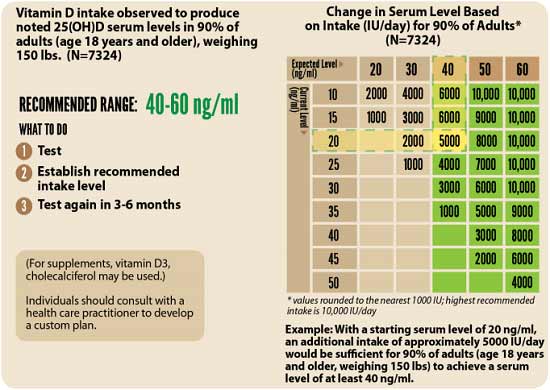
3. Retest in three to six months — Lastly, you’ll need to remeasure your vitamin D level in three to six months, to evaluate how your sun exposure and/or supplement dose is working for you.
Take Your Vitamin D With Magnesium and K2
It’s strongly recommended to take magnesium and K2 concomitant with oral vitamin D. Data from nearly 3,000 individuals reveal you need 244% more oral vitamin D if you’re not also taking magnesium and vitamin K2.9
What this means in practical terms is that if you take all three supplements in combination, you need far less oral vitamin D in order to achieve a healthy vitamin D level.

Frequently Asked Questions (FAQs) About Vitamin D Toxicity
Q: Why is vitamin D important for health, and how is it processed in my body?
A: Vitamin D supports your mental, skeletal, and immune health. It is produced in the skin from sun exposure, then converted in the liver to 25-hydroxyvitamin D, and finally activated in the kidneys as 1,25-dihydroxyvitamin D. This final form helps absorb calcium and maintain cell function.
Q: Can I have too much vitamin D? If so, what are the risks?
A: Yes, excessive vitamin D leads to hypercalcemia (high blood calcium), causing symptoms like fatigue, thirst, digestive issues, and even kidney failure. This occurs with chronic supplementation or consuming fortified foods unknowingly.
Q: How does vitamin D interact with calcium and other nutrients?
A: Vitamin D enhances calcium absorption from food, which is essential for bone, muscle, and nerve health. It works with parathyroid hormone (PTH) to maintain calcium balance. Supplementing with magnesium and vitamin K2 is also crucial, as they help vitamin D function efficiently and safely.
Q: What’s the safest way for me to get vitamin D from sunlight?
A: Expose your skin to sun just before it turns pink to avoid sunburn. If your diet is high in linoleic acid (from vegetable oils), it increases sun sensitivity. Reducing LA intake over time and using protective strategies (like astaxanthin or niacinamide) help your skin handle UV exposure better.
Q: How should I supplement vitamin D correctly?
A: Start by testing your blood levels. The optimal range is 60 to 80 ng/mL (150 to 200 nmol/L). Use tools like the DMinder app or GrassrootsHealth’s calculator to assess sun exposure and determine the right supplement dose. Retest every three to six months to adjust intake as needed.
The Hidden Link Between Vitamins and Hair Loss
Reproduced from original article:
https://articles.mercola.com/sites/articles/archive/2025/03/29/vitamin-deficiencies-hair-loss.aspx
Analysis by Dr. Joseph Mercola March 29, 2025

STORY AT-A-GLANCE
- Vitamin deficiencies often cause hair loss, with nutrients like vitamins D, B12, A and iron playing key roles in maintaining healthy hair growth cycles
- Vitamin D supports hair follicle development, while B vitamins help produce keratin — the protein that forms hair structure
- Iron carries oxygen to hair follicles; deficiency causes poor growth, but too much is also harmful, with ideal serum ferritin levels being 20 to 40 ng/mL
- Vitamin A regulates scalp health through sebum production; both deficiency and excess cause hair problems like dryness or thinning
- A nutrient-rich whole foods diet is the best approach to addressing vitamin-related hair loss, with supplement used only when necessary after proper testing
Picture this: you step out of the shower, glance down and see a tangle of hair circling the drain — more than usual. You wonder, “What’s happening to my hair?” While stress or family genes could be culprits, there’s another common reason you might not have thought of: vitamin deficiencies. Your hair acts like a window to your overall health, dropping hints about what’s going on inside your body.
When it thins or falls out more than normal, it’s often a signal that you’re low on some key nutrients. Nutrients like vitamins D, B12 and A, and iron, keep your hair looking full and healthy. If you don’t get enough, your locks could suffer as a result.
Understanding Your Hair’s Growth Cycle
Hair growth follows a structured cycle with three phases: the growth phase (anagen), the transition phase (catagen) and the resting phase (telogen). This process ensures continuous renewal, much like a plant that sprouts, matures and eventually sheds to allow new growth. Vitamins are indispensable to this cycle, acting as nutrients that sustain hair health.
For example, vitamin D supports healthy hair follicles, while B vitamins contribute to keratin production — the protein that forms your hair’s structure. Insufficient levels of these nutrients disrupt the cycle, causing hair to enter the resting phase prematurely and resulting in increased shedding or thinning. In short, vitamins are the foundation for robust hair growth; without them, the cycle falters.
What Are the Benefits of Vitamin D for Your Hair?
Widely recognized for supporting bone health, vitamin D also plays a key role in hair vitality. It promotes the development of cells that develop into hair follicles, the microscopic structures responsible for hair growth. Vitamin D levels are often low in people with hair loss.1 A deficiency impairs follicle production, leading to thinning hair. Consider vitamin D as the catalyst that initiates healthy hair renewal.
• Identifying a vitamin D deficiency — Signs like excessive hair loss, along with fatigue or muscle pain, may point to low vitamin D levels. The best way to know if you’re in an optimal or deficient state is to get your vitamin D level tested. I recommend doing this twice a year. Regular monitoring is key to keeping your vitamin D level in a healthy range, ideally between 60 to 80 ng/mL (150 to 200 nmol/L).
• Strategies to boost vitamin D — The best way to boost your vitamin D level is with regular sun exposure. However, I must caution you about sun exposure during peak hours if you consume dietary seed oils. These oils, which are rampant in processed and fast food, are packed with linoleic acid (LA). When LA accumulated in your skin interacts with the sun’s ultraviolet (UV) rays, it triggers inflammation and DNA damage.
It’s best to avoid direct sunlight during peak hours (10 a.m. to 4 p.m.) until you’ve cut back on seed oils for six months. This gives your body time to clear some of the accumulated LA. If sun exposure isn’t possible, vitamin D3 supplementation is often necessary. Remember to have your vitamin D levels tested and, based on your test results, adjust your sun exposure or your supplement dose accordingly to maintain a vitamin D level between 60 to 80 ng/mL (150 nmol/L to 200 nmol/L).
Save This Article for Later – Get the PDF Now
How Do B Vitamins Help Your Hair Stay Strong?
Biotin (B7) helps make keratin, the protein your hair’s built from. Not enough, and your hair might get brittle or thin.2 Think of biotin as the builder stacking strong bricks for your hair. Vitamin B12 keeps your blood cells healthy, carrying oxygen to your hair roots. When these vitamins are low, hair becomes weak, grows slowly or break easily.
• Signs of deficiency — Thinning hair with symptoms like fatigue or pale skin often signal a B vitamin shortage. Hair loss and red, scaly rashes (especially on your face) are common signs you may need more biotin.
• Increasing B Vitamin Intake — To increase vitamin B12, incorporating B12-rich foods, such as grass fed meat and dairy, into your diet. Vegetarians and vegans, who are particularly at risk for vitamin B12 deficiency due to dietary restrictions, should consider supplements. Oral or intramuscular vitamin B12 supplements help restore adequate levels, depending on the severity of the deficiency.
Oral supplementation is generally effective for mild to moderate deficiency, with dosages ranging from 1,000 to 2,000 micrograms daily. Intramuscular injections, typically administered monthly, may be more suitable for patients with severe deficiency or those with malabsorption issues. Pastured egg yolk is one of the best sources of biotin. Since biotin is water-soluble, your body will not store it. Hence, your intake must remain consistent.
Why Does Iron Matter for Your Hair Growth?
Iron is essential for hair health, helping red blood cells carry oxygen to hair follicles.3 Without enough iron, follicles can’t get the oxygen they need, slowing growth and causing shedding. While too little iron contributes to thinning hair, having either too much or too little iron has serious repercussions. The fact is, iron deficiency is commonly checked for, but iron overload is a far more common problem, yet is often overlooked or ignored.
• Detecting low iron — Hair loss along with fatigue, memory problems or pale skin could mean an iron deficiency.4 A straightforward blood test, known as a serum ferritin test, provides valuable insights into your iron status. You want your ferritin level below 100 ng/mL; the ideal range is 20 to 40 ng/mL. Below 20 ng/mL is an indicator that you are iron deficient, while a level above100 ng/mL indicates inflammation, high iron or both.
• Boosting iron levels — Eat iron-rich foods like grass fed red meat and pair them with vitamin C from citrus fruits to improve absorption.5 Before considering supplements, be sure to get a serum ferritin blood test, as too much iron is harmful.
How Does Vitamin A Help or Hurt Your Hair?
Vitamin A keeps your scalp healthy by controlling sebum production, the natural oil that moisturizes hair.6 Too little dries out hair, while too much contributes to thinning and scalp issues. Vitamin A is like a dial that needs to be set just right for optimal hair growth.
• Recognizing vitamin A imbalance — Dry, brittle hair is often a sign you need more vitamin A, but thinning or scalp irritation could signal excess.7
• How to boost vitamin A levels — To optimize your vitamin A status, focus on consuming a variety of vitamin A-rich foods. Good sources include liver, egg yolks, butter from grass fed cows, and orange and yellow vegetables like sweet potatoes and carrots. These foods provide either preformed vitamin A or provitamin A carotenoids that your body can convert to active vitamin A.
While supplementation can be beneficial if you’re deficient, it’s important not to exceed recommended intake levels, due to negative effects from excessive supplementation. If you’re concerned about your vitamin A status, consider having your levels tested before starting any supplementation regimen.
Supporting Hair Health Through Nutrition
Your hair often reflects your inner health, with thinning or shedding hinting at deficiencies in vitamins D, B12 or A, and possibly iron. Each plays a unique role in hair growth, strength and scalp health. The good news? Fixing these gaps is simple. A diet rich in nutrients, based on a variety of whole foods, gives your hair what it needs.
If hair loss continues, however, see a holistic doctor, as a blood test is useful for spotting nutrient deficiencies, leading to targeted treatments like diet changes or supplements to get your hair growth and overall health back on track.
Frequently Asked Questions About Vitamins and Hair Loss
Q: Which vitamins are most important for hair growth?
A: Vitamin D, vitamin B12, biotin, iron and vitamin A are key nutrients for hair health, supporting follicles, hair strength and scalp health.
Q: How should I get these vitamins?
A: Sunlight is the best source of vitamin D. Vitamin B12-rich foods include grass fed meat and dairy. Pastured egg yolk is one of the best sources of biotin, while iron-rich foods include grass fed red meat. Good sources of vitamin A include liver, egg yolks, butter from grass fed cows, and orange and yellow vegetables like sweet potatoes and carrots.
Q: How can I identify a vitamin deficiency?
A: Watch for thinning hair, fatigue or pale skin. Ask for a blood test from your doctor to confirm any nutrient deficiencies.
Q: Can supplements quickly fix hair loss?
A: While certain supplements support hair health, diet comes first. Be sure to prioritize dietary changes, with a focus on fresh, whole foods, and use supplements only when needed.
Q: What causes excessive hair shedding?
A: Some shedding is normal, but nutrient deficiencies often worsen it. Hair shedding is also triggered by hormonal changes (pregnancy, menopause, thyroid disorders), stress, certain medications, sudden weight loss, seasonal changes and underlying health conditions like autoimmune disorders.
Mitochondrial Dysfunction Sparks Inflammation and Cancer Risk
Reproduced from original article:
https://articles.mercola.com/sites/articles/archive/2025/03/19/mitochondrial-dysfunction.aspx
Analysis by Dr. Joseph Mercola March 19, 2025

STORY AT-A-GLANCE
- Suppression of mitochondrial ATP production prevents apoptosis and activates the NLRP3 inflammasome, a key player in inflammation and disease
- Inhibitors of oxidative phosphorylation (OXPHOS) lead to changes in mitochondrial cristae structure and retention of cytochrome c, which is necessary for NLRP3 activation but not sufficient on its own
- Activation of the NLRP3 inflammasome requires two signals, one of which is mitochondrial, highlighting the complexity of its regulation
- Diverse NLRP3 activators share the ability to suppress apoptosis, allowing damaged cells to survive and contributing to chronic inflammation and cancer
- Mitochondrial dysfunction is closely linked to inflammation and various diseases, emphasizing the importance of understanding these mechanisms for optimal health
Chronic inflammation and cancer are health challenges that affect millions of individuals worldwide. Inflammation is your body’s natural response to injury or infection, characterized by redness, swelling and pain. However, when inflammation becomes persistent, it leads to tissue damage and contributes to the development of various diseases, including cancer.
Cancer itself is marked by the uncontrolled growth and spread of abnormal cells, which invade surrounding tissues and form harmful tumors. If left untreated, these conditions significantly impair quality of life and increase mortality rates. Mitochondria, often referred to as the powerhouses of the cell, play a key role in producing adenosine triphosphate (ATP), the energy currency essential for numerous cellular functions.
When mitochondrial function is compromised, ATP production decreases, leading to cellular energy deficits. This reduction in energy impairs the cell’s ability to regulate normal processes, fostering an environment ripe for chronic inflammation.
According to research published in Immunity,1 impaired mitochondrial function directly activates the NOD-like receptor protein 3 (NLRP3) inflammasome, a key component in the inflammatory response. This activation not only sustains inflammation but also creates conditions that promote cancer development by enabling cancer cells to thrive and evade your immune system.
Mitochondrial Function Is Intricately Involved in Inflammation and Cancer
The impact of mitochondrial dysfunction on inflammation and cancer is significant. Studies show that approximately 20% of all cancers are linked to chronic inflammation, highlighting the strong connection between these conditions.2 Additionally, individuals with mitochondrial disorders are at a higher risk of developing inflammatory diseases compared to the general population.3
• Millions of Americans are affected by mitochondrial dysfunction — In the U.S. alone, chronic inflammatory conditions affect close to 125 million adults,4 while cancer remains the second leading cause of death, accounting for more than 608,000 fatalities each year.5 Moreover, research indicates that mitochondrial dysfunction contributes to the resistance of cancer cells to conventional therapies, making treatment more challenging.6
• A call to action for better treatment for mitochondrial health — The statistics underscore the urgent need to address mitochondrial health as a strategy to combat both inflammation and cancer effectively.
Beyond their direct effects, chronic inflammation and cancer driven by mitochondrial dysfunction leads to a cascade of additional health problems. Persistent inflammation is associated with cardiovascular diseases, diabetes and neurodegenerative disorders, further compounding the burden on affected individuals.
• Cancer stems largely from mitochondrial dysfunction — Cancer progression often results in debilitating symptoms such as pain, fatigue and loss of organ function, which drastically reduces life expectancy and quality of life.
Understanding the role of mitochondrial dysfunction in driving inflammation and cancer not only illuminates therapeutic targets but also emphasizes the importance of maintaining mitochondrial health to prevent a wide array of serious health issues.
• Mitochondrial dysfunction is a key player in the development of NLRP3-related conditions — When mitochondria fail to produce adequate ATP, it sets off a cascade of cellular stress signals.
These signals activate the NLRP3 inflammasome, a protein complex that plays a significant role in the body’s inflammatory response.7 The activation of this inflammasome is linked to various diseases, including chronic inflammation and cancer, as it leads to uncontrolled cell death and tissue damage.
Study Reveals How Mitochondrial Dysfunction Fuels Inflammation and Cancer
A recent study investigated the intricate relationship between mitochondrial function and the activation of the NLRP3 inflammasome. The research focused on understanding how the inhibition of oxidative phosphorylation (OXPHOS), the process by which mitochondria produce ATP, affects cell death and inflammation.
The study employed various cell types, including myeloid cells, primary murine microglia, human monocyte-derived macrophages, HCT116 and HeLa cells, as well as conducted in vivo experiments using Xenopus laevis tadpoles.8
• The NLRP3 inflammasome negatively impacts mitochondrial health — The population studied encompassed a diverse range of cells to mimic different physiological conditions. The findings revealed that activators of NLRP3 significantly hinder mitochondrial ATP production, which in turn suppresses apoptosis, the process of programmed cell death.
This suppression allows damaged cells to survive longer than they should, contributing to inflammation and leading to cancer development. The study demonstrated that when OXPHOS is inhibited, mitochondrial cristae — the inner folds of mitochondria — undergo structural changes that trap cytochrome c, a molecule essential for apoptosis.9
• Other factors that diminish apoptosis — The research also showed that various NLRP3 activators, such as nigericin, imiquimod and extracellular ATP, inhibit apoptosis not by activating the inflammasome directly, but through their disruptive effects on mitochondrial function. These compounds cause the closure of crista junctions, preventing cytochrome c from being released into the cytoplasm, which is a necessary step for apoptosis to proceed.
• The impact of viral infections on mitochondrial function and apoptosis — It was observed that infections like SARS-CoV-2 could strongly suppress apoptosis by inhibiting the cleavage of caspase-3, an enzyme involved in the execution of apoptosis. This suppression not only hinders the removal of infected cells but also facilitates the activation of the NLRP3 inflammasome, thereby promoting an inflammatory response.10
Save This Article for Later – Get the PDF Now
Mitochondrial Dysfunction Is at the Root of Most Chronic Disease
Biologically, the mechanism at play involves the inhibition of mitochondrial ATP production by NLRP3 activators. When OXPHOS is blocked, mitochondria cannot produce sufficient ATP, leading to the rearrangement of cristae and retention of cytochrome c within the mitochondria. This retention prevents apoptosis, allowing damaged cells to survive and multiply unchecked.
• The process of NLRP3 signaling and activation — The suppression of ATP production provides a necessary signal for the activation of NLRP3. However, full activation of NLRP3 requires a second signal, highlighting the complexity of the inflammasome’s regulation.11
The study also compared the effects of different NLRP3 activators and OXPHOS inhibitors, revealing that while all these agents suppress apoptosis, only certain ones could activate NLRP3 without an additional signal.
• A way to control mitochondrial processes to facilitate healing — This comparison highlights the intricate relationship between mitochondrial function and inflammasome activation, suggesting that modulating mitochondrial processes could be an effective strategy for managing inflammation and reducing cancer risk.12
The research provides compelling evidence that mitochondrial dysfunction, specifically through the inhibition of OXPHOS, plays a pivotal role in suppressing apoptosis and activating the NLRP3 inflammasome.
This dual action not only fosters a proinflammatory environment but also allows for the survival of malignant cells, thereby linking reduced mitochondrial function to the progression of inflammation and cancer.13 As noted on Georgi Dinkov’s blog, the study demonstrates that mitochondrial dysfunction is a key player in both cancer and inflammation:14
“Yet another study, which demonstrates the inseparable link between metabolism and ‘structural’ problems such as cellular integrity and lifecycle (e.g. apoptosis), as well as mysterious processes of systemic inflammation, often occurring without any cause that medicine can identify.
Both of these processes are highly visible in cancer — i.e., lack of apoptosis in ‘cancer’ cells despite their wrecked genome and metabolic dysfunction, as well as their highly inflamed nature that ‘recruits’ nearby cells to the ‘cancer’ process through the cytokines the ‘cancer’ cells produce and releases in the blood.
In other words, all that takes for systemic inflammation and even cancer (i.e., lack of apoptosis in damaged cells) to form is reduced mitochondrial function, resulting in a prolonged drop of ATP levels.
Thus, chronic stress, inflammatory diet (PUFA anyone?), endocrine disruptors, and the ‘modern’ life characterized by never-ending soul-crushing routines are all direct causes of all our ailments as the one thing all those pathological processes have in common is their profoundly suppressive effects on mitochondria/OXPHOS.
Conversely, simply restoring/improving mitochondrial function may be enough to ameliorate/cure virtually all chronic diseases known to medicine.”
How to Address Mitochondrial Dysfunction and Reduce Inflammation
Your mitochondria power every cell in your body. When they don’t work properly, inflammation rises and damaged cells multiply instead of dying off naturally. Here’s how to support your mitochondrial function and restore cellular energy:
1. Eliminate processed foods and vegetable oils — The modern diet is rife with processed foods and vegetable oils rich in linoleic acid (LA) that damage your gut microbiome and promote harmful bacteria.
LA is a mitochondrial poison that compromises your cellular energy production. In addition to processed foods, avoid nuts and seeds as well to reduce LA intake. It’s also advisable to avoid dining out, since most restaurants use vegetable oils in their cooking, sauces and dressings.
Additionally, limit your consumption of chicken and pork, which are typically high in LA. Replace processed foods with whole, unprocessed foods and healthy fats such as grass fed butter, tallow and ghee. It’s wise to keep your LA intake below 5 grams from all sources. If you can get it below 2 grams, that’s even better. To help track your LA intake, enter all your daily meals into an online nutrition tracker.
2. Optimize carbohydrate intake — Carbohydrates play an important role in supporting mitochondrial function since glucose is the preferred fuel for energy production at the cellular level. Tailor your carbohydrate consumption to support cellular energy by aiming for at least 250 grams of targeted carbohydrates daily for most adults. Individuals with higher activity levels typically require more.
Introduce carbohydrates gradually to allow your gut to adapt, thereby minimizing digestive issues and endotoxin levels. Begin with white rice and whole fruits to nourish beneficial bacteria before considering vegetables, whole grains and starches. Avoiding high-fiber diets initially is important if your gut microbiome is compromised, as excessive fiber will increase endotoxin levels.
If your gut health is severely compromised, focus on easily digestible carbohydrates like dextrose water for the first week or two. Sip it slowly throughout the day to support gradual gut healing.
3. Reduce exposure to environmental toxins — Exposure to synthetic endocrine-disrupting chemicals (EDCs), estrogen and pervasive electromagnetic fields (EMFs) further impairs your cells’ ability to generate energy efficiently. This energy deficit makes it challenging to sustain the oxygen-free gut environment necessary for beneficial bacteria like Akkermansia to flourish.
Further, a lack of cellular energy creates an environment in your gut that favors endotoxin-producing bacteria, damaging mitochondria and creating a vicious cycle of worsening health. By tackling excess LA, estrogens (xenoestrogens found in everyday items like plastic), EDCs and EMFs, you restore your cellular energy and start down the path toward optimal mitochondrial function and health.
4. Get proper sun exposure and boost NAD+ levels — Take niacinamide (50 milligrams three times daily) to increase NAD+ production, which helps your mitochondria generate more energy. NAD+ enables proper cell death signaling and supports your immune system’s ability to identify and remove damaged cells.
Daily sun exposure is also important as it promotes cellular energy production by stimulating mitochondrial melatonin, offering powerful antioxidant protection. Start with brief morning exposures and gradually increase tolerance. It’s important to avoid direct sunlight during peak hours (from 10 a.m. to 4 p.m. in most U.S. regions) until you’ve eliminated vegetable oils from your diet for at least six months to reduce sunburn risk associated with stored linoleic acid.
Frequently Asked Questions About Mitochondrial Dysfunction and Inflammation
Q: What is the connection between mitochondrial dysfunction and chronic inflammation?
A: When mitochondria underproduce ATP, the body perceives this energy deficit as cellular stress. This stress triggers the NLRP3 inflammasome, a protein complex that amplifies inflammation. Over time, chronic inflammation damages tissues and can set the stage for serious conditions like cancer.
Q: How does mitochondrial dysfunction contribute to cancer development?
A: Damaged mitochondria hinder the cell’s ability to undergo apoptosis (programmed cell death). When apoptosis is suppressed, abnormal cells survive longer than they should, accumulating more mutations and fueling tumor growth. Additionally, chronic inflammation driven by impaired mitochondrial function creates an environment that supports cancer progression.
Q: Why is linoleic acid problematic for mitochondrial health?
A: LA, found in most vegetable oils and many processed foods, is considered a mitochondrial poison because it impairs cellular energy production. Consuming high amounts of LA results in both gut dysbiosis (an imbalance of gut bacteria) and heightened inflammation, further undermining mitochondrial function and overall health.
Q: Can improving carbohydrate intake help restore mitochondrial function?
A: Yes. Glucose is a key fuel for energy production (via oxidative phosphorylation) in the mitochondria. By incorporating adequate, easily digestible carbohydrates — such as white rice or whole fruits — you’ll be able to support cellular energy and encourage healthier gut bacteria. This approach is especially important if your gut microbiome is already compromised.
Q: What lifestyle strategies can support better mitochondrial function?
A: Key strategies include eliminating processed foods (especially those high in vegetable oils), optimizing carbohydrate intake, reducing exposure to toxins like endocrine disruptors and heavy electromagnetic fields, getting regular sun exposure, and boosting NAD+ levels (e.g., via niacinamide supplementation). These measures help reduce inflammation, restore proper cell death signaling, and protect against chronic diseases linked to mitochondrial dysfunction.
- 1, 8, 9, 10, 11, 12, 13 Immunity, November 20, 2024
- 2 Signal Transduction and Targeted Therapy volume 6, Article number: 263 (2021)
- 3 J Clin Med. 2020 Mar 9;9(3):740
- 4 National Institutes of Health, November 3, 2020
- 5 CDC, Cancer Data and Statistics, December 3, 2024
- 6 J Hematol Oncol. 2022 Jul 18;15:97
- 7 Annual Review of Immunology April 2023, 41:301-316
- 14 To Extract Knowledge from Matter, December 7, 2024
Probiotics Offer New Hope for Alzheimer’s and Other Neurodegenerative Diseases
Reproduced from original article:
https://articles.mercola.com/sites/articles/archive/2025/03/20/probiotics-alzheimers-neurodegenerative-diseases.aspx
Analysis by Dr. Joseph Mercola March 20, 2025

STORY AT-A-GLANCE
- Probiotics may influence brain function through the gut-brain axis, offering benefits for neurodegenerative diseases like Alzheimer’s and Parkinson’s by improving gut health and reducing inflammation
- Studies suggest a link between gut health and cognitive function, indicating that an improved gut microbiome enhances mental health and may reduce inflammation associated with neurodegeneration
- Research indicates that probiotics could play a role in managing Alzheimer’s and Parkinson’s, offering a noninvasive treatment option that helps slow or reverse cognitive decline
- Biologically, the action of probiotics involves several interconnected pathways. By restoring a healthy balance of gut bacteria, probiotics reduce the production of harmful substances like lipopolysaccharides (LPS) that trigger inflammation in the brain
- Further studies are needed to confirm the long-term benefits of probiotics in neurodegenerative disease management, emphasizing the importance of continued research in this area
Neurodegenerative diseases (NDs) are disorders characterized by the progressive loss of structure or function of neurons, leading to their eventual death. This decline disrupts essential brain functions, resulting in symptoms that significantly impair daily living and overall quality of life.
Common neurodegenerative diseases include Alzheimer’s disease, Parkinson’s disease, and amyotrophic lateral sclerosis (ALS).1 Individuals affected by these conditions often experience memory loss, impaired movement, and difficulties with speech and coordination. As the diseases advance, patients may face severe cognitive and physical challenges, necessitating extensive care and support.
Neurodegenerative diseases now affect millions worldwide, with Alzheimer’s alone responsible for approximately 60% to 70% of dementia cases.2 On the other hand, the prevalence of Parkinson’s disease is expected to double by 2040, underscoring a growing public health concern.3 Additionally, ALS affects about 2 per 100,000 people in Europe and the U.S., with no known cure currently available.4
Risk Factors Associated with Neurodegenerative Diseases
Understanding the underlying causes of NDs is complex, as they often involve a combination of factors. For example, genetic mutations can predispose individuals to conditions like Alzheimer’s and Parkinson’s. According to a review published in the NPJ Parkinson’s Disease journal, genetic factors are linked to 56% to 79% of the risk for developing Alzheimer’s disease.5
• Environmental toxins — Exposure to heavy metals and pesticides, may contribute to neuronal damage. According to the aforementioned study:
“Several studies have suggested that lead (Pb), arsenic (As), and methyl mercury (MeHg) are also neurotoxins and can disrupt brain function, cause cognitive dysfunction, and increase the risk of AD and PD by disrupting mRNA splicing, the ubiquitin-proteasome system, the electron transport chain, and oxidative stress.”6
• Lifestyle factors — Poor diet, lack of exercise and chronic stress exacerbate NDs by affecting overall brain health and function. The progression from these underlying causes to full-blown NDs involves a cascade of biological events. Neurons, the brain’s communication cells, begin to lose their structure and function due to factors like oxidative stress and inflammation.
This deterioration disrupts the brain’s ability to process information, leading to symptoms such as memory loss and impaired motor skills. Over time, the cumulative effect of these disruptions results in the characteristic decline associated with NDs.
• Diagnosing NDs can be challenging — Often, symptoms overlap with other conditions, making it difficult to pinpoint the exact disease. For instance, memory loss could be attributed to normal aging, stress, or other medical issues, leading to potential misdiagnosis. Additionally, the lack of definitive biomarkers for many NDs means that diagnosis often relies on clinical assessments and patient history, which can be subjective and vary between practitioners.
• Limitations in diagnostic tools complicate the process — Imaging techniques like MRI and CT scans show brain changes, but these are not always specific to NDs. Blood tests and genetic screenings may offer clues but are not conclusive.
As a result, patients may undergo a lengthy diagnostic journey, experiencing uncertainty and anxiety while seeking answers. This underscores the need for more precise and reliable diagnostic methods to improve early detection and treatment outcomes.
Beyond the immediate symptoms, neurodegenerative diseases lead to profound emotional and financial burden for patients and their families. The progressive nature of these disorders often results in loss of independence, increased healthcare costs and emotional stress. Addressing these multifaceted challenges is crucial in improving patient outcomes and enhancing the quality of life for those affected.
Breakthrough Research Shows Probiotics’ Role in Alzheimer’s Disease Management
A recent study published in the journal Nutrients7 investigated the impact of probiotics on individuals diagnosed with Alzheimer’s disease. The research, conducted by Taiwanese researchers, aimed to determine whether introducing specific beneficial bacteria could influence cognitive function and reduce disease markers.
• Investigating the effects of probiotics — The study involved 60 elderly participants who were diagnosed with Alzheimer’s disease. They were assigned to either a probiotic treatment group or a placebo group over a 12-week period.
The probiotic group consumed a milk product containing strains such as Lactobacillus acidophilus, Lactobacillus casei, Bifidobacterium bifidum, and Lactobacillus fermentum. The primary goal was to assess any changes in cognitive function and Alzheimer’s disease markers as a result of the probiotic intervention.
• Probiotics improve cognitive function — Significant improvements were seen in the group that received probiotics compared to the placebo group. The probiotic group showed a notable increase in their Mini-Mental State Examination (MMSE) scores, indicating enhanced cognitive function. In contrast, those who took the placebo experienced a decline in their MMSE scores over the same period.
• Probiotics’ positive effects against inflammation — The probiotic group demonstrated a reduction in inflammation markers, which are often elevated in Alzheimer’s patients. “Considering these findings, specific probiotics demonstrate robust and effective antioxidant capabilities,” the study authors report.8
Save This Article for Later – Get the PDF Now
Probiotics Reduce Inflammation and Oxidative Stress
The mechanism behind these improvements is believed to involve the gut-brain axis, a communication network between the gastrointestinal tract and the brain.
• Probiotics balance your gut health — Probiotics help maintain a healthy balance of gut microbiota, which in turn influence brain function. By promoting a diverse and balanced microbiome, probiotics may reduce systemic inflammation and oxidative stress, both of which are implicated in the progression of Alzheimer’s disease. For more information on how gut health affects your mental health, read “Gut Health’s Impact on Mental Well-Being.”
• Benefits of SCFAs — Short-chain fatty acids (SCFAs) are metabolites produced by beneficial bacteria in the gut. SCFAs have been shown to support brain health by providing energy to neurons and reducing inflammation. The increased abundance of Lactobacillus species in the probiotic group likely contributed to higher SCFA levels, fostering an environment conducive to cognitive stability and improvement.
• Benefits are observed in a short amount of time — Cognitive improvements were observed after just 12 weeks of probiotic supplementation, suggesting that the benefits of probiotics can manifest relatively quickly. This rapid response underscores the potential of probiotics as a viable intervention for slowing or even reversing aspects of cognitive decline in Alzheimer’s patients.
Comparing the effects of probiotics to other variables in the study, probiotics consistently outperformed the placebo in all measured outcomes. While both groups participated in the same environment and received similar care apart from the probiotic intervention, only the probiotic group showed meaningful improvements in physiological markers related to brain health. This stark contrast emphasizes the specific role that probiotics play in enhancing brain health.
Probiotics’ Mechanisms of Action Against Neurodegenerative Disorders
Biologically, the action of probiotics involves several interconnected pathways. By restoring a healthy balance of gut bacteria, probiotics reduce the production of harmful substances like lipopolysaccharides (LPS) that trigger inflammation in the brain.9
• Enhancing the integrity of the gut barrier — They prevent the leakage of proinflammatory agents into the bloodstream, which could otherwise reach the brain and exacerbate neuroinflammation.10
• Affecting neurotransmitter production — Probiotics influence the production of neurotransmitters such as dopamine and gamma-aminobutyric acid (GABA).11 These chemicals are crucial for mood regulation and cognitive function. By increasing the levels of these neurotransmitters, probiotics contribute to improved mental health and cognitive resilience against the degenerative processes of Alzheimer’s disease.
Four Ways to Support Your Brain Through Gut Health
Your gut microbiome acts as a second brain, directly influencing your cognitive function and neurological health, and how you nourish it plays a significant role in your risk for neurodegenerative conditions. For example, a study published in Scientific Reports showed that certain bacterial strains in the gut, such as Collinsella, Lachnospira, and Veillonella, increased the risk of Alzheimer’s disease.12
However, they also identified strains that provide protective benefits against Alzheimer’s disease, such as Eubacterium nodatum and Eisenbergiella. These produce short-chain fatty acids (SCFAs), particularly butyrate, from dietary carbohydrates.
Butyrate nourishes your colonic epithelial cells, reinforcing the intestinal barrier. SCFAs also stimulate mucin production, creating a protective shield against harmful bacteria. Akkermansia muciniphila also produces SCFAs, and research has shown that Alzheimer’s patients tend to have very low levels of this important keystone species.13 Here are four proven strategies geared toward optimizing the Akkermansia in your gut to enhance your brain health:
1. Increase Akkermansia through targeted diet and supplementation — Prioritize foods and supplements that promote the growth of Akkermansia. Include well-cooked, prebiotic-rich vegetables and fiber sources that nourish this crucial bacterium.
Consider high-quality supplements specifically designed to enhance Akkermansia levels, supporting gut barrier integrity and reducing brain inflammation. Avoid processed foods and high-fat diets that can hinder Akkermansia growth.
2. Enhance beneficial short-chain fatty acids — Focus on increasing butyrate-producing bacteria alongside Akkermansia to protect against amyloid buildup in the brain. Incorporate fermented foods and resistant starches that feed these beneficial microbes.
Tailor your carbohydrate intake to support your unique microbiome, aiming for at least 250 grams of targeted carbs daily, and adjust based on your activity level to optimize Akkermansia and short-chain fatty acid production.
3. Support your gut-brain connection — Optimize your gut barrier integrity to prevent inflammatory compounds from reaching your brain. This includes removing inflammatory processed foods and supporting the growth of protective bacteria like Akkermansia, which strengthens your intestinal lining.
Maintain a balanced diet with sufficient carbohydrates tailored to your microbiome, and use pharmaceutical-grade supplements as needed to sustain Akkermansia levels.
4. Address systemic inflammation by promoting Akkermansia diversity — Lower inflammation throughout your body by enhancing microbiome diversity, with a specific emphasis on increasing Akkermansia.
Eliminate vegetable oils and other proinflammatory fats that damage gut bacteria, and incorporate foods that reduce inflammatory markers while supporting Akkermansia growth. Regularly monitor biomarkers such as C-reactive protein levels to track your progress in reducing systemic inflammation and maintaining a healthy gut microbiome.
Additional Strategies to Prevent Alzheimer’s Disease
Aside from optimizing your gut health and Akkermansia levels, there’s a plethora of helpful Alzheimer’s prevention strategies, several of which are outlined below:
• Avoid gluten and casein (primarily wheat and pasteurized dairy, but not dairy fat, such as butter) — As noted in a 2022 study,14 your blood-brain barrier is negatively affected by gluten. When bacteria enter your bloodstream, the risk of Alzheimer’s disease increases. Other cognitive disorders are linked to a weakened blood-brain barrier as well, such as Parkinson’s disease, anxiety and depression.
• Make sure you’re getting enough animal-based omega-3 fats — Omega-3 fats, namely EPA and DHA, help by preventing cell damage caused by Alzheimer’s disease, thereby slowing down its progression and lowering your risk of developing the disorder. That said, omega-3s are PUFAs, so don’t overdo it.
• Optimize your vitamin D level with safe sun exposure — Strong links between low levels of vitamin D in Alzheimer’s patients and poor outcomes on cognitive tests have been revealed. In a 2023 study,15 increasing vitamin D reduced dementia risk by 40%.
The best way to get vitamin D is through sensible sun exposure, aiming for a blood level between 60 and 80 ng/mL. However, you need to purge vegetable oils from your body before going into the sun at solar noon. The LA in your skin oxidizes when exposed to sunlight, causing inflammation and skin damage.
To protect your skin, avoid sun exposure during solar noon for four to six months as you work on removing LA from your body. Stick to sunlight during early morning and late afternoon in the meantime. For additional skin defense, you can take astaxanthin, a low-dose aspirin or molecular hydrogen. Niacinamide cream will also lower your risk of skin damage.
• Keep your fasting insulin levels below 3 — Insulin resistance is linked to accelerated brain aging, as well as neurodegeneration.16
• Eat a nutritious diet, rich in folate — Vegetables, without question, are your best form of folate. Avoid supplements like folic acid, which is the inferior synthetic version of folate. Research shows that folate is a protective factor against Alzheimer’s disease.17
• Avoid and eliminate mercury and aluminum from your body — Dental amalgam fillings, which are 50% mercury by weight, are one of the major sources of heavy metal toxicity. Make sure you use a biological dentist to have your amalgams removed. Sources of aluminum include antiperspirants, nonstick cookware and vaccine adjuvants.
• Make sure your iron isn’t elevated and donate blood if it is — A study18 published in Aging Medicine shows that excess iron increases your risk of Alzheimer’s disease by initiating the Fenton reaction, leading to increased oxidative stress.
• Exercise regularly — Exercise triggers beneficial changes that support cognitive function. Particularly, it’s been shown to improve blood flow to the brain, leading to an increase in biomarkers related to improved neuronal plasticity and better cell survival.19
• Eat blueberries and other antioxidant-rich foods — Wild blueberries, which have high anthocyanin and antioxidant content, are known to guard against neurological diseases.
• Challenge your mind daily — Mental stimulation, such as learning to play a musical instrument, is associated with a decreased risk of Alzheimer’s.20
• Avoid anticholinergics and statin drugs — Drugs that block acetylcholine, a nervous system neurotransmitter, have been shown to increase your risk of dementia. These drugs include certain nighttime pain relievers, antihistamines, sleep aids, certain antidepressants, medications to control incontinence, and certain narcotic pain relievers.
Statin drugs are particularly problematic because they suppress the synthesis of cholesterol, deplete your brain of CoQ10 and neurotransmitter precursors, and prevent adequate delivery of essential fatty acids and fat-soluble antioxidants to your brain by inhibiting the production of the indispensable carrier biomolecule known as low-density lipoprotein.
Frequently Asked Questions (FAQs) About Probiotics and Neurodegenerative Diseases
Q: How do probiotics impact neurodegenerative diseases like Alzheimer’s?
A: Probiotics influence brain function through the gut-brain axis by restoring gut microbiome balance, reducing inflammation, and improving cognitive function. Studies suggest that beneficial bacteria can slow or even reverse aspects of cognitive decline in conditions like Alzheimer’s and Parkinson’s.
Q: What research supports the benefits of probiotics for Alzheimer’s patients?
A: A study published in Nutrients found that Alzheimer’s patients who took probiotics for 12 weeks showed improved cognitive function and reduced inflammation compared to a placebo group. These improvements were measured using the Mini-Mental State Examination (MMSE) and inflammatory markers.
Q: What mechanisms make probiotics beneficial for brain health?
A: Probiotics help reduce inflammation by lowering harmful substances like lipopolysaccharides (LPS), enhancing gut barrier integrity, and influencing neurotransmitter production (e.g., dopamine and GABA). They also promote the production of short-chain fatty acids (SCFAs), which support brain health.
Q: Can improving gut health help prevent neurodegenerative diseases?
A: Yes. A diverse gut microbiome, particularly the presence of beneficial bacteria like Akkermansia muciniphila, plays a crucial role in reducing inflammation and protecting against neurodegeneration. Consuming prebiotic-rich foods, fermented foods, and maintaining a fiber-rich diet can support gut health.
Q: What other lifestyle changes can support brain health and reduce Alzheimer’s risk?
A: In addition to probiotics, strategies like avoiding processed foods, eliminating heavy metal exposure (e.g., mercury and aluminum), maintaining optimal vitamin D and omega-3 levels, exercising regularly, and engaging in mental stimulation activities can help prevent cognitive decline and support brain health.
- 1 Int J Mol Sci. 2020 Dec 14;21(24):9500
- 2, 7, 8 Nutrients. 2023 Dec 20;16(1):16
- 3 Clinical Medicine, Volume 20, Issue 4, July 2020, Pages 393-398
- 4 Amyotrophic Lateral Sclerosis and Frontotemporal Degeneration, 20(3–4), 264–274
- 5, 6 NPJ Parkinsons Dis. 2021 Aug 11;7:70
- 9 Front Immunol. 2021 Feb 26;12:578386
- 10 Front Immunol. 2023 Apr 24;14:1143548
- 11 Front Microbiol. 2023 Aug 17;14:1216674
- 12 Scientific Reports, Volume 13, Article number: 5258 (2023)
- 13 Critical Reviews in Microbiology, 49(2), 151–176
- 14 Am J Lifestyle Med. 2022 Jan 11;16(1):32–35, Abstract
- 15 Alzheimers Dement (Amst). 2023 Mar 1;15(1):e12404, Highlights
- 16 Int J Mol Sci. 2021 Sep 15;22(18):9987, Abstract
- 17 Front Neurosci. 2021 Apr 14;15:661198, Abstract
- 18 Aging Med (Milton). 2024 Oct 22;7(5):649–667, Iron Overload/Accumulation and Increase Risk of AD
- 19 Front Aging Neurosci. 2023 Aug 4;15:1243869
- 20 Aging Ment Health. 2021 Apr;25(4):593-601
Unveiling Vitamin K’s Underrecognized Benefits for Bone Health
Reproduced from original article:
https://articles.mercola.com/sites/articles/archive/2025/03/12/vitamin-k-bone-health.aspx
Analysis by Dr. Joseph Mercola March 12, 2025

STORY AT-A-GLANCE
- Vitamin K is important for bone health beyond its role in blood clotting, with deficiencies linked to increased fracture risk and osteoporosis
- Vitamin K works synergistically with vitamin D to enhance bone mineral density. This highlights the importance of maintaining adequate levels through diet or supplementation
- The gut microbiome plays a significant role in vitamin K production, which impacts bone health by supporting the formation and maintenance of bone tissue
- Vitamin K influences energy metabolism through osteocalcin, affecting insulin sensitivity and glucose metabolism, which are essential for preventing osteoporosis and diabetes
- Strategies to help optimize vitamin K levels including consuming more food sources of both vitamin K1 and K2, strategically timing your vitamin K2 supplement intake and nourishing your gut microbiome with probiotics and prebiotics
Vitamin K is a fat-soluble vitamin that occurs in two forms, K1 (phylloquinone), found abundantly in green leafy vegetables, and K2. Vitamin K2 has several subforms, known as menaquinones, which are designated MK-4 through MK-13. These subforms differ primarily in the length of their side chains, which affects their distribution in the body, sources in the diet and specific biological functions.
MK-7 through MK-13 are produced by various strains of bacteria in the gut, while MK-4 is primarily found in animal-based foods such as eggs, dairy products (like cheese and butter) and certain meats (especially liver).
Vitamin K1 and K2 play pivotal roles in various body functions, including bone health. In fact, being deficient in vitamin K is closely linked to an increased risk of fractures and osteoporosis, a condition characterized by weakened bones and a heightened susceptibility to breaks.
Recent studies highlight that individuals with optimal vitamin K levels exhibit greater bone mass and a lower incidence of fractures. For instance, research demonstrates that vitamin K is essential for the production of a protein that’s vital for bone mineralization.1
Additionally, vitamin K2 has been shown to work synergistically with vitamin D, enhancing bone mineral density and overall bone quality. These findings underscore the importance of maintaining adequate vitamin K levels through diet or supplementation to support bone health effectively.
Understanding Vitamin K’s Bone-Strengthening Benefits
Vitamin K is a key player in maintaining bone strength and preventing arterial calcification, ensuring that calcium is directed to where it’s needed most — your bones and teeth, not your arteries.
• Vitamin K1 (phylloquinone) supports vascular health and bone retention — This form of vitamin K reduces calcification in blood vessels while helping bones retain calcium, preventing unnecessary calcium buildup in the arteries.
• Vitamin K2 (menaquinone) activates vitamin D and directs calcium — K2 works by activating vitamin D-dependent proteins, allowing calcium to be efficiently transported out of the arterial system and into bones and teeth.2
• Vitamin K is vital for the carboxylation of osteocalcin — This is a process that enhances bone strength. It might sound complex, but it’s actually a straightforward process. To understand this, you must first know what osteocalcin is — this is a small protein produced by osteoblasts during bone formation.3
• Carboxylation transforms osteocalcin into a functional hormone — Carboxylation is a process wherein osteocalcin is transformed from a simple protein into a sophisticated hormone involved in bone metabolism and broader metabolic regulation.
When your body lacks sufficient vitamin K, the carboxylation of osteocalcin is impaired. This means that osteocalcin cannot effectively bind calcium to the bone matrix. Over time, this contributes to osteoporosis, where bones become porous and more prone to fractures.
• Vitamin K deficiency increases cardiovascular risk — When calcium is not directed to your bones where it’s needed most, it not only decreases bone mineral density and increases fragility, but also puts you at risk of cardiovascular conditions. This is because calcium accumulates in places like your blood vessels and arteries instead.
• Common causes of vitamin K deficiency — The underlying causes of vitamin K deficiency often stem from poor dietary intake, certain medical conditions or the use of medications that interfere with vitamin K absorption. For instance, individuals with gastrointestinal disorders struggle to absorb this nutrient effectively. Additionally, long-term use of antibiotics disrupts gut bacteria, reducing your body’s natural production of vitamin K.
To learn more about the differences between vitamin K1 and K2, read “Vitamin K1 vs. K2 — Understanding Their Distinct Roles in Your Health.”
Vitamin K Plays an Essential Role in Enhancing Bone Health
A recent study investigated the multifaceted roles of both vitamin K1 and K2, as well as vitamin K-dependent proteins, in maintaining bone health, particularly emphasizing their interaction with vitamin D and their effects on calcium metabolism. The research aimed to determine how these vitamins work together to support bone integrity and prevent conditions like osteoporosis.4
• Vitamin K activates key bone-building proteins — Vitamin K facilitates the carboxylation of osteocalcin and matrix Gla-protein (MGP), which are essential for binding calcium within the bone matrix. MGP is also a potent inhibitor of arterial calcification.
• Vitamin K enables calcium-binding through carboxylation — One of the researchers’ key findings was that vitamin K acts as a necessary cofactor for enzymes that convert glutamic acid residues in proteins into gamma-carboxyglutamic acid residues. This chemical transformation is important for osteocalcin to effectively bind calcium. According to the researchers:
“The important effects of vitamin K on Ca and skeletal homeostasis are known to be mediated through its role as a cofactor for the γ-glutamyl carboxylase enzyme that promotes conversion of glutamate (Glu) residues to gamma-carboxyglutamic (Gla) residues in the post-translational carboxylation of osteocalcin (OC) and matrix Gla protein (MGP). This may have a significant impact on osteogenesis.”5
• Vitamin K2 plays a key role in bone remodeling — The study also revealed that vitamin K2 plays a significant role in bone remodeling. It assists in directing calcium to the bones and prevents its deposition in soft tissues, which leads to vascular calcification. This dual action supports both bone and cardiovascular health.
• Vitamin K and vitamin D work together to regulate calcium balance — The research highlighted the synergistic relationship between vitamins K and D in regulating calcium homeostasis.
Vitamin D enhances calcium absorption in the gut, while vitamin K ensures that the absorbed calcium is appropriately utilized by the bones. This collaboration between the two vitamins is essential for maintaining optimal bone density and reducing the risk of fractures.
“Vitamin D exerts effects directly on osteoblasts by promoting osteoblast maturation and OC synthesis. The multiple effects of vitamin D on bone are associated with a high expression of the vitamin D receptor in several types of bone cells.”6
• Researchers identified biomarkers for vitamin K status — The ratio of carboxylated osteocalcin (cOC) to undercarboxylated osteocalcin (ucOC) was used to assess functional vitamin K levels in the body. A lower cOC-to-ucOC ratio indicated an inadequate vitamin K status, which was associated with increased bone loss and a higher risk of hip fractures.
The biological mechanisms underlying these findings involve vitamin K’s role in activating proteins that regulate calcium placement in the body. By enabling osteocalcin and MGP to bind calcium effectively, vitamin K ensures that calcium is deposited in the bones rather than in the arteries or other soft tissues.
Save This Article for Later – Get the PDF Now
How Does Vitamin K’s Influence Energy Metabolism and Blood Sugar Levels?
Another recent study published in the journal Nutrients explored the role of vitamin K in both bone health and energy metabolism.The research aimed to determine how vitamin K interacts with proteins involved in bone formation and how its deficiency could impact overall metabolic processes.7
• Vitamin K is essential for both bone integrity and metabolic health — The study focused on individuals with varying levels of vitamin K intake, analyzing their bone integrity and insulin sensitivity. Findings revealed that adequate vitamin K levels are essential for maintaining strong bones and regulating blood sugar levels, highlighting this nutrient’s importance in inhibiting conditions like osteoporosis and diabetes.
• Osteocalcin links vitamin K to both bone and glucose metabolism — Activated osteocalcin contributes to bone strength and influences how the body regulates insulin and blood sugar levels. This dual function means that vitamin K deficiency could lead to both weakened bones and impaired glucose metabolism, heightening the risk of developing diabetes.
“[V]itamin K deficiency in the bone results in a lower production of OC and a low serum level of OC, predisposing to a state of glucose intolerance and diabetes mellitus that may then enhance bone matrix deterioration via the production of cross-linked advanced glycation end products (AGE), which have been further associated with bone fractures after adjustment of confounders.
We believe that as a whole, vitamin K deficiency plays an important role in glucose metabolism, ultimately leading to a disturbance of bone quality,” the researchers reported.8
• Vitamin K prevents arterial calcification and cardiovascular disease — In addition to its effects on bone and energy metabolism, vitamin K was found to play a role in preventing the calcification of soft tissues, such as blood vessels. This prevents the hardening of arteries, which is a significant risk factor for cardiovascular diseases. The research emphasized that vitamin K ensures calcium is directed to the bones rather than accumulating in the arteries
• Uncarboxylated osteocalcin (ucOC) is a marker of vitamin K deficiency — The study identified specific markers for vitamin K deficiency, such as elevated levels of uncarboxylated osteocalcin (ucOC) in the blood. Monitoring these markers will help in early detection of vitamin K insufficiency, allowing for timely interventions to maintain bone density and metabolic health.
• Low vitamin K intake is linked to a higher risk of fractures — Individuals with low vitamin K intake were found to have a higher incidence of hip and vertebral fractures across different populations. The research underscored the importance of maintaining adequate vitamin K levels through diet or supplementation to significantly reduce the risk of such fractures.
Your Microbiome Also Influences Your Bone Health
In related news, a recent review published in the Gut Microbes journal explored how the gut microbiome, which is the community of microorganisms living in your intestines, works in collaboration with vitamin K2 to maintain strong and healthy bones, highlighting how these microorganisms contribute to bone density and the overall integrity of your skeletal system.9
• Gut bacteria naturally produce vitamin K2 — The research focused on understanding how variations in the gut microbiome affect the risk of developing bone-related conditions such as osteoporosis. It was found that certain beneficial gut bacteria species, like Bacteroides and Lactococcus lactis, produce vitamin K2 and help maintain adequate levels of this nutrient.
• Dysbiosis lowers vitamin K2 levels, weakening bones — One of the key findings was that disruptions in the gut microbiome, a condition known as dysbiosis, decreases your vitamin K2 production,10 compromising bone strength and increasing your risk of fractures and osteoporosis.
Vitamin K2 produced by gut bacteria activates proteins that are essential for bone formation and mineralization, and maintaining a balanced and healthy gut microbiome is essential for ensuring the proper synthesis of vitamin K2.
• Probiotics and prebiotics enhance bone health —The review also discussed the role of probiotics and prebiotics in enhancing bone health. Probiotics are live beneficial bacteria that are consumed through fermented foods or supplements, while prebiotics are non-digestible food ingredients that promote the growth of these healthy bacteria.11
Incorporating probiotics and prebiotics into your diet supports your gut microbiome, leading to increased production of vitamin K2 and improved bone density.
• The gut microbiome supports calcium and vitamin D absorption — The study also highlighted the interconnectedness between the gut microbiome and other aspects of bone metabolism, such as hormone regulation and immune system function.
Your gut bacteria influence the absorption of calcium and vitamin D as well, both of which are vital for bone health. Additionally, a healthy gut microbiome helps modulate the immune system, reducing inflammation that negatively impacts bone integrity.
• Antibiotics disrupt vitamin K2 production — The review also pointed out that antibiotic treatments, which disrupt the gut microbiome, lead to a significant reduction in vitamin K2 production.12 This not only affects bone health but also emphasizes the importance of being cautious when taking antibiotics, to preserve the beneficial bacteria responsible for maintaining bone integrity.
For a deeper dive into vitamin K’s role in bone and heart health, read “Vitamin K 101 — Essential Basics for Bone and Heart Health.”
Egg Yolks Are Your Best Source of Vitamin K2 as MK-4
Egg yolks are among the highest dietary sources of MK-4, a vital form of vitamin K2 that plays an important role in bone health, cardiovascular function and calcium regulation. Including egg yolks in your diet significantly contributes to your MK-4 intake, supporting various aspects of your health.
• Egg quality is important — You need to be careful about your egg sources as most commercial egg sources — even free-range organic — have high PUFA levels as they are fed grains like soy and corn.
• Chickens should be fed low-PUFA grains for optimal egg quality — Ideally, chickens should be fed rice, barley and split peas. I personally eat six egg yolks a day from chickens who are fed this and have 80% less linoleic acid than regular chickens.
Below is a comprehensive overview of egg yolks as a top source of MK-4, along with additional dietary sources and considerations.

Six Key Steps to Enhance Bone Health Through Vitamin K
To optimize your vitamin K levels and support optimal bone and overall health, consider the following recommendations:
1. Add more green leafy veggies into your diet — They are some of the best sources of vitamin K1. Collard and turnip greens, kale, spinach, broccoli, Brussels sprouts, cabbage and lettuces are good choices.13
2. Boost your natural vitamin K2 production — Enhance your body’s natural vitamin K2 levels by incorporating fermented foods such as natto and fermented vegetables cultivated with specific bacterial cultures. Additionally, consume grass fed animal products like egg yolks, liver and grass-fed dairy to support K2 production.
3. Optimize your vitamin K2 supplementation — Supplement with 180 to 200 micrograms of vitamin K2, ideally in the MK-7 form, daily, combined with 1,000 IU of vitamin D3 and 400 mg of magnesium. This combination ensures optimal absorption and effectiveness, working synergistically to strengthen bones while protecting against arterial calcification.
You should also time your vitamin K2 intake strategically. Take vitamin K2 supplements with your fattiest meal of the day since it’s fat-soluble. This maximizes absorption and allows vitamin K2 to activate osteocalcin. Consistent timing helps maintain steady levels of vitamin K2 for optimal bone and heart protection.
4. Balance key nutrient cofactors — Ensure sufficient intake of calcium, magnesium, vitamin D3 and vitamin K2 through both diet and supplementation. These nutrients collaborate to build and maintain bone density, promoting proper calcium utilization and bone matrix formation for optimal bone health.
5. Support your gut microbiome — Maintain a healthy intestinal environment to naturally boost vitamin K2 production. Focus on whole, unprocessed foods and consider targeted probiotic supplementation tailored to your microbiome.
6. Focus on bone-building exercises — Regular weight-bearing movement stimulates bone formation while K2 ensures proper calcium deposition. Walking, resistance training and bodyweight exercises work together with K2 to strengthen your skeleton. The combination reduces fracture risk far more than supplementation alone.
Frequently Asked Questions (FAQs) About Vitamin K and Bone Health
Q: What’s the main role of vitamin K in bone health?
A: Vitamin K helps direct calcium into bones, making them stronger and reducing the risk of fractures. It also prevents calcium from building up in arteries, which protects heart health.
Q: Do I need both vitamin K2 and vitamin D?
A: Yes, they work together. Vitamin D helps your body absorb calcium, and vitamin K2 ensures it gets used for bone-building instead of accumulating in arteries.
Q: Can vitamin K2 help prevent osteoporosis?
A: Studies show that vitamin K2 improves bone mineral density and lowers fracture risk by activating osteocalcin, a key protein for bone formation.
Q: How much vitamin K2 should I take daily?
A: For optimal bone and heart health, aim for 180 to 200 mcg of vitamin K2 (MK-7) daily. Taking it with a meal that contains healthy fats improves absorption.
The Transformative Effects of High-Dose Thiamine Therapy: Dr Derrick Lonsdale’s Legacy
Reproduced from original OMNS article (OrthoMolecular News Service):
http://orthomolecular.org/
Subscribe to the free Orthomolecular Newsletter: http://orthomolecular.org/subscribe.html
Go to the OMNS Archive: http://orthomolecular.org/resources/omns/index.shtml
Orthomolecular Medicine News Service, March 4, 2025
by Elliot Overton
Dr. Derrick Lonsdale, a true luminary in the field of nutritional medicine, passed away last year at the ripe old age of 100 years old. Beginning his career as a pediatrician at the Cleveland clinic, he dedicated nearly five decades to uncovering the profound impact of high-dose thiamine (vitamin B1) on chronic disease. He was a vocal proponent of the orthomolecular approach to medicine, and tirelessly sought to raise awareness of what he referred to as high-calorie malnutrition-a state where an abundance of processed foods depletes essential micronutrients, impairing metabolism at a fundamental level and setting the stage for chronic health conditions.
Furthermore, his pioneering work challenged conventional medicine’s narrow view of vitamin deficiency, revealing that pharmacological doses of thiamine may achieve far more than merely prevent deficiency-they can actively restore energy metabolism. Dr. Lonsdale often referred to this nutrient as the “spark of life” and “gateway to energy metabolism,” emphasising its vital role in counteracting the metabolic stressors of modern life when administered in appropriate amount. He introduced the concept of thiamine dependency-the state where some individuals require supraphysiological doses on a consistent basis to maintain health-an insight that may be highly relevant to many modern chronic diseases. Remarkably, much of what he hypothesized decades ago is now being validated by cutting edge research, demonstrating that his intuitive sense was accurate.
Yet, despite his extensive research and clinical contributions, thiamine remains one of the most overlooked nutrients in medicine today. Even after seven years of applying these principles, I am still astounded by the transformative effects this vitamin can have in such a broad range of health problems.
In the sections that follow, we will explore thiamine’s essential role in bioenergetics, its unique anti-stress properties, and the overlooked concept of organ-specific localized deficiency-an emerging phenomenon that may be crucial for specific health conditions, particularly neurodegenerative disease. We will also examine the rationale behind using pharmacological doses as a powerful therapeutic intervention for a wide array of modern diseases.
Thiamine: A Universal “Anti-Stress” Molecule
Thiamine (also known as vitamin B1) is an essential vitamin naturally found in a variety of whole foods, especially meats and organs, legumes and whole grains. Due to its hydrophilic nature and short half-life, it requires continuous dietary replenishment. Its widespread role in human physiology centres on its participation as an essential cofactor for enzymes involved in various biochemical pathways. Among these, thiamine-dependent dehydrogenases are uniquely positioned at key metabolic cross-points, enabling cellular metabolic flexibility and modulating the global rate of energy metabolism.
Pyruvate Dehydrogenase (PDH), the rate-limiting enzyme for mitochondrial glucose oxidation, bridges glycolysis and the TCA cycle, rendering thiamine indispensable for efficient utilization of carbohydrate. Alpha-Ketoglutarate Dehydrogenase (KGDH), another rate-limiting enzyme of the TCA cycle, links bioenergetics, amino acid metabolism, and neurotransmitter synthesis. Beyond the generation of NADH and disposal of glutamate, KGDH acts as a metabolic “signaling hub,” influencing redox balance, growth, the hypoxic response, protein signaling, and calcium homeostasis (1).
The unique positioning of these rate-limiting thiamine-dependent enzymes at critical junctures in metabolism means that they are tasked with governing the global rate of ATP production. For this reason, intracellular thiamine levels are also tightly coupled to mitochondrial oxygen utilization. Insufficient thiamine levels disrupt this process, leading to pseudohypoxia-a state in which cells cannot utilize oxygen despite adequate availability, leading to widespread energy deficit.
This connection is evidenced by the striking resemblance between the histological changes observed in thiamine-deficient brains and those seen in hypoxic injury (2-4). Both conditions activate the same coordinated cellular response to stress, marked by the stabilization and activation of Hypoxia-Inducible Factor 1-alpha (HIF-1α) (5-7). Additionally, both hypoxia and thiamine deficiency were shown to upregulate SLC19A3, a membrane-bound thiamine transporter, presumably as a compensatory mechanism to enhance thiamine uptake into the cell for the purpose of stress mitigation. Consistent with these findings, a decline in thiamine status correlates with hypothermia and and a marked reduction in oxidative metabolic rate in animals (8). Furthermore, in was shown to produce identical symptoms those found in Hans Selye’s model of “The General Adaptation Syndrome” (9). Reflecting this tight-knit relationship between thiamine, bioenergetics, and adaptation to stress, Dr. Lonsdale aptly referred to thiamine as “the spark of life” and “the gateway to energy metabolism.”
Thiamine’s anti-stress properties also appear to be conserved across plants, fungi, and bacteria. The nutrient has been referred to as an “environmental stress protectant” and “stress alarmone” (10) in plants. Under conditions of biotic and abiotic stress, plants are known to increase their production of thiamine and upregulate thiamine-dependent enzymes as a means of improving resilience in the face of unfavorable environmental conditions. Furthermore, exogenous thiamine confers resistance against many different types of disease (11).
https://www.sciencedirect.com/science/article/abs/pii/B9780123864796000044
For bacteria, similar upregulation of genes involved in thiamine biosynthesis occurs in cyanobacteria under stress (12), and E. coli were found to “accumulate large amounts of thiamine triphosphate, a thiamine derivative, under severe energy stress (13). In yeast, thiamine also confers protection against oxidative, osmotic, and thermal stress (14).
From a broad perspective, it would appear that thiamine exhibits distinctive “anti-stress” properties and can enhance cellular resilience across diverse biological systems. Recognizing these qualities can provide a solid foundation for understanding how and why thiamine might be so useful in mitigating the impact of chronic health conditions which are characterized by metabolic, oxidative, and environmental stressors.
“The Great Imitator”: The Many Faces of Deficiency
A severe deficiency in this nutrient is classically know to result in thiamine deficiency disorders (TDDs), which predominantly affect one or more of three major body systems: The central nervous system (causing Wernicke encephalopathy), the cardiovascular system (causing Wet Beriberi), and the peripheral nerves (leading to Dry Beriberi). The classic pattern of symptoms depends on the subtype and can include, but is not limited to, ataxia, opthalmoplegia, peripheral neuropathy, paresthesia, vertigo, vascular insufficiency, edema, and even heart failure.
However, micronutrient status should best be understood as existing on a continuum, rather than as a clear-cut binary divide between ‘sufficiency’ and ‘deficiency.’ The latter view is overly simplistic and fails to capture the complex and progressive effects that insufficiency can exert on human physiology. Evidence shows that even marginal deficits in thiamine can manifest in subtle yet significant ways, negatively impacting metabolism and organ function long before overt deficiency disorders are diagnosed. Given the ubiquity of thiamine-dependent enzymes and their governing role in energy metabolism, it is conceivable that sufficiency and/or impaired utilization of thiamine could therefore impact any cell, tissue and organ which requires ATP.
Indeed, nearly a century’s worth of research has demonstrated that even mild insufficiency can manifest in a broad array of non-specific symptoms that extent well beyond the classic spectrum of TDDs. Given thiamine’s central role in energy metabolism, organs with the highest demand for energy are the most vulnerable, with many associated symptoms reflecting some degree of autonomic dysfunction.
Lonsdale himself spearheaded the treatment of dysautonomia (of various kinds) with thiamine derivatives (15-17), based on the principle that it could address disturbed bioenergetics in the limbic/brainstem regions of the brain involved in modulation of the autonomic nervous system. In his own words: “Beriberi is the prototype for functional dysautonomia in its early stages.” (16). He described the brain as becoming “hyper-irritable” under oxidative stress, leading to exaggerated autonomic nervous system responses to even minor stimuli, such as changes in weather or exposure to air conditioning. Indeed, signs and symptoms of dysautonomia are remarkably prevalent among all of the established TDDs (18-21), and in my own experience, thiamine can be the single most effective treatment for such conditions. Clinical manifestations range from mild to severe, and can also fluctuate based on season, physical activity level, and other environmental factors. Furthermore, they can be non-specific and therefore difficult to pinpoint in clinical practice.
The general and non-specific nature of symptoms was illustrated as early as 1940 in one of the first ever studies on experimental deficiency in human subjects (22). Remarkably, classical signs of beriberi were virtually absent for the majority of the study period (88 days). Instead, the researchers documented a broad range of symptoms pertaining to every body system, many of which fall under the modern diagnostic umbrella of dysautonomia.
These symptoms included:
Fatigue upon mild exertion, tachycardia, pseudoangina, irregular heart rate, pallor, blushing, hyperhidrosis, temperature dysregulation, genitourinary paresthesia, frequency of micturition, shortness of breath, vertigo, impaired glucose tolerance, insomnia, along with mood disturbances and poor concentration. General malaise, heavy sensation in the lower extremities, loss of strength, chest tightness, diminished visual acuity and restlessness. Abdominal distension, belching, and alternating constipation and diarrhea, achlorhydria or hypochlorhydria, delayed gastric emptying, and reduced intestinal motility.
These broad and often nebulous symptoms observed in thiamine insufficiency highlight the challenges involved in accurate identification, and no doubt contributes to its misdiagnosis. Many of these manifestations are subtle, easily overlooked, and commonly misattributed to other conditions, which leads to widespread under recognition. In reality, thiamine insufficiency develops insidiously, raising an important question: Could a significant portion of the population be affected without realizing it?
Thiamine Insufficiency: A Hidden Epidemic
Outside of the context of specific comorbidities such as alcoholism (23), malabsorptive syndromes (24), and eating disorders (25), frank deficiency widely considered rare in developed nations, with the prevailing assumption that food fortification has largely eradicated it. A closer examination of the evidence, however, suggests otherwise.
Insufficiency is, in fact, far more prevalent than commonly recognized (26), with some studies showing occurrence between 20-50% in psychiatric (27) and elderly populations (28,29). Plasma thiamine levels were up to 76% lower in type 1 and type 2 diabetics (30). Even severe cases of Wernicke Encephalopathy are frequently underdiagnosed (31). Furthermore, testing methods are woefully inadequate for assessing intracellular thiamine status (32).
Perhaps the primary driver of widespread deficiency is the occurrence of “high-calorie malnutrition”, a term popularized by Lonsdale describing the overconsumption of processed foods rich in calories but devoid of micronutrients, which progressively depletes the body’s reserves. This is especially relevant in the modern world, given that consumption of ultra-processed foods has risen substantially in recent decades (33) and now accounts for 50-60% daily caloric intake in some high-income countries (34).
Thiamine status is largely influenced by carbohydrate intake, which necessitates a proportional dietary supply to match metabolic demands (35). In other words, the overconsumption of sugar-laden foods and refined starches naturally places a large burden on the body’s thiamine reserves. One of the most striking examples of thiamine’s importance comes from the historical epidemic of beriberi in Asia, where populations who were reliant on polished white rice (devoid of thiamine-rich husks) were the first to suffer from severe deficiency.
However, another insidious driver is the widespread consumption of oxidized, PUFA-rich industrial seed oils, as demonstrated by a recent study effect (36). Although the exact mechanism wasn’t elucidated, research suggests that oxidized breakdown products of PUFAs, such as malondialdehyde, can negatively impact thiamine status in several ways (37,38). Additionally, there are many pharmaceutical drugs which also display potent anti-thiamine effects, such as metformin (39), metronidazole (40), diuretics (41), and omeprazole (42) among several others (43). Dysbiotic changes to the gut microbiome (44), along with stressors of various kinds can also be responsible for reducing thiamine status (45).
However, the complexity of this issue extends beyond simple deficiency. In fact, a significant portion of patients who present with thiamine-responsive health conditions may not even have insufficient systemic levels of thiamine. That is, despite “normal” circulating thiamine levels, functional impairment at the cellular or enzymatic level may still justify supplementation with high doses. This emerging area of research, which I find particularly fascinating, challenges the conventional paradigms of nutritional status and highlights the need for more nuanced understanding of nutrient handling and nutritional therapy as a whole.
Beyond Addressing Deficiency: Localized or Functional Impairments in Thiamine Utilization
Dr. Derrick Lonsdale often emphasized that high-dose thiamine functions as a pharmacological agent rather than merely a nutritional supplement. The doses required to achieve therapeutic effects-often hundreds or even thousands of times the Recommended Dietary Allowance (RDA)-far exceed what is needed to correct a simple nutritional deficiency, and for prolonged periods of time. In his own words, high-dose thiamine could “coerce energy metabolism back to life,” acting as a metabolic stimulant of sorts.
The Pharmacological Action of Thiamine
This concept is perhaps best illustrated in examples of inborn errors of metabolism, such as thiamine-responsive maple syrup urine disease, Leigh’s disease, methylmalonic academia, homocysteinuria, and other hereditary vitamin-responsive disorders. In these conditions, genetic mutations reduce the affinity of vitamin-dependent enzymes for their respective vitamin cofactor, leading to severe metabolic dysfunction. Mega-doses of the nutrient are required to saturate cells, and compensate for these enzymatic defects, effectively restoring function despite the genetic abnormality (46).
While rare, I suspect that similar principles may apply to a much broader range of modern diseases involving enzymatic inactivation or inhibition of vitamin-dependent pathways. This inactivation can occur independently of genetic predisposition and may result from environmental toxins, xenobiotics, chronic inflammation, or oxidative stress.
In the context of thiamine-responsive conditions, the issue is not a simple deficiency of thiamine intake but rather defective intracellular handling of thiamine or a functional blockade of thiamine-dependent enzymes, which happen to be uniquely sensitive to such insults (47).
There are numerous factors unrelated to dietary intake that have this effect, including:
- Heavy metals (e.g., aluminum, arsenic) (48,49)
- Neuroinflammatory molecules (50)
- Mycotoxins (51)
- Pro-oxidants (52)
- Xenobiotics (53)
Although temporary enzymatic inhibition may serve normal physiological roles, chronic inhibition is increasingly recognized to be a key driver of pathology (54,55), particularly in the context of neurodegenerative disease. The cumulative effect is mitochondrial dysfunction and a resulting bioenergetic deficit, which triggers a cascade of metabolic impairments that ultimately contribute toward tissue and organ dysfunction.
Localized “Deficiency” in Specific Organs & Tissues
The inhibition or “blockade” of thiamine-dependent, rate-limiting enzymes of involved in energy metabolism can mimic the consequences of a systemic nutritional deficiency, but may instead be restricted to specific tissues. Emerging evidence supports the concept of localized thiamine deficiency various tissues and organs, including the brain, heart, pancreas, and even intestine.
For this reason, thiamine can be considered a metabolic stimulant, as high-dose administration has the capacity to restore energy metabolism when cells become saturated, via bypassing or overriding metabolic blocks imposed by enzymatic inhibition. In essence, this perspective shifts the focus beyond from merely correcting dietary deficiency to addressing localized dysfunction and enzyme inhibition, potentially offering new therapeutic possibilities for a wide range of chronic conditions.
Another important consideration is that tissues with high metabolic demand -particularly in the context of chronic injury or infection- can rapidly deplete their thiamine reserves in response to stress. Such is another scenario that can potentially lead to localized deficiency, in the absence of systemic signs, symptoms, and/or diagnostic tests. Transcriptomic data (forthcoming publication) supports this notion, revealing distinct metabolic abnormalities, including long-term alterations thiamine-dependent enzymes, in enterocytes up to 9 months after gastric bypass surgery. It is conceivable that such changes could occur in practically any cell, tissue or organ exposed to chronic injury, and should therefore be considered in our approach to managing chronic illness.
Furthermore, genetic factors undoubtedly play an important role. Abnormalities in genes encoding proteins involved in the transport, activation, and utilization of thiamine could render an individual more susceptible even to minor fluctuations in thiamine status. Such genetic differences may also help to account for some individuals respond favorably to this therapy, while others do not.
Keys to Neurodegeneration: Untapped potential for the aging brain?
These mechanisms are particularly relevant in the field of neurodegeneration, where decades of research highlight a strong link between disrupted thiamine homeostasis and neurodegenerative processes. Notably, dysfunction of thiamine-dependent enzymes appears to be a hallmark feature of several neurodegenerative conditions, suggesting that localized thiamine dysfunction may be a key contributor to CNS vulnerability and disease progression.
Alzheimer’s Dementia (AD)
A “localized deficiency” of thiamine in the brain has been proposed as a defining characteristic of AD (56), and is known to independently cause many of the key pathological changes including: impaired glucose metabolism (57), neuroinflammation (58), neuron loss (59), impaired cholinergic function (60) and greater presence of amyloid plaques and tangles (61). Disruptions of cerebral thiamine homeostasis and glucose metabolism have been identified (62), and activity of three thiamine-dependent enzymes are reduced in the brain (63,64). One of those is KGDH, whose activity is reduced by as much as 57% (65,66) which occurs in both genetic (67) and sporadic (68) forms of AD. Furthermore, Thiamine transporters decline (69) and metabolic impairments in the brain are closely related to TPP levels (70). Furthermore, levels of active TPP were significantly reduced in the frontal, temporal, parietal and occipital cortex in frontotemporal dementia (71) at autopsy. The synthetic thiamine derivative benfotiamine has been shown to counteract many of the pathological drivers of AD and cognitive decline (72), and a $45 milllon trial is currently underway in AD patients (73).
Parkinson’s Disease
Endogenous neurotoxic metabolites associated with PD, such as MPP+ and isoquinolones, are potent inhibitors of thiamine-dependent enzymes (74), Numerous studies have identified KGDH as a key pathological target in Parkinson’s disease (PD) (75). KGDH activity is greatly reduced in the substantia nigra (76), with the degree of inhibition correlating with the severity of neurodegeneration (77). Additionally, PD is associated with significant reductions in PDH activity (78,79). Free thiamine concentrations in cerebrospinal fluid were also found to be lower than controls (80).
Although there are only two small trials using pharmacological doses of thiamine for PD, the results are extremely promising (81,82). In one of those reports, patients with a milder phenotype achieved complete clinical remission with thiamine therapy. Today, there is an online network of thousands who are self-treating with this approach and are witnessing great benefits.
According to the author of the case reports:
“It is reasonable to infer that a focal, severe thiamine deficiency due to a dysfunction of thiamine metabolism could cause selective neuronal damage in the centres that are typically hit in this disease. Injection of high doses of thiamine was effective in reversing the symptoms, suggesting that the abnormalities in thiamine-dependent processes could be overcome by diffusion-mediated transport at supranormal thiamine concentrations.”
The Cholinergic System
Although widespread deficits to the cholinergic system are a well-recognized pathological hallmark of AD, similar -or even more severe- deficits may also occur in PD, albeit in different regions of the brain (83,84). These shared mechanisms may help to further shed light on why thiamine holds significant therapeutic potential. Thiamine is inherently pro-cholinergic, playing a crucial role in acetylcholine (ACh) function at multiple levels. An intimate relationship between thiamine and cholinergic neurotransmission stems from both its coenzyme and non-coenzyme roles. A well-documented consequence of thiamine deficiency is reduced ACh synthesis (60), partly due to the direct role of pyruvate dehydrogenase in supplying acetyl-CoA, the essential precursor for ACh. Additionally, the distribution of acetyl-CoA and its supply for ACh synthesis is also governed by KGDH through its regulation of the TCA cycle, which further links thiamine homeostasis with cholinergic function.
However, beyond its metabolic role as a coenzyme, thiamine is essential for axonal membrane excitability and neuronal action potentials (85). Phosphorylated thiamine derivatives regulate ACh neurotransmission (86) and are involved in synaptic function (87,88). Synaptic co-release of thiamine and ACh, with thiamine facilitating neurotransmission, has also been found (89-92). At high concentration, thiamine binds to nicotine ACh receptors (93), while the synthetic thiamine derivatives TTFD and sulbutiamine demonstrate pro-cholinergic effects in multiple studies (94,95). Given these diverse mechanisms, thiamine likely exerts its neuroprotective action through a combination of coenzyme and non-coenzyme effects, further cementing its role as a promising candidate for supporting neurodegenerative states which involve cholinergic decline.
Huntington’s Disease
Inhibition of pyruvate dehydrogenase (96,97) and KGDH (98) have been found. A decrease in thiamine content of the cerebrospinal fluid was shown to precede the onset of motor symptoms (99). Abnormal thiamine metabolism has been found in HD animal models (100), and a recent study identified impaired thiamine transport as a potentially treatable cause of HD. More specifically, abberant polyadenylation of thiamine transporters (SLC19A3) were identified in HD, accompanied by low striatal content of active thiamine in both humans and animals. Supplementation with high dose thiamine combined with biotin improved radiological, motor and neuropathological phenotypes in the animal model (101), and human studies are currently underway (102).
Amyotrophic Lateral Sclerosis
The frontal cortext in ALS displays substantially lower levels of TPPase, the enzyme which activates thiamine to thiamine pyrophosphate (103), and patients exhibit decreased thiamine derivatives in the cerebrospinal fluid (104). The morphological signatures of Wernicke Encephalopathy have been reported in ALS (105). A case study using high dose benfotiamine (a thiamine derivative) showed promising results in ALS patient (106). Likewise, high doses of another derivative named dibenzoylthiamine normalized the metabolome and lead to improvements in all physiological parameters, motor function and muscle atrophy in an animal model of ALS (107).
A series of case reports by Dr Antonio Costantini showed positive responses to high-dose thiamine (HDT) treatment in various other neurological conditions such as dystonia and spinocerebellar ataxia (108), along with a case report on fibromyalgia which showed up to 70% improvement in fatigue and 80% reduction in pain scores (109). Such findings could potentially be explained by dysfunctional thiamine utilization reported in this condition, evidenced by a significantly elevated TPP effect for transketolase (110), and low TPP levels (111), along with low thiamine binding affinity was observed for PDH (112) and transketolase (113).
Collectively, these findings suggest that dysfunction of thiamine-dependent enzymes and/or defects in thiamine metabolism, utilization, or transport may extend far beyond conventional associations, playing a significant role in the pathophysiology of diseases which not traditionally associated with TD.
Leveraging Thiamine as a “Cell Protectant”
As discussed thus far, evidence strongly supports thiamine’s role as a protective agent against mitochondrial impairment. Notably, these benefits are not strictly limited to the correction of a deficiency in the traditional sense, but rather through optimizing key metabolic pathways to maintain energy metabolism. However, it’s important to mention that these effects are not limited to neurodegeneration but may also apply to injury various kinds.
Dr Victoria Bunik and her team at the Belozersky Institute of Physicochemical Biology in Moscow have extensively studied the effects thiamine in various models. Their research demonstrated that pre-treatment with pharmacological doses of thiamine provided significant neuroprotection and largely mitigated damage in an animal model of traumatic brain injury (114). Following injury, mitochondrial ATP production was preserved, accompanied by vast reductions in glutamate-mediated excitotoxicity along with improvements in neuroinflammation. The proposed mechanism of protection was pharmacological activation of KGDH by thiamine.
To quote the authors:
“The impairment of OGDHC [KGDH] plays a key role in the glutamate mediated neurotoxicity in neurons during TBI; pharmacological activation of OGDHC [KGDH] may thus be of neuroprotective potential. “
The same team later reported similar results in model on spinal trauma, where thiamine was shown to preserve glutathione levels and ameliorate the effects of excess nitric oxide (115). Once again, the mechanism of neuroprotection was thiamine’s activation of KGDH, which preserved mitochondrial function and maintained generation of ATP.
Indeed, many other studies have highlighted similar protective effects of thiamine (116). Pre-treatment with thiamine pyrophosphate preserved cardiac function via maintaining ATP production in another model of ischemia (117). Thiamine preserved PDH activity in animals post-cardiac arrest (118). Likewise thiamine was also protective in a model of copper toxicity, once again through preventing the inactivation of PDH (119). Beyond these protective roles, thiamine also improves markers of metabolic dysfunction and glucose metabolism improves markers of metabolic dysfunction (120) while exerting anti-fatigue effects, largely by attenuating ATP depletion in skeletal muscle during workload-induced fatigue (121).
Beyond Metabolic Support: Thiamine’s Non-Coenzyme Protective Effects
The benefits extend beyond thiamine’s function as a coenzyme, because there are a range of non-coenzyme effects which may further contribute to its ability to protect cells and tissues from damage.For example, thiamine pyrophosphate has been shown to shield the liver from cisplatin toxicity protected rat liver (122), and prevented ischemia-related infertility prevented infertility (123).
While thiamine itself has demonstrated comparable efficacy to N-acetylcysteine (NAC) (123) in protecting against acetaminophen-induced liver damage. Additionally, cells were safeguarded radiation-induced genetic damage (124), and both thiamine and benfotiamine could counteract ultrasound-induced aggression and oxidative stress while normalizing AMPA receptor expression and plasticity markers in animal studies (125). High-dose thiamine also alleviates biomarkers of oxidative stress and inflammation in models of lead toxicity (126), and pretreatment protected cardiomyocytes from hypoxia-induced apoptosis and DNA fragementation (127).
Honoring Dr. Lonsdale’s Legacy by Advancing Thiamine Therapy: A New Frontier
Many of Dr. Lonsdale’s theories on thiamine’s mechanisms and therapeutic potential continue to be validated by cutting-edge research. As someone deeply inspired by his work, I have spent the past seven years studying and applying high-dose thiamine in clinical practice, and its effects continue to astound me. I have witnessed remarkable improvements in patients-many without classical risk factors for deficiency-experiencing chronic fatigue, brain fog, fibromyalgia, autonomic dysfunction, functional gut disorders, mood imbalances, and more.
Yet, despite its profound impact, thiamine remains largely overlooked, even within functional and alternative medicine circles. In the time I have spent using and learning about this nutrient, my observations have aligned with those of Dr. Derrick Lonsdale, who long recognized that high-dose thiamine does more than correct a dietary deficiency-it reactivates energy metabolism in ways conventional medicine has yet to fully grasp. For decades, Lonsdale tirelessly worked to raise awareness, but conventional medicine failed to listen.
Though I was never fortunate enough to meet him in person, we exchanged many emails, and I can attest that he closely followed the scientific research right into his final year. His dedication is evident in his extensive body of work, including Thiamine Deficiency Disease, Dysautonomia, and High-Calorie Malnutrition, co-authored with Dr. Chandler Marrs. So as we continue to explore the implications of his research, we honor his legacy-a testament to the profound effects that nutrients can have in restoring health. In his own words:
“We’re using the vitamin as a drug and coercing the energy metabolism back into life.”
About the author:
Elliot is a naturopathic nutritionist with additional training through A4M’s fellowship program in Anti-Aging, Metabolic, and Functional Medicine. His primary research area focuses on the underlying mechanisms and clinical application of high-dose thiamine (vitamin B1) derivatives. As a speaker, he regularly lectures at professional events to advance the understanding and therapeutic use of thiamine in pharmacological doses for a host of chronic disease states including neurodegeneration, functional GI disorders, and complex conditions such as CFS and fibromyalgia. Through education and advocacy, Elliot aims to make this knowledge accessible to both practitioners and the general public. He is the co-founder of the nutraceutical company Objective Nutrients, runs the YouTube channel “EONutrition” and also manages the website thiamineprotocols.com.
Elliot is a member of the Board of Editors of the Orthomolecular Medicine News Service.
References:
1. Hansen GE, Gibson GE. The α-Ketoglutarate Dehydrogenase Complex as a Hub of Plasticity in Neurodegeneration and Regeneration. Int J Mol Sci (2022) 23:12403. https://doi.org/10.3390/ijms232012403
2. Vortmeyer AO, Hagel C, Laas R. Hypoxia-ischemia and thiamine deficiency. Clin Neuropathol (1993) 12:184-190.
3. Johkura K, Naito M. Wernicke’s encephalopathy-like lesions in global cerebral hypoxia. J Clin Neurosci Off J Neurosurg Soc Australas (2008) 15:318-319. https://doi.org/10.1016/j.jocn.2006.10.022
4. Vortmeyer AO, Colmant HJ. Differentiation between brain lesions in experimental thiamine deficiency. Virchows Arch A (1988) 414:61-67. https://doi.org/10.1007/BF00749739
5. Valle ML, Anderson YT, Grimsey N, Zastre J. Thiamine insufficiency induces Hypoxia Inducible Factor-1α as an upstream mediator for neurotoxicity and AD-like pathology. Mol Cell Neurosci (2022) 123:103785. https://doi.org/10.1016/j.mcn.2022.103785
6. Zera K, Zastre J. Thiamine deficiency activates hypoxia inducible factor-1α to facilitate pro-apoptotic responses in mouse primary astrocytes. PLOS ONE (2017) 12:e0186707. https://doi.org/10.1371/journal.pone.0186707
7. Sweet RL, Zastre JA. HIF1-α-mediated gene expression induced by vitamin B1 deficiency. Int J Vitam Nutr Res Int Z Vitam- Ernahrungsforschung J Int Vitaminol Nutr (2013) 83:188-197. https://doi.org/10.1024/0300-9831/a000159
8. Langlais PJ, Hall T. Thiamine deficiency-induced disruptions in the diurnal rhythm and regulation of body temperature in the rat. Metab Brain Dis (1998) 13:225-239. https://doi.org/10.1023/a:1023276009477
9. Some Specific and Non-Specific Effects of Thiamine Deficiency in the Rat. – Floyd R. Skelton, 1950. https://journals.sagepub.com/doi/abs/10.3181/00379727-73-17729 [Accessed February 20, 2025]
10. Rapala-Kozik M. “Vitamin B1 (Thiamine).,” In: Rébeillé F, Douce R, editors. Advances in Botanical Research. Biosynthesis of Vitamins in Plants Part A. Academic Press (2011). p. 37-91 https://doi.org/10.1016/B978-0-12-386479-6.00004-4
11. Ahn I-P, Kim S, Lee Y-H. Vitamin B1 functions as an activator of plant disease resistance. Plant Physiol (2005) 138:1505-1515. https://doi.org/10.1104/pp.104.058693
12. Upregulation of thiamine (vitamin B1) biosynthesis gene upon stress application in Anabaena sp. and Nannochloropsis oculata. https://www.kspbtjpb.org/journal/view.html?volume=44&number=4&spage=462 [Accessed February 20, 2025]
13. Gigliobianco T, Lakaye B, Makarchikov AF, Wins P, Bettendorff L. Adenylate kinase-independent thiamine triphosphate accumulation under severe energy stress in Escherichia coli. BMC Microbiol (2008) 8:16. https://doi.org/10.1186/1471-2180-8-16
14. Wolak N, Kowalska E, Kozik A, Rapala-Kozik M. Thiamine increases the resistance of baker’s yeast Saccharomyces cerevisiae against oxidative, osmotic and thermal stress, through mechanisms partly independent of thiamine diphosphate-bound enzymes. FEMS Yeast Res (2014) 14:1249-1262. https://doi.org/10.1111/1567-1364.12218
15. Lonsdale D, Nodar RH, Orlowski JP. The Effects of Thiamine on Abnormal Brainstem Auditory Evoked Potentials. J Adv Med (1998) 11:199-207. https://doi.org/10.1023/A:1023344513264
16. Lonsdale D. Exaggerated Autonomic Asymmetry: A Clue to Nutrient Deficiency Dysautonomia. (2011) https://www.semanticscholar.org/paper/Exaggerated-Autonomic-Asymmetry%3A-A-Clue-to-Nutrient-Lonsdale/2f66f5baa669933fed0b4e23fb2bac616f2c558e [Accessed February 20, 2025]
17. Lonsdale D, Nodar RH, Orlowski JP. Brainstem dysfunction in infants responsive to thiamine disulfide: preliminary studies in four patients. Clin EEG Electroencephalogr (1982) 13:82-88. https://doi.org/10.1177/155005948201300203
18. Shible AA, Ramadurai D, Gergen D, Reynolds PM. Dry Beriberi Due to Thiamine Deficiency Associated with Peripheral Neuropathy and Wernicke’s Encephalopathy Mimicking Guillain-Barré syndrome: A Case Report and Review of the Literature. Am J Case Rep (2019) 20:330-334. https://doi.org/10.12659/AJCR.914051
19. Lonsdale D, Marrs C. Thiamine-Deficient Dysautonomias. Elsevier (2018). p. 161-211 https://doi.org/10.1016/B978-0-12-810387-6.00005-8
20. Reuler JB, Girard DE, Cooney TG. Current concepts. Wernicke’s encephalopathy. N Engl J Med (1985) 312:1035-1039. https://doi.org/10.1056/NEJM198504183121606
21. Branco de Oliveira MV, Irikura S, Lourenço FH de B, Shinsato M, Irikura TCDB, Irikura RB, Albuquerque TVC, Shinsato VN, Orsatti VN, Fontanelli AM, et al. Encephalopathy responsive to thiamine in severe COVID-19 patients. Brain Behav Immun – Health (2021) 14:100252. https://doi.org/10.1016/j.bbih.2021.100252
22. Williams RD. Observations on induced thiamine (vitamin B1) deficiency in man. Arch Intern Med (1940) 66:785. https://doi.org/10.1001/archinte.1940.00190160002001
23. Laforenza U, Patrini C, Gastaldi G, Rindi G. Effects of acute and chronic ethanol administration on thiamine metabolizing enzymes in some brain areas and in other organs of the rat. Alcohol Alcohol Oxf Oxfs (1990) 25:591-603. https://doi.org/10.1093/oxfordjournals.alcalc.a045055
24. Oudman E, Wijnia JW, Oey MJ, van Dam M, Postma A. Wernicke’s encephalopathy in Crohn’s disease and ulcerative colitis. Nutrition (2021) 86:111182. https://doi.org/10.1016/j.nut.2021.111182
25. Winston AP, Jamieson CP, Madira W, Gatward NM, Palmer RL. Prevalence of thiamin deficiency in anorexia nervosa. Int J Eat Disord (2000) 28:451-454. https://doi.org/10.1002/1098-108x(200012)28:4<451::aid-eat14>3.0.co;2-i
26. Marrs C, Lonsdale D. Hiding in Plain Sight: Modern Thiamine Deficiency. Cells (2021) 10:2595. https://doi.org/10.3390/cells10102595
27. Carney MW, Ravindran A, Rinsler MG, Williams DG. Thiamine, riboflavin and pyridoxine deficiency in psychiatric in-patients. Br J Psychiatry J Ment Sci (1982) 141:271-272. https://doi.org/10.1192/bjp.141.3.271
28. Kwok T, Falconer-Smith JF, Potter JF, Ives DR. Thiamine status of elderly patients with cardiac failure. Age Ageing (1992) 21:67-71. https://doi.org/10.1093/ageing/21.1.67
29. Nichols HK, Basu TK. Thiamin status of the elderly: dietary intake and thiamin pyrophosphate response. J Am Coll Nutr (1994) 13:57-61. https://doi.org/10.1080/07315724.1994.10718372
30. Thornalley PJ, Babaei-Jadidi R, Al Ali H, Rabbani N, Antonysunil A, Larkin J, Ahmed A, Rayman G, Bodmer CW. High prevalence of low plasma thiamine concentration in diabetes linked to a marker of vascular disease. Diabetologia (2007) 50:2164-2170. https://doi.org/10.1007/s00125-007-0771-4
31. Kohnke S, Meek CL. Don’t seek, don’t find: The diagnostic challenge of Wernicke’s encephalopathy. Ann Clin Biochem (2021) 58:38-46. https://doi.org/10.1177/0004563220939604
32. Whitfield KC, Bourassa MW, Adamolekun B, Bergeron G, Bettendorff L, Brown KH, Cox L, Fattal‐Valevski A, Fischer PR, Frank EL, et al. Thiamine deficiency disorders: diagnosis, prevalence, and a roadmap for global control programs. Ann N Y Acad Sci (2018) 1430:3-43. https://doi.org/10.1111/nyas.13919
33. Monteiro CA, Moubarac J-C, Cannon G, Ng SW, Popkin B. Ultra-processed products are becoming dominant in the global food system. Obes Rev Off J Int Assoc Study Obes (2013) 14 Suppl 2:21-28. https://doi.org/10.1111/obr.12107
34. Cordova R, Viallon V, Fontvieille E, Peruchet-Noray L, Jansana A, Wagner K-H, Kyrø C, Tjønneland A, Katzke V, Bajracharya R, et al. Consumption of ultra-processed foods and risk of multimorbidity of cancer and cardiometabolic diseases: a multinational cohort study. Lancet Reg Health – Eur (2023) 35: https://doi.org/10.1016/j.lanepe.2023.100771
35. Elmadfa I, Majchrzak D, Rust P, Genser D. The Thiamine Status of Adult Humans Depends on Carbohydrate Intake. Int J Vitam Nutr Res (2001) 71:217-221. https://doi.org/10.1024/0300-9831.71.4.217
36. Leano J, Raut G, Martinez-Lomeli J, Chen G, Luna C, Degnan P, Curras-Collazo M, Sladek F, Deol P. Vitamin B1 Supplementation Ameliorates Obesogenic Effects of a High Fat Diet in Male Mice. Physiology (2024) 39:1230. https://doi.org/10.1152/physiol.2024.39.S1.1230
37. Vuorinen PJ, Rokka M, Ritvanen T, Käkelä R, Nikonen S, Pakarinen T, Keinänen M. Changes in thiamine concentrations, fatty acid composition, and some other lipid-related biochemical indices in Baltic Sea Atlantic salmon (Salmo salar) during the spawning run and pre-spawning fasting. Helgol Mar Res (2020) 74:10. https://doi.org/10.1186/s10152-020-00542-9
38. Lukienko PI, Mel’nichenko NG, Zverinskii IV, Zabrodskaya SV. Antioxidant properties of thiamine. Bull Exp Biol Med (2000) 130:874-876. https://doi.org/10.1007/BF02682257
39. Liang X, Chien H-C, Yee SW, Giacomini MM, Chen EC, Piao M, Hao J, Twelves J, Lepist E-I, Ray AS, et al. Metformin Is a Substrate and Inhibitor of the Human Thiamine Transporter, THTR-2 (SLC19A3). Mol Pharm (2015) 12:4301-4310. https://doi.org/10.1021/acs.molpharmaceut.5b00501
40. Hassan M, Awadalla E, Ali R, Fouad S, Abdel-Kahaar E. Thiamine deficiency and oxidative stress induced by prolonged metronidazole therapy can explain its side effects of neurotoxicity and infertility in experimental animals: Effect of grapefruit co-therapy. Hum Exp Toxicol (2020) 39:834-847. https://doi.org/10.1177/0960327119867755
41. Katta N, Balla S, Alpert MA. Does Long-Term Furosemide Therapy Cause Thiamine Deficiency in Patients with Heart Failure? A Focused Review. Am J Med (2016) 129:753.e7-753.e11. https://doi.org/10.1016/j.amjmed.2016.01.037
42. Nixon PF, Diefenbach RJ, Duggleby RG. Inhibition of transketolase and pyruvate decarboxylase by omeprazole. Biochem Pharmacol (1992) 44:177-179. https://doi.org/10.1016/0006-2952(92)90053-l
43. Karadima V, Kraniotou C, Bellos G, Tsangaris GTh. Drug-micronutrient interactions: food for thought and thought for action. EPMA J (2016) 7:10. https://doi.org/10.1186/s13167-016-0059-1
44. Costliow ZA, Degnan PH. Thiamine Acquisition Strategies Impact Metabolism and Competition in the Gut Microbe Bacteroides thetaiotaomicron. mSystems (2017) 2:e00116-17. https://doi.org/10.1128/mSystems.00116-17
45. Attaluri P, Castillo A, Edriss H, Nugent K. Thiamine Deficiency: An Important Consideration in Critically Ill Patients. Am J Med Sci (2018) 356:382-390. https://doi.org/10.1016/j.amjms.2018.06.015
46. Ames BN, Elson-Schwab I, Silver EA. High-dose vitamin therapy stimulates variant enzymes with decreased coenzyme binding affinity (increased K(m)): relevance to genetic disease and polymorphisms. Am J Clin Nutr (2002) 75:616-658. https://doi.org/10.1093/ajcn/75.4.616
47. Tretter L, Adam-Vizi V. Alpha-ketoglutarate dehydrogenase: a target and generator of oxidative stress. Philos Trans R Soc B Biol Sci (2005) 360:2335-2345. https://doi.org/10.1098/rstb.2005.1764
48. Mailloux RJ, Bériault R, Lemire J, Singh R, Chénier DR, Hamel RD, Appanna VD. The Tricarboxylic Acid Cycle, an Ancient Metabolic Network with a Novel Twist. PLOS ONE (2007) 2:e690. https://doi.org/10.1371/journal.pone.0000690
49. Bergquist ER, Fischer RJ, Sugden KD, Martin BD. Inhibition by methylated organo-arsenicals of the respiratory 2-oxo-acid dehydrogenases. J Organomet Chem (2009) 694:973-980. https://doi.org/10.1016/j.jorganchem.2008.12.028
50. Inhibition of the α‐ketoglutarate dehydrogenase complex by the myeloperoxidase products, hypochlorous acid and mono‐N‐chloramine – Jeitner – 2005 – Journal of Neurochemistry – Wiley Online Library. https://onlinelibrary.wiley.com/doi/ftr/10.1111/j.1471-4159.2004.02868.x [Accessed February 20, 2025]
51. Pirrung MC, Nauhaus SK, Singh B. Cofactor-Directed, Time-Dependent Inhibition of Thiamine Enzymes by the Fungal Toxin Moniliformin. J Org Chem (1996) 61:2592-2593. https://doi.org/10.1021/jo950451f
52. Humphries KM, Yoo Y, Szweda LI. Inhibition of NADH-linked mitochondrial respiration by 4-hydroxy-2-nonenal. Biochemistry (1998) 37:552-557. https://doi.org/10.1021/bi971958i
53. Park LC, Gibson GE, Bunik V, Cooper AJ. Inhibition of select mitochondrial enzymes in PC12 cells exposed to S-(1,1,2,2-tetrafluoroethyl)-L-cysteine. Biochem Pharmacol (1999) 58:1557-1565. https://doi.org/10.1016/s0006-2952(99)00247-6
54. Tretter L, Adam-Vizi V. Alpha-ketoglutarate dehydrogenase: a target and generator of oxidative stress. Philos Trans R Soc B Biol Sci (2005) 360:2335-2345. https://doi.org/10.1098/rstb.2005.1764
55. Gibson GE, Park LC, Sheu KF, Blass JP, Calingasan NY. The alpha-ketoglutarate dehydrogenase complex in neurodegeneration. Neurochem Int (2000) 36:97-112. https://doi.org/10.1016/s0197-0186(99)00114-x
56. Gibson GE, Feldman HH, Zhang S, Flowers SA, Luchsinger JA. Pharmacological thiamine levels as a therapeutic approach in Alzheimer’s disease. Front Med (2022) 9:1033272. https://doi.org/10.3389/fmed.2022.1033272
57. Hakim AM, Carpenter S, Pappius HM. Metabolic and histological reversibility of thiamine deficiency. J Cereb Blood Flow Metab Off J Int Soc Cereb Blood Flow Metab (1983) 3:468-477. https://doi.org/10.1038/jcbfm.1983.73
58. Hazell AS, Rao KV, Danbolt NC, Pow DV, Butterworth RF. Selective down-regulation of the astrocyte glutamate transporters GLT-1 and GLAST within the medial thalamus in experimental Wernicke’s encephalopathy. J Neurochem (2001) 78:560-568. https://doi.org/10.1046/j.1471-4159.2001.00436.x
59. Ke Z-J, Gibson GE. Selective response of various brain cell types during neurodegeneration induced by mild impairment of oxidative metabolism. Neurochem Int (2004) 45:361-369. https://doi.org/10.1016/j.neuint.2003.09.008
60. Barclay LL, Gibson GE, Blass JP. Impairment of behavior and acetylcholine metabolism in thiamine deficiency. J Pharmacol Exp Ther (1981) 217:537-543.
61. Karuppagounder SS, Xu H, Shi Q, Chen LH, Pedrini S, Pechman D, Baker H, Beal MF, Gandy SE, Gibson GE. Thiamine deficiency induces oxidative stress and exacerbates the plaque pathology in Alzheimer’s mouse model. Neurobiol Aging (2009) 30:1587-1600. https://doi.org/10.1016/j.neurobiolaging.2007.12.013
62. Gibson GE, Feldman HH, Zhang S, Flowers SA, Luchsinger JA. Pharmacological thiamine levels as a therapeutic approach in Alzheimer’s disease. Front Med (2022) 9: https://doi.org/10.3389/fmed.2022.1033272
63. Gibson GE, Hirsch JA, Cirio RT, Jordan BD, Fonzetti P, Elder J. Abnormal thiamine-dependent processes in Alzheimer’s Disease. Lessons from diabetes. Mol Cell Neurosci (2013) 55:17-25. https://doi.org/10.1016/j.mcn.2012.09.001
64. Héroux M, Raghavendra Rao VL, Lavoie J, Richardson JS, Butterworth RF. Alterations of thiamine phosphorylation and of thiamine-dependent enzymes in Alzheimer’s disease. Metab Brain Dis (1996) 11:81-88. https://doi.org/10.1007/BF02080933
65. Butterworth RF, Besnard AM. Thiamine-dependent enzyme changes in temporal cortex of patients with Alzheimer’s disease. Metab Brain Dis (1990) 5:179-184. https://doi.org/10.1007/BF00997071
66. Mastrogiacoma F, Lindsay JG, Bettendorff L, Rice J, Kish SJ. Brain protein and alpha-ketoglutarate dehydrogenase complex activity in Alzheimer’s disease. Ann Neurol (1996) 39:592-598. https://doi.org/10.1002/ana.410390508
67. Gibson GE, Zhang H, Sheu KF, Bogdanovich N, Lindsay JG, Lannfelt L, Vestling M, Cowburn RF. Alpha-ketoglutarate dehydrogenase in Alzheimer brains bearing the APP670/671 mutation. Ann Neurol (1998) 44:676-681. https://doi.org/10.1002/ana.410440414
68. Albers DS, Augood SJ, Park LC, Browne SE, Martin DM, Adamson J, Hutton M, Standaert DG, Vonsattel JP, Gibson GE, et al. Frontal lobe dysfunction in progressive supranuclear palsy: evidence for oxidative stress and mitochondrial impairment. J Neurochem (2000) 74:878-881. https://doi.org/10.1046/j.1471-4159.2000.740878.x
69. Alzheimer’s disease is associated with disruption in thiamin transport physiology: A potential role for neuroinflammation – PubMed. https://pubmed.ncbi.nlm.nih.gov/35750148/ [Accessed February 20, 2025]
70. Gibson GE, Feldman HH, Zhang S, Flowers SA, Luchsinger JA. Pharmacological thiamine levels as a therapeutic approach in Alzheimer’s disease. Front Med (2022) 9: https://doi.org/10.3389/fmed.2022.1033272
71. Rao VLR, Richardson JS, Butterworth RF. Decreased activities of thiamine diphosphatase in frontal and temporal cortex in Alzheimer’s disease. Brain Res (1993) 631:334-336. https://doi.org/10.1016/0006-8993(93)91554-6
72. Gibson GE, Feldman HH, Zhang S, Flowers SA, Luchsinger JA. Pharmacological thiamine levels as a therapeutic approach in Alzheimer’s disease. Front Med (2022) 9:1033272. https://doi.org/10.3389/fmed.2022.1033272
73. Burke Neurological Institute Receives a $45 Million NIH Grant to Study a Vitamin B1 Precursor for Treatment of Alzheimer’s Disease in Multi-center Clinical Trial. Burke Neurol Inst https://burke.weill.cornell.edu/gibson-lab/impact/news-articles/burke-neurological-institute-receives-45-million-nih-grant-study [Accessed February 20, 2025]
74. Shen XM, Li H, Dryhurst G. Oxidative metabolites of 5-S-cysteinyldopamine inhibit the alpha-ketoglutarate dehydrogenase complex: possible relevance to the pathogenesis of Parkinson’s disease. J Neural Transm Vienna Austria 1996 (2000) 107:959-978. https://doi.org/10.1007/s007020070045
75. Schmidt S, Stautner C, Vu DT, Heinz A, Regensburger M, Karayel O, Trümbach D, Artati A, Kaltenhäuser S, Nassef MZ, et al. A reversible state of hypometabolism in a human cellular model of sporadic Parkinson’s disease. Nat Commun (2023) 14:7674. https://doi.org/10.1038/s41467-023-42862-7
76. Gibson GE, Kingsbury AE, Xu H, Lindsay JG, Daniel S, Foster OJF, Lees AJ, Blass JP. Deficits in a tricarboxylic acid cycle enzyme in brains from patients with Parkinson’s disease. Neurochem Int (2003) 43:129-135. https://doi.org/10.1016/s0197-0186(02)00225-5
77. Mizuno Y, Matuda S, Yoshino H, Mori H, Hattori N, Ikebe S. An immunohistochemical study on alpha-ketoglutarate dehydrogenase complex in Parkinson’s disease. Ann Neurol (1994) 35:204-210. https://doi.org/10.1002/ana.410350212
78. Zagare A, Preciat G, Nickels SL, Luo X, Monzel AS, Gomez-Giro G, Robertson G, Jaeger C, Sharif J, Koseki H, et al. Omics data integration suggests a potential idiopathic Parkinson’s disease signature. Commun Biol (2023) 6:1-14. https://doi.org/10.1038/s42003-023-05548-w
79. Schmidt S, Stautner C, Vu DT, Heinz A, Regensburger M, Karayel O, Trümbach D, Artati A, Kaltenhäuser S, Nassef MZ, et al. A reversible state of hypometabolism in a human cellular model of sporadic Parkinson’s disease. Nat Commun (2023) 14:7674. https://doi.org/10.1038/s41467-023-42862-7
80. Jiménez-Jiménez FJ, Molina JA, Hernánz A, Fernández-Vivancos E, de Bustos F, Barcenilla B, Gómez-Escalonilla C, Zurdo M, Berbel A, Villanueva C. Cerebrospinal fluid levels of thiamine in patients with Parkinson’s disease. Neurosci Lett (1999) 271:33-36. https://doi.org/10.1016/s0304-3940(99)00515-7
81. Costantini A, Fancellu R. An open-label pilot study with high-dose thiamine in Parkinson’s disease. Neural Regen Res (2016) 11:406-407. https://doi.org/10.4103/1673-5374.179047
82. Costantini A, Pala MI, Grossi E, Mondonico S, Cardelli LE, Jenner C, Proietti S, Colangeli M, Fancellu R. Long-Term Treatment with High-Dose Thiamine in Parkinson Disease: An Open-Label Pilot Study. J Altern Complement Med N Y N (2015) 21:740-747. https://doi.org/10.1089/acm.2014.0353
83. Bohnen NI, Albin RL. The Cholinergic System and Parkinson Disease. Behav Brain Res (2011) 221:564-573. https://doi.org/10.1016/j.bbr.2009.12.048
84. Bohnen NI, Yarnall AJ, Weil RS, Moro E, Moehle MS, Borghammer P, Bedard M-A, Albin RL. Cholinergic system changes in Parkinson’s disease: emerging therapeutic approaches. Lancet Neurol (2022) 21:381-392. https://doi.org/10.1016/S1474-4422(21)00377-X
85. Sasa M, Takemoto I, Nishino K, Itokawa Y. The Role of Thiamine on Excitable Membrane of Crayfish Giant Axon. J Nutr Sci Vitaminol (Tokyo) (1976) 22:21-24. https://doi.org/10.3177/jnsv.22.Supplement_21
86. Nghiem H-O, Bettendorff L, Changeux J-P. Specific phosphorylation of Torpedo 43K rapsyn by endogenous kinase(s) with thiamine triphosphate as the phosphate donor. FASEB J (2000) 14:543-554. https://doi.org/10.1096/fasebj.14.3.543
87. Parkhomenko IM, Strokina AA, Pilipchuk SI, Stepanenko SP, Chekhovskaia LI, Donchenko GV. Existence of two different active sites on thiamine binding protein in plasma membranes of synaptosomes. Ukr Biokhimichnyi Zhurnal 1999 (2010) 82:34-41.
88. Parkhomenko YuM, Vovk AI, Protasova ZS, Pylypchuk SY, Chorny SA, Pavlova OS, Mejenska OA, Chehovska LI, Stepanenko SP. Thiazolium salt mimics the non-coenzyme effects of vitamin B1 in rat synaptosomes. Neurochem Int (1999) 178:105791. https://doi.org/10.1016/j.neuint.2024.105791
89. Parkhomenko YuM, Pavlova AS, Mezhenskaya OA. Mechanisms Responsible for the High Sensitivity of Neural Cells to Vitamin B1 Deficiency. Neurophysiology (2016) 48:429-448. https://doi.org/10.1007/s11062-017-9620-3
90. Von Muralt A. The role of thiamine (vitamin B1) in nervous excitation. Exp Cell Res (1958) 14:72-79.
91. Von Muralt-(Bern) A. “Thiamine and Peripheral Neurophysiology.,” In: Harris RS, Thimann KV, editors. Vitamins & Hormones. Academic Press (1947). p. 93-118 https://doi.org/10.1016/S0083-6729(08)60807-9
92. Minz, B. Sur la liberation de la vitamine B1 par le trone isole de nerf pneumogastrique soumis a l’exitation electrique. CRSoc Biol (1938) 127:1251-1253.
93. Waldenlind L, Elfman L, Rydqvist B. Binding of thiamine to nicotinic acetylcholine receptor in Torpedo marmorata and the frog end plate. Acta Physiol Scand (1978) 103:154-159. https://doi.org/10.1111/j.1748-1716.1978.tb06202.x
94. Ollat H, Laurent B, Bakchine S, Michel B-F, Touchon J, Dubois B. Effets de l’association de la Sulbutiamine à un inhibiteur de l’acétylcholinestérase dans les formes légères à modérées de la maladie d’Alzheimer. L’Encéphale (2007) 33:211-215. https://doi.org/10.1016/S0013-7006(07)91552-3
95. Nagata M, Sugimoto J. The Influences of Drugs on the Experimental Constipation of Mice treated with Atropine and Papaverine. J Kansai Med Univ (1973) 25:300-321. https://doi.org/10.5361/jkmu1956.25.3_300
96. Sorbi S, Bird ED, Blass JP. Decreased pyruvate dehydrogenase complex activity in Huntington and Alzheimer brain. Ann Neurol (1983) 13:72-78. https://doi.org/10.1002/ana.410130116
97. Naia L, Cunha-Oliveira T, Rodrigues J, Rosenstock TR, Oliveira A, Ribeiro M, Carmo C, Oliveira-Sousa SI, Duarte AI, Hayden MR, et al. Histone Deacetylase Inhibitors Protect Against Pyruvate Dehydrogenase Dysfunction in Huntington’s Disease. J Neurosci Off J Soc Neurosci (2017) 37:2776-2794. https://doi.org/10.1523/JNEUROSCI.2006-14.2016
98. Chang C-P, Wu C-W, Chern Y. Metabolic dysregulation in Huntington’s disease: Neuronal and glial perspectives. Neurobiol Dis (2024) 201:106672. https://doi.org/10.1016/j.nbd.2024.106672
99. Pose-Utrilla J, Martínez-Horta S, Macías-García D, Vázquez-Oliver A, Picó S, Ojeda-Lepe E, Castro M, Rivas-Asensio E, Pérez B, Iglesias T, et al. D013 Thiamine deficiency in CSF of Huntington’s disease presymptomatic carriers evidences etiological relevance and value as predictive biomarker. J Neurol Neurosurg Psychiatry (2024) 95:A39-A40. https://doi.org/10.1136/jnnp-2024-EHDN.88
100. Lim RG, Al-Dalahmah O, Wu J, Gold MP, Reidling JC, Tang G, Adam M, Dansu DK, Park H-J, Casaccia P, et al. Huntington disease oligodendrocyte maturation deficits revealed by single-nucleus RNAseq are rescued by thiamine-biotin supplementation. Nat Commun (2022) 13:7791. https://doi.org/10.1038/s41467-022-35388-x
101. Picó S, Parras A, Santos-Galindo M, Pose-Utrilla J, Castro M, Fraga E, Hernández IH, Elorza A, Anta H, Wang N, et al. CPEB alteration and aberrant transcriptome-polyadenylation lead to a treatable SLC19A3 deficiency in Huntington’s disease. Sci Transl Med (2021) 13:eabe7104. https://doi.org/10.1126/scitranslmed.abe7104
102. Study on the Use of Thiamine and Biotin for Patients with Huntington’s Disease. Eur Clin Trials Inf Netw https://clinicaltrials.eu/trial/study-on-the-use-of-thiamine-and-biotin-for-patients-with-huntingtons-disease/ [Accessed February 20, 2025]
103. Laforenza U, Patrini C, Poloni M, Mazzarello P, Ceroni M, Gajdusek DC, Garruto RM. Thiamin mono- and pyrophosphatase activities from brain homogenate of Guamanian amyotrophic lateral sclerosis and parkinsonism-dementia patients. J Neurol Sci (1992) 109:156-161. https://doi.org/10.1016/0022-510x(92)90162-e
104. Poloni M, Patrini C, Rocchelli B, Rindi G. Thiamin monophosphate in the CSF of patients with amyotrophic lateral sclerosis. Arch Neurol (1982) 39:507-509. https://doi.org/10.1001/archneur.1982.00510200049009
105. Jesse S, Thal DR, Ludolph AC. Thiamine deficiency in amyotrophic lateral sclerosis. J Neurol Neurosurg Psychiatry (2015) 86:1166-1168. https://doi.org/10.1136/jnnp-2014-309435
106. Mann RH. Impaired Thiamine Metabolism in Amyotrophic Lateral Sclerosis and Its Potential Treatment With Benfotiamine: A Case Report and a Review of the Literature. Cureus 15:e40511. https://doi.org/10.7759/cureus.40511
107. Probert F, Gorlova A, Deikin A, Bettendorff L, Veniaminova E, Nedorubov A, Chaprov KD, Ivanova TA, Anthony DC, Strekalova T. In FUS[1−359]‐tg mice O,S-dibenzoyl thiamine reduces muscle atrophy, decreases glycogen synthase kinase 3 beta, and normalizes the metabolome. Biomed Pharmacother (2022) 156:113986. https://doi.org/10.1016/j.biopha.2022.113986
108. Antonio Costantini’s research works | American University of Rome and other places. ResearchGate https://www.researchgate.net/scientific-contributions/Antonio-Costantini-2004482521 [Accessed February 20, 2025]
109. Costantini A, Pala MI, Tundo S, Matteucci P. High-dose thiamine improves the symptoms of fibromyalgia. BMJ Case Rep (2013) 2013:bcr2013009019. https://doi.org/10.1136/bcr-2013-009019
110. Eisinger J, Ayavou T. Transketolase stimulation in fibromyalgia. J Am Coll Nutr (1990) 9:56-57. https://doi.org/10.1080/07315724.1990.10720350
111. Eisinger J, Plantamura A, Ayavou T. Glycolysis abnormalities in fibromyalgia. J Am Coll Nutr (1994) 13:144-148. https://doi.org/10.1080/07315724.1994.10718387
112. Eisinger J. Metabolic Abnormalities in Fibromyalgia. Clin Bull Myofascial Ther (1998) 3:3-21. https://doi.org/10.1300/J425v03n01_02
113. Vitamin B1 abnormalities in persons with fibromyalgia, myofascial pain syndrome and chronic alcoholism. ResearchGate https://www.researchgate.net/publication/297350573_Vitamin_B1_abnormalities_in_persons_with_fibromyalgia_myofascial_pain_syndrome_and_chronic_alcoholism [Accessed February 20, 2025]
114. Mkrtchyan GV, Üçal M, Müllebner A, Dumitrescu S, Kames M, Moldzio R, Molcanyi M, Schaefer S, Weidinger A, Schaefer U, et al. Thiamine preserves mitochondrial function in a rat model of traumatic brain injury, preventing inactivation of the 2-oxoglutarate dehydrogenase complex. Biochim Biophys Acta Bioenerg (2018) 1859:925-931. https://doi.org/10.1016/j.bbabio.2018.05.005
115. Boyko A, Ksenofontov A, Ryabov S, Baratova L, Graf A, Bunik V. Delayed Influence of Spinal Cord Injury on the Amino Acids of NO• Metabolism in Rat Cerebral Cortex Is Attenuated by Thiamine. Front Med (2018) 4: https://doi.org/10.3389/fmed.2017.00249
116. Voloboueva LA, Lee SW, Emery JF, Palmer TD, Giffard RG. Mitochondrial Protection Attenuates Inflammation-Induced Impairment of Neurogenesis In Vitro and In Vivo. J Neurosci (2010) 30:12242-12251. https://doi.org/10.1523/JNEUROSCI.1752-10.2010
117. Yamada Y, Kusakari Y, Akaoka M, Watanabe M, Tanihata J, Nishioka N, Bochimoto H, Akaike T, Tachibana T, Minamisawa S. Thiamine treatment preserves cardiac function against ischemia injury via maintaining mitochondrial size and ATP levels. J Appl Physiol (2021) 130:26-35. https://doi.org/10.1152/japplphysiol.00578.2020
118. Ikeda K, Liu X, Kida K, Marutani E, Hirai S, Sakaguchi M, Andersen LW, Bagchi A, Cocchi MN, Berg KM, et al. Thiamine as a neuroprotective agent after cardiac arrest. Resuscitation (2016) 105:138-144. https://doi.org/10.1016/j.resuscitation.2016.04.024
119. Sheline CT, Choi EH, Kim-Han J-S, Dugan LL, Choi DW. Cofactors of mitochondrial enzymes attenuate copper-induced death in vitro and in vivo. Ann Neurol (2002) 52:195-204. https://doi.org/10.1002/ana.10276
120. Tanaka T, Sohmiya K, Kono T, Terasaki F, Horie R, Ohkaru Y, Muramatsu M, Takai S, Miyazaki M, Kitaura Y. Thiamine attenuates the hypertension and metabolic abnormalities in CD36-defective SHR: Uncoupling of glucose oxidation from cellular entry accompanied with enhanced protein O-GlcNAcylation in CD36 deficiency. Mol Cell Biochem (2007) 299:23-35. https://doi.org/10.1007/s11010-005-9032-3
121. Nozaki S, Mizuma H, Tanaka M, Jin G, Tahara T, Mizuno K, Yamato M, Okuyama K, Eguchi A, Akimoto K, et al. Thiamine tetrahydrofurfuryl disulfide improves energy metabolism and physical performance during physical-fatigue loading in rats. Nutr Res (2009) 29:867-872. https://doi.org/10.1016/j.nutres.2009.10.007
122. Turan MI, Siltelioglu Turan I, Mammadov R, Altınkaynak K, Kisaoglu A. The Effect of Thiamine and Thiamine Pyrophosphate on Oxidative Liver Damage Induced in Rats with Cisplatin. BioMed Res Int (2013) 2013:783809. https://doi.org/10.1155/2013/783809
123. Yapca OE, Turan MI, Cetin N, Borekci B, Gul MA. Use of thiamine pyrophosphate to prevent infertility developing in rats undergoing unilateral ovariectomy and with ischemia reperfusion induced in the contralateral ovary. Eur J Obstet Gynecol Reprod Biol (2013) 170:521-525. https://doi.org/10.1016/j.ejogrb.2013.07.027
124. Konopacka M, Rogoliński J. Thiamine prevents X-ray induction of genetic changes in human lymphocytes in vitro. Acta Biochim Pol (2004) 51:839-843.
125. Gorlova A, Pavlov D, Anthony DC, Ponomarev ED, Sambon M, Proshin A, Shafarevich I, Babaevskaya D, Lesсh K-P, Bettendorff L, et al. Thiamine and benfotiamine counteract ultrasound-induced aggression, normalize AMPA receptor expression and plasticity markers, and reduce oxidative stress in mice. Neuropharmacology (2019) 156:107543. https://doi.org/10.1016/j.neuropharm.2019.02.025
126. Nourian K, Shahsavani D, Baghshani H. Effects of lead (Pb) exposure on some blood biochemical indices in Cyprinus carpio: potential alleviative effects of thiamine. Comp Clin Pathol (2019) 28:189-194. https://doi.org/10.1007/s00580-018-2814-2
127. Shin BH, Choi SH, Cho EY, Shin M-J, Hwang K-C, Cho HK, Chung JH, Jang Y. Thiamine attenuates hypoxia-induced cell death in cultured neonatal rat cardiomyocytes. Mol Cells (2004) 18:133-140.
Orthomolecular Medicine
Orthomolecular medicine uses safe, effective nutritional therapy to fight illness. For more information: http://www.orthomolecular.org
Find a Doctor
To locate an orthomolecular physician near you: http://orthomolecular.org/resources/omns/v06n09.shtml
The peer-reviewed Orthomolecular Medicine News Service is a non-profit and non-commercial informational resource.
Editorial Review Board:
Jennifer L. Aliano, M.S., L.Ac., C.C.N. (USA)
Albert G. B. Amoa, MB.Ch.B, Ph.D. (Ghana)
Seth Ayettey, M.B., Ch.B., Ph.D. (Ghana)
Ilyès Baghli, M.D. (Algeria)
Greg Beattie, Author (Australia)
Barry Breger, M.D. (Canada)
Ian Brighthope, MBBS, FACNEM (Australia)
Gilbert Henri Crussol, D.M.D. (Spain)
Carolyn Dean, M.D., N.D. (USA)
Ian Dettman, Ph.D. (Australia)
Susan R. Downs, M.D., M.P.H. (USA)
Ron Ehrlich, B.D.S. (Australia)
Hugo Galindo, M.D. (Colombia)
Gary S. Goldman, Ph.D. (USA)
William B. Grant, Ph.D. (USA)
Claus Hancke, MD, FACAM (Denmark)
Patrick Holford, BSc (United Kingdom)
Ron Hunninghake, M.D. (USA)
Bo H. Jonsson, M.D., Ph.D. (Sweden)
Dwight Kalita, Ph.D. (USA)
Felix I. D. Konotey-Ahulu, M.D., FRCP (Ghana)
Peter H. Lauda, M.D. (Austria)
Fabrice Leu, N.D., (Switzerland)
Alan Lien, Ph.D. (Taiwan)
Homer Lim, M.D. (Philippines)
Stuart Lindsey, Pharm.D. (USA)
Pedro Gonzalez Lombana, M.D., Ph.D. (Colombia)
Victor A. Marcial-Vega, M.D. (Puerto Rico)
Juan Manuel Martinez, M.D. (Colombia)
Mignonne Mary, M.D. (USA)
Dr.Aarti Midha M.D., ABAARM (India)
Jorge R. Miranda-Massari, Pharm.D. (Puerto Rico)
Karin Munsterhjelm-Ahumada, M.D. (Finland)
Sarah Myhill, MB, BS (United Kingdom)
Tahar Naili, M.D. (Algeria)
Zhiyong Peng, M.D. (China)
Pawel Pludowski, M.D. (Poland)
Isabella Akyinbah Quakyi, Ph.D. (Ghana)
Selvam Rengasamy, MBBS, FRCOG (Malaysia)
Jeffrey A. Ruterbusch, D.O. (USA)
Gert E. Schuitemaker, Ph.D. (Netherlands)
Thomas N. Seyfried, Ph.D. (USA)
Han Ping Shi, M.D., Ph.D. (China)
T.E. Gabriel Stewart, M.B.B.CH. (Ireland)
Jagan Nathan Vamanan, M.D. (India)
Dr. Sunil Wimalawansa, M.D., Ph.D. (Sri Lanka)
Andrew W. Saul, Ph.D. (USA), Founding Editor
Richard Cheng, M.D., Ph.D. (USA), Editor-In-Chief
Associate Editor: Robert G. Smith, Ph.D. (USA)
Editor, Japanese Edition: Atsuo Yanagisawa, M.D., Ph.D. (Japan)
Editor, Chinese Edition: Richard Cheng, M.D., Ph.D. (USA)
Editor, Norwegian Edition: Dag Viljen Poleszynski, Ph.D. (Norway)
Editor, Arabic Edition: Moustafa Kamel, R.Ph, P.G.C.M (Egypt)
Editor, Korean Edition: Hyoungjoo Shin, M.D. (South Korea)
Editor, Spanish Edition: Sonia Rita Rial, PhD (Argentina)
Editor, German Edition: Bernhard Welker, M.D. (Germany)
Associate Editor, German Edition: Gerhard Dachtler, M.Eng. (Germany)
Assistant Editor: Michael Passwater (USA)
Contributing Editor: Thomas E. Levy, M.D., J.D. (USA)
Contributing Editor: Damien Downing, M.B.B.S., M.R.S.B. (United Kingdom)
Contributing Editor: W. Todd Penberthy, Ph.D. (USA)
Contributing Editor: Ken Walker, M.D. (Canada)
Contributing Editor: Michael J. Gonzalez, N.M.D., Ph.D. (Puerto Rico)
Technology Editor: Michael S. Stewart, B.Sc.C.S. (USA)
Associate Technology Editor: Robert C. Kennedy, M.S. (USA)
Legal Consultant: Jason M. Saul, JD (USA)
Comments and media contact: editor@orthomolecular.org OMNS welcomes but is unable to respond to individual reader emails. Reader comments become the property of OMNS and may or may not be used for publication.
Riordan Clinic | Orthomolecular.org
3100 N Hillside Ave
Wichita, Kansas 67219
United States
Lemon: 12-Evidence Based Health Benefits
© GreenMedInfo LLC. This work is reproduced and distributed with the permission of GreenMedInfo LLC.
Want to learn more from GreenMedInfo? Sign up for the newsletter here:
www.greenmedinfo.com/greenmed/newsletter
Reproduced from original article:
https://greenmedinfo.com/blog/lemon-rx-12-evidence-based-reasons-why-it-powerful-medicine
Posted on: Wednesday, July 31st 2019 at 4:30 pm

You know lemon, right? It is a tart, exciting, delicious and indispensable ingredient in recipes and beverages. But did you know that scientists have identified it is as a powerful medicine as well?
The most powerful healing substances on this planet are masquerading as foods, and this is all the more true for fruits. I’ve already written about The Amazing Healing Properties of 13 Common Fruits , including lemons, but it deserves its own mini-article for a number of reasons.
Here are 12 reasons why it deserves respect as a cutting-edge medicine, and not just something found in refreshing beverages or as an indispensable ingredient in a number of culinary recipes.
Lemon’s Evidence-Based Healing Properties Revealed:
- It Can Lower Blood Pressure: Simply smelling this fruit has been found in lower blood pressure.[1]
- It Can Lessen Constipation: In combination with rosemary and peppermint essential oil, the smell of lemon has been found to relieve constipation in the elderly.[2]
- It Can Remedy Bad Breath: In combination with tea tree and peppermint essential oil, it can reduce malodour and sulphur compounds in the breath of intensive care patients.[3]
- It Can Reduce Esophageal Cancer Risk: Lemon, like all citrus fruits, contains physiological significant levels of flavones; Flavone intake has been found to be inversely associated with esophageal cancer risk. [4]
- Oral Yeast Infection: Lemon juice has therapeutic value in the treatment of oral thrush (oral candida infection) in HIV/AIDS patients. [5]
- It Can Dissolve Kidney Stones: Lemonade therapy appears to be a reasonable alternative for patients with hypocitraturic nephrolithiasis (a type of kidney stone).[6] [7]
- It Can Reduce Inflammation: Lemon mucilage has significant in vivo and in vitro anti-inflammatory effects.[8]
- It Can Protect Your Heart: Lemon juice antioxidant and cardioprotective properties. [9]
- It Can Protect Against Cholera: Lemon juice is a biocide against Vibrio cholerae, the pathogen that can cause cholera.[10]
- It Is A Powerful Antioxidant and Prevents LDL Oxidation: Excessive oxidative stress is a contributing factor to accelerating aging, inflammation and a wide range of acute and chronic health conditions. One of the primary reasons why LDL cholesterol can cause heart disease is that it oxidizes LDL cholesterol, resulting in artherogenicity (the ability to damage the inner lining of the arteries). Lemon is a powerful antioxidant and is capable of preventing LDL oxidation.[11]
- It Can Help Lift Your Mood and Reduce Anxiety: Lemon oil possesses anxiety relieving (anxiolytic), antidepressant-like via modulation of neurotransmitters.[12]
- It Can Help You Maintain An Ideal Weight: Lemon peel polyphenols suppress diet-induced obesity, hyperlipidemia, hyperglycemia and insulin resistance.[13
In addition, lemon is also one of the most concentrated sources of food vitamin C on the planet. Unlike ascorbic acid – which is the synthetic form of vitamin C, commonly derived from GMO corn – lemon contains all the essential cofactors needed to provide the body the means to utilize it, and maximize its effectiveness in disease prevention and treatment (Pssssst. Please don’t tell the FDA, since its not approved to ‘prevent, treat, or cure any disease,’ and they wouldn’t’ look kindly on this statement).
For more information on the healing properties of lemon, view our lemon database and share this information far and wide. Also, learn more about the amazing healing properties of vitamin C by reading Research Proving Vitamin C’s Therapeutic Value in 200+ Conditions.
REFERENCES t[1] Jung Hee Cha, Sun Hee Lee, Yang Sook Yoo. [Effects of aromatherapy on changes in the autonomic nervous system, aortic pulse wave velocity and aortic augmentation index in patients with essential hypertension]. J Korean Acad Nurs. 2010 Oct;40(5):705-13. PMID: 21157172 [2] Myung-Ae Kim, Jung-Kyu Sakong, Eun-Jin Kim, Eun-Ha Kim, Eun-Ha Kim. [Effect of aromatherapy massage for the relief of constipation in the elderly]. Taehan Kanho Hakhoe Chi. 2005 Feb;35(1):56-64. PMID: 15778557 [3] Myung-Haeng Hur, Joohyang Park, Wendy Maddock-Jennings, Dong Oak Kim, Myeong Soo Lee. Reduction of mouth malodour and volatile sulphur compounds in intensive care patients using an essential oil mouthwash. J Nutr. 2002 Apr;132(4):703-7. PMID: 17380550 [4] Marta Rossi, Werner Garavello, Renato Talamini, Carlo La Vecchia, Silvia Franceschi, Pagona Lagiou, Paola Zambon, Luigino Dal Maso, Cristina Bosetti, Eva Negri. Flavonoids and risk of squamous cell esophageal cancer. Arch Intern Med. 2000 Apr 10;160(7):1009-13. PMID: 17192901 [5] S C Wright, J E Maree, M Sibanyoni. Treatment of oral thrush in HIV/AIDS patients with lemon juice and lemon grass (Cymbopogon citratus) and gentian violet. Phytomedicine. 2009 Mar;16(2-3):118-24. Epub 2008 Dec 23. PMID: 19109001 [6] David E Kang, Roger L Sur, George E Haleblian, Nicholas J Fitzsimons, Kristy M Borawski, Glenn M Preminger. Long-term lemonade based dietary manipulation in patients with hypocitraturic nephrolithiasis. J Urol. 2007 Apr;177(4):1358-62; discussion 1362; quiz 1591. PMID: 17382731 [7] Mohammed Touhami, Amine Laroubi, Khadija Elhabazi, Farouk Loubna, Ibtissam Zrara, Younes Eljahiri, Abdelkhalek Oussama, Félix Grases, Abderrahman Chait. Lemon juice has protective activity in a rat urolithiasis model. BMC Urol. 2007 Oct 5;7:18. PMID: 17919315 [8] Enza Maria Galati, Antonia Cavallaro, Tommaso Ainis, Maria Marcella Tripodo, Irene Bonaccorsi, Giuseppe Contartese, Maria Fernanda Taviano, Vincenzo Fimiani. Anti-inflammatory effect of lemon mucilage: in vivo and in vitro studies. Immunopharmacol Immunotoxicol. 2005;27(4):661-70. PMID: 16435583 [9] Yoshiaki Miyake, Mika Mochizuki, Miki Okada, Masanori Hiramitsu, Yasujiro Morimitsu, Toshihiko Osawa. Isolation of antioxidative phenolic glucosides from lemon juice and their suppressive effect on the expression of blood adhesion molecules. Biosci Biotechnol Biochem. 2007 Aug;71(8):1911-9. Epub 2007 Aug 7. PMID: 17690486 [10] M C de Castillo, C G de Allori, R C de Gutierrez, O A de Saab, N P de Fernandez, C S de Ruiz, A P Holgado, O M de Nader. Bactericidal activity of lemon juice and lemon derivatives against Vibrio cholerae. Biol Pharm Bull. 2000 Oct;23(10):1235-8. PMID: 11041258 [11] J Grassmann, D Schneider, D Weiser, E F Elstner. Antioxidative effects of lemon oil and its components on copper induced oxidation of low density lipoprotein. Arzneimittelforschung. 2001 Oct;51(10):799-805. PMID: 11715632 [12] Migiwa Komiya, Takashi Takeuchi, Etsumori Harada. Lemon oil vapor causes an anti-stress effect via modulating the 5-HT and DA activities in mice. Behav Brain Res. 2006 Sep 25;172(2):240-9. Epub 2006 Jun 15. PMID: 16780969 [13] Yoshiko Fukuchi, Masanori Hiramitsu, Miki Okada, Sanae Hayashi, Yuka Nabeno, Toshihiko Osawa, Michitaka Naito. Lemon Polyphenols Suppress Diet-induced Obesity by Up-Regulation of mRNA Levels of the Enzymes Involved in beta-Oxidation in Mouse White Adipose Tissue. J Clin Biochem Nutr. 2008 Nov;43(3):201-9. Epub 2008 Oct 31. PMID: 19015756
Article updated: 2019-07-30

Acid reflux medication interferes with vitamin and mineral absorption
Reproduced from original article:
https://www.naturalhealth365.com/acid-reflux-medication-interferes-with-vitamin-and-mineral-absorption.html
by: March 2, 2025
 (NaturalHealth365) Gastroesophageal reflux disease, or acid reflux as it is often known, is one of the most common health complaints among adults. So much so that medications to treat the symptoms make up the third highest-selling class of drugs in the United States.
It is estimated that a staggering 20% of the the U.S. population suffer with symptoms of acid reflux. While proton-pump inhibiting (PPI) drugs, such as Prilosec, Prevacid, and Nexium, bring welcomed relief from the symptoms of acid reflux, habitual use of these pharmaceuticals over time can be a health risk, increasing the likelihood of nutritional deficiencies, bone fractures, and heart disease.
Understanding the cause of acid reflux
Acid reflux is caused by the incomplete closure of the sphincter valve – located between the stomach and the esophagus. When this occurs, harsh stomach contents can splash back up across the delicate lining of the esophagus and the throat, nose, and other vulnerable tissues.
Most commonly, acid reflux can cause pain in the center of the chest and, at times, an acidic taste in the mouth. While uncomfortable, the true concern is that – left untreated – symptoms of acid reflux will lead to esophageal cancer and other related changes down the road.
But one thing should be made perfectly clear: toxic medications will never cure this problem.
The dangers of PPI drugs and acid reflux
What many acid reflux sufferers don’t realize is that medications to treat their heartburn and other symptoms come at a hefty price. There are Prilosec side effects, for example, that may go unnoticed for months or even years.
Using PPIs to gain some control over symptoms may seem helpful, but these medications don’t stop the physical reflux of digestive enzymes, bile, and corrosive food/drinks flowing into the delicate esophageal lining. Continued exposure to the acids and protein-digesting enzymes in stomach fluid can bring about precancerous changes, such as the development of the condition known as Barrett’s esophagus.
Reducing stomach acid is also likely to interfere with your body’s ability to extract vitamin B12 from foods. Research shows that most oral acid-suppressing medications, including PPIs, decrease the absorption of vitamin B12 from foods. In one study, 75 percent of PPI users were deficient in vitamin B12, compared with just 11 percent among non-users.
Gut problems can be life-threatening
Regular use of PPIs may also lead to low blood magnesium levels. Symptoms include fatigue, unsteadiness, numbness/tingling, seizures, and heart rhythm disturbances. In severe cases, hospitalization may be necessary. However, magnesium levels can return to normal when PPI medications are halted.
PPI use can also lead to poor calcium absorption, increasing the risk of bone fractures. These medications are also associated with elevated risk for cardiovascular disease and even risk of death due to heart attack. PPIs inhibit the normal production of nitric oxide, a signaling molecule vital for normal blood vessel function.
Lower your risk of disease with natural solutions
Avoiding the use of PPIs in favor of natural solutions to acid reflux and heartburn results in fewer unwanted impacts on your health. Often, dietary and lifestyle changes hold the key to diminishing occurrence.
Avoid processed and “rich” foods, particularly desserts. Do not overeat, and chew your food – very well. In terms of a plant remedy, check out this article about how slippery elm can help you.
If you are a smoker, acid reflux is yet another reason to quit. If you still have acid reflux symptoms, consult a trusted holistic physician with experience in natural remedies – including essential oils and herbs – to help guide you toward a full recovery.
Sources for this article include: