Glaucoma
now browsing by category
Statins Raise Glaucoma Risk
Reproduced from original article:
https://articles.mercola.com/sites/articles/archive/2025/02/10/statins-raise-glaucoma-risk.aspx
Analysis by Dr. Joseph Mercola February 10, 2025

STORY AT-A-GLANCE
- Research indicates statin use is associated with a 13% increased risk of glaucoma, with a greater risk for those aged 60 to 69
- Studies suggest that longer-term statin use (over three years) poses an increased risk for developing glaucoma compared to shorter durations of use
- Beyond glaucoma, statins have been linked to other side effects, including muscle pain (myopathy), an increased risk of Type 2 diabetes, cataracts, neurological issues and CoQ10 depletion
- Cholesterol plays essential roles in your body, such as building cell membranes and producing hormones; focusing on optimizing cholesterol levels rather than solely lowering them is important for overall health
- A comprehensive approach to heart health, including dietary changes, regular exercise, stress reduction and addressing root causes like insulin resistance, is a beneficial long-term strategy
Glaucoma is a serious eye condition that damages your optic nerve, the connection between your eye and your brain. This damage leads to irreversible vision loss and even blindness. Often, glaucoma is linked to increased pressure inside your eye, known as intraocular pressure (IOP).
This pressure harms the delicate nerve fibers of your optic nerve. Think of the optic nerve like a cable carrying visual information to your brain; if that cable is damaged, the signal gets lost, resulting in vision problems. There are different types of glaucoma, but the most common is open-angle glaucoma.
This type often develops slowly and painlessly, with no noticeable symptoms in the early stages. This makes it particularly dangerous, as people may not realize they have it until significant vision loss has occurred. Other, less common types of glaucoma exist, but open-angle glaucoma accounts for the vast majority of cases.
Several factors increase your risk of developing glaucoma. These include age (being older increases your risk), family history of glaucoma, certain ethnicities and high IOP. Now, research has revealed another risk factor: the use of statin medications.
The Link Between Statins and Glaucoma
Statins are a widely prescribed class of medications used to lower cholesterol levels in the blood. They work by blocking an enzyme in your liver that’s responsible for producing cholesterol. Recent research uncovered a link between statin use and an increased risk of developing glaucoma.
A 2024 study published in Ophthalmology Glaucoma examined data from the All of Us (AoU) Research Program, a large-scale research effort aimed at understanding health and disease.1 This study looked at 79,742 adults aged 40 and older who had high cholesterol (hyperlipidemia) and available electronic health record (EHR) data. The researchers analyzed the relationship between statin use and the occurrence of glaucoma within this group.
The AoU study found a notable association between statin use and an increased likelihood of having glaucoma.2 Specifically, they found that statin users had a 13% higher chance of having glaucoma compared to those who did not use statins, after adjusting for confounding variables.
The researchers also looked at how cholesterol levels and age play a role in this association.3 They found that the link between statin use and glaucoma was stronger in people with optimal or high low-density lipoprotein (LDL) cholesterol levels.
Those with optimal LDL who used statins had a 39% increased likelihood of glaucoma and those with high LDL had a 37% increased likelihood. Additionally, they found a stronger association in people aged 60 to 69, with a 28% increased likelihood of glaucoma for statin users in this age group. This indicates that age may be a factor in the risk associated with statin use and glaucoma.
Statin Use Duration and Glaucoma Risk
A key question in the statin-glaucoma connection is whether the duration of statin use plays a role. A 2023 study published in Scientific Reports investigated this using a large Japanese claims database.4 This study specifically looked at the relationship between statin use and open-angle glaucoma in Japanese patients with high cholesterol. The researchers used a nested case-control study design, comparing individuals who developed OAG with matched controls who did not.
The study examined statin exposure over two different timeframes: 12 months (Model 1) and 24 months (Model 2) prior to the diagnosis of OAG.5 The researchers found no significant association between statin use and the development of OAG in either model. However, other studies have suggested that longer-term statin use is associated with an increased risk of glaucoma.
For instance, a 10-year cohort study conducted in Australia investigated the long-term effects of statin use on glaucoma onset.6 This study used data from a large cohort of Australians aged over 45 and examined medication use through pharmaceutical claims records between 2009 and 2016.
This research differed from the previously discussed Japanese study by focusing on a longer follow-up period and a different population. The researchers defined glaucoma onset as three or more claims for anti-glaucoma medications.
They then compared these individuals with matched controls who had not been prescribed such medications. The study found that while overall statin use was not significantly associated with glaucoma onset, a different picture emerged when looking at the duration of use.
Specifically, the Australian study found that individuals who had used statins for more than three years had a 12% higher risk of developing glaucoma compared to those who had used them for less than one year.7 This finding suggests that the risk of glaucoma associated with statins may become more apparent with longer-term use.
Regular eye exams are important for detecting glaucoma early. Because the early stages of open-angle glaucoma are often symptom-free, a comprehensive eye exam is the best way to catch it before significant damage occurs.
During an exam, an eye doctor measures your IOP, examines your optic nerve and performs other tests to assess your eye health. Early detection is key to managing glaucoma and preserving your vision, however avoiding unnecessary statin usage is also important.

Save This Article for Later – Get the PDF Now
A Broader Look at Statin Risks
While this article focuses primarily on the link between statins and glaucoma, it’s important to acknowledge other side effects associated with these medications. Some individuals taking statins experience muscle pain or weakness, a condition known as myopathy. These muscular issues are thought to stem from mitochondrial dysfunction and alterations in muscle protein metabolism.
Statins are also linked to an increased risk of Type 2 diabetes, cataracts, neurological issues and CoQ10 depletion. For instance, a 2024 Lancet study confirmed that statins increase diabetes risk, with high-intensity statins raising the risk by 36%.8
Further, statins increase cataract risk by interfering with cholesterol biosynthesis in the lens epithelium. One study found that 1.9% of patients underwent cataract surgery during a three-year follow-up period.9
Notably, rosuvastatin was associated with a 1% higher incidence of cataract surgery compared to atorvastatin, likely due to its greater LDL cholesterol-lowering capacity. This suggests that more potent statins like rosuvastatin carry an increased risk of cataract formation.
In addition, statins are associated with an increased risk of hemorrhagic stroke,10 while research published in Scientific Reports also found a significant association between long-term use of anticholesterol drugs (primarily statins) and an increased risk of pancreatic cancer.11 This effect was particularly pronounced in individuals who had been using these drugs for more than five years.
Further, as mentioned, statin use depletes CoQ10, so, for individuals taking statins, supplementing with CoQ10 or its more bioavailable form, ubiquinol, is important. The recommended dosage varies from 100 to 200 milligrams (mg) daily for statin users to 30 mg to 1,200 mg for others, depending on health status and lifestyle factors. Consult with your health care provider to determine the appropriate dosage.
Cholesterol Is Your Body’s Essential Building Block
Before deciding to take statin drugs to lower cholesterol, it’s important to understand that cholesterol is actually a very important substance in your body. Think of cholesterol like tiny building blocks. They help form the walls of your cells, keeping them strong and flexible.
Cholesterol also helps make hormones your body needs, and it even plays a role in creating vitamin D from sunlight, which keeps your bones strong and your immune system healthy. In your gut, cholesterol helps make bile acids. These are like little helpers that absorb fats and fat-soluble vitamins from your food. Plus, cholesterol is important for creating a protective sheath around your nerves, which helps them send signals quickly throughout your body.
Having the right amount of cholesterol is essential for good health, especially as you age.12 Instead of just trying to lower your cholesterol as much as possible, it’s better to focus on keeping your levels in a healthy range. Further, heart disease often occurs when the lining of your arteries gets damaged by unhealthy food, smoking, pollution and stress.
When this happens, your body sends cholesterol to help repair the damage. So, cholesterol is often found in areas where arteries are damaged — it’s actually there to help heal, not cause harm.13 Remember, the goal is overall health, not achieving a particular number on a test result. That said, you get a more accurate idea of your risk of heart disease with the following tests:
| Omega-3 index | HDL/total cholesterol ratio | Fasting insulin level |
| Fasting blood sugar level | Triglyceride/HDL ratio | Iron level |
How to Protect Your Heart Naturally
While statins are primarily prescribed to lower LDL cholesterol, some experts argue that insulin resistance, not high LDL, is the primary driver of atherosclerosis, which underlies many heart diseases. Insulin resistance leads to a decline in your mitochondrial energy production, and this poor mitochondrial health underlies heart disease and many other chronic conditions.
Dietary factors, particularly the excessive consumption of linoleic acid (LA) in seed oils, are also intricately involved. The primary reason why excess LA is harmful to your health is because it disrupts your mitochondria — the tiny energy factories in your cells that produce adenosine triphosphate (ATP), the essential fuel that keeps your cells running and repairing themselves.
Without energy, your cells can’t repair and regenerate themselves. So, the fundamental issue underlying most chronic disease is that your cells are not producing enough energy.
In addition to LA, exposure to synthetic endocrine-disrupting chemicals (EDCs), estrogen and pervasive electromagnetic fields (EMFs) also impair your cells’ ability to generate energy efficiently. This energy deficit makes it challenging to sustain the oxygen-free gut environment necessary for beneficial bacteria like Akkermansia to flourish.
Meanwhile, your gut microbiome significantly impacts cholesterol levels, but a lack of cellular energy creates an environment in your gut that favors endotoxin-producing bacteria, further damaging mitochondria, triggering insulin resistance and creating a vicious cycle of worsening health.
By tackling the “Four E’s” — excess LA, estrogens (xenoestrogens found in everyday items like plastic), EMFs and endotoxins — you restore your cellular energy and start down the path toward optimal health.
Additionally, ensure you get quality sleep and effectively manage stress, as both play significant roles in your cardiovascular health. Getting regular sun exposure is another foundational aspect of health, but avoid high-intensity sun exposure until you’ve been off seed oils for about six months, as these oils significantly raise your risk of sunburn.
Avoiding a sedentary lifestyle is equally important. Simple activities like taking regular walks significantly enhance your health, supporting not only your heart but your entire body’s functionality. Incorporating these movements helps maintain flexibility, improve circulation and reduce your risk of chronic diseases.
This integrated strategy not only promotes a healthier heart but also allows you to live a longer, healthier life without relying on pharmaceutical interventions like statins.
- 1, 2, 3 Ophthalmology Glaucoma November-December 2024, Volume 7, Issue 6, Pages 563-571
- 4, 5 Scientific Reports volume 13, Article number: 11677 (2023)
- 6, 7 British Journal of Ophthalmology 2023;107:66-71
- 8 The Lancet Diabetes & Endocrinology March 27, 2024
- 9 BMJ 2023;383:e075837
- 10 Pharmaceutics 2024, 16(2), 214
- 11 Scientific Reports February 5, 2024
- 12 Front. Endocrinol., 12 June 2024, Discussion
- 13 YouTube, The Primal Podcast October 6, 2024, 0:21
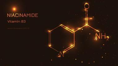
Even More Health Benefits of Niacinamide
Reproduced from original article:
https://articles.mercola.com/sites/articles/archive/2024/02/27/more-health-benefits-of-niacinamide.aspx
The original Mercola article may not remain on the original site, but I will endeavor to keep it on this site as long as I deem it to be appropriate.
Analysis by Dr. Joseph Mercola February 27, 2024
STORY AT-A-GLANCE
- Niacinamide, also known as nicotinamide (a form of vitamin B3), is important for healthy mitochondrial function and cellular energy production
- Patients with established glaucoma scored significantly higher on visual field tests after taking niacinamide and calcium pyruvate daily for about two months
- Niacinamide can be used to treat declining testosterone levels in men and women, and protects against stress by lowering cortisol
- Niacinamide has anticancer and antiaging effects, and can be useful in the treatment of Alzheimer’s disease and COVID-19
- It may also be valuable in the treatment of both alcoholic liver disease and nonalcoholic liver disease (NAFLD), as low NAD is involved in the disease process
I recently posted an article detailing the importance of niacinamide (aka, nicotinamide, a form of vitamin B3 or niacin) for healthy mitochondrial function and cellular energy production, and how it can help reverse obesity and leaky gut, and prevent neurodegeneration, kidney disease and heart failure. Here, I’ll review several additional conditions that can be prevented and/or treated with this inexpensive and readily available supplement.
More Niacinamide Is NOT Better, There’s a Goldilocks’ Dose
Keep in mind that the dosages used in the research studies discussed below do vary widely, but as a rule, I only recommend taking small doses of 50 milligrams of niacinamide three times a day.
This dosage has been shown to optimize energy metabolism and boost NAD+ levels, which are foundational for everything else to work. It can be taken four times a day if you space them out. Take a dose as soon as you get up, before going to bed, and twice evenly spaced between those times.
The problem with taking too much vitamin B3, whether in the form of niacin or niacinamide, is that it might backfire and contribute to cardiovascular disease as documented by the Cleveland Clinic. ZeroHedge reports:1
“The new study out of the Cleveland Clinic, published in Nature Medicine,2 determined there is a delicate balance between too much niacin and just enough — a sort of Goldilocks effect … [As] observed by the Cleveland Clinic team, too much niacin creates a byproduct known as 4PY.
This product circulates within the bloodstream and is associated with a higher risk of heart attack, stroke, and other cardiac events. Additionally, 4PY was shown in preclinical studies to trigger vascular inflammation, damaging blood vessels and eventually leading to atherosclerosis.
The researchers discovered this by examining data from 1,162 patients who had experienced major cardiovascular events. Just under half of the patients (442) were female. Initially, the team sought common markers that could lead to cardiovascular events. The most common factor within the pool of patients was excess levels of niacin.
The findings led to additional studies to validate the initial research. Both cohort studies, conducted in the United States and Europe, confirmed that niacin breakdown predicted an individual’s future risk of heart attack, stroke, and death from cardiovascular disease.”
Niacinamide Limits Damage From PUFA Peroxidation
With that caveat out of the way, let’s take a look at some of the health benefits associated with niacinamide supplementation, starting with its ability to ameliorate damage caused by linoleic acid (LA) consumption.
One of the most toxic metabolic byproducts of LA is 4 HNE, a toxic aldehyde that appears to play a causative role in heart failure.3 Fortunately, there is an enzyme system called aldehyde dehydrogenases that deactivates 4 HNE. The best way to increase the activity of this enzyme system is to make sure you have sufficient NAD+, and the most efficient way to optimize your NAD+ level is to make sure you’re getting 50 mg of niacinamide three times a day.
Download this Article Before it Disappears
Raise Your Testosterone Naturally With Niacinamide
Niacinamide can be used to treat problems associated with declining testosterone levels due to aging, as demonstrated in a February 2022 Nature Aging study.4 This study also involved eye health, specifically evaporative dry eye disease, but with a focus on the effects of androgens.
They found that by raising NAD+, local testosterone distribution in the meibomian gland in the eyelids, which produces oil to prevent the evaporation of tears, was improved. As a result, age-related atrophy of the gland was reduced, which alleviated the dry eye condition. In this case, they used a 5% solution of the NAD+ precursors nicotinamide mononucleotide (NMN) or nicotinamide riboside (NR). The two regimens assessed were as follows:
- Chronic — 2 mg NAD+ precursor four times a day in each eye for 90 days.
- Acute — 5 mg NAD+ precursor six times a day in each eye for 14 days.
However, as noted by biohacker Georgi Dinkov,5 plain niacinamide has been shown to be just as effective as NMN and NR for raising NAD+, so there’s no reason why it wouldn’t work as well. Moreover, these findings are also applicable for other tissues and organs affected by declining testosterone, not just your eyes.
It confirms that niacinamide is a necessary cofactor to address the nearly universal decline in testosterone in both males and females, so this is yet another reason that most people need it. Dinkov writes:6
“… the findings that androgen (in this case, testosterone) deficiency seen almost universally with advancing age is apparently due primarily to deficiency of the co-factor NAD+, which can apparently be remediated by supplementing NAD+ precursors such as niacinamide.
It appears that the rate-limiting step for androgen (testosterone) synthesis is the enzyme 3b-HSD, for which NAD+ is the main cofactor. Thus, a decline in NAD+ seen in aging (and disease) leads to decline in 3b-HSD activity and thus reduced androgen (testosterone) levels.
Conversely, the study found that restoring NAD+ levels almost fully remediated the reduced function of 3b-HSD, the androgen (testosterone deficiency), and thus was basically curative for the specific condition (dry-eye) the study investigated…
As far as the systemic steroidogenic deficiency seen with aging that I mentioned earlier, oral dosing of an NAD+ precursor such as niacinamide would likely be needed, and human studies have demonstrated that daily doses in the 300 mg-500 mg daily range optimally stimulate NAD+ synthesis. (It would be better to start with 50 mg three times a day (150 mg) and increase if necessary.)
While most studies found that higher daily oral doses are unable to further raise NAD+ levels through the precursor pathway, higher doses of niacinamide specifically have been shown to inhibit the enzyme PARP-1, which is a ‘consumer’ of NAD+ and inhibiting it also raises NAD+ levels.
So, experimenting with the niacinamide with doses in the 500mg-1,500mg daily range would probably identify an optimal daily dose regimen for most people looking to increase steroidogenesis, especially if precursors such as pregnenolone and/or DHEA are taken as well.”
To address declining testosterone, I recommend taking:
- 50 mg of niacinamide three times a day
- 5 mg to 10 mg of DHEA orally once a day
- 50 mg of oral pregnenolone once a day
Ideally, take the hormone supplements DHEA and pregnenolone with a saturated fat, like a teaspoon of butter, to make sure they bypass metabolism in the liver, which will radically decrease their effectiveness.
Niacinamide Protects Against Stress
Another phenomenally important human study7,8,9 confirms that niacinamide lowers cortisol by increasing NAD+. NAD+ is the cofactor for an enzyme called 11b-HSD2, which deactivates cortisol by turning it into cortisone. So, increasing NAD+ with niacinamide will help lower cortisol, and this can benefit just about everyone, seeing how most people are plagued by stress and have elevated cortisol, which in turn drives inflammation and inhibits fat loss.
Raising NAD+ with niacinamide supplementation was also shown to improve glucose metabolism and insulin sensitivity. It also increased ATP levels, which is your cellular energy currency. As such, niacinamide may be one of the most important supplements you can take.
Importantly, this study used plain niacinamide rather than the more expensive NMN or NR, so we don’t have to infer these benefits. They took place using plain niacinamide and D-ribose. The dosages used were 240 mg of niacinamide twice a day, and 1,280 mg of D-ribose twice a day, for one week.
Anticancer Effects
Niacinamide can also help prevent and treat cancer. As noted in one 2022 study in Nature Metabolism,10,11 limiting fat availability to cancer cells may be one of the keys to curing cancer. NAD+ also plays an important role, and niacinamide raises NAD+:
“Production of oxidized biomass, which requires regeneration of the cofactor NAD+, can be a proliferation bottleneck that is influenced by environmental conditions … Here, we show that de novo lipid biosynthesis can impose a substantial NAD+ consumption cost in proliferating cancer cells.
When electron acceptors are limited, environmental lipids become crucial for proliferation because NAD+ is required to generate precursors for fatty acid biosynthesis. We find that both oxidative and even net reductive pathways for lipogenic citrate synthesis are gated by reactions that depend on NAD+ availability.”
An earlier study,12 published in 2020, found that niacin (vitamin B3) could control the growth of brain tumors by reactivating myeloid cells (a type of bone marrow cell). As explained by the authors:
“Although innate immune cells are typically present inside tumors, they often have an inactive phenotype such that they are ineffective at killing the cancer cells or even promote tumor growth. Sarkar et. al. discovered that it may be possible to reprogram these cells to a more active type using niacin (vitamin B3).
The authors showed that niacin-exposed monocytes can inhibit the growth of brain tumor-initiating cells. Moreover, niacin treatment of intracranial mouse models of glioblastoma increased monocyte and macrophage infiltration into the tumors, stimulated antitumor immune responses, and extended the animals’ survival …”
Here, a mere 7 mg of niacin per kilo of bodyweight per day halted the growth of brain tumors and nearly doubled survival times. The niacin group also survived longer than those receiving chemotherapy alone. While this study used niacin, niacinamide would work just as well. Tying the results back to the study above, Dinkov notes:13
“The proposed mechanism of action was immune system activation. I, personally, do not buy this explanation as a main mechanism of action. IMHO vitamin B3’s known effects on inhibiting lipolysis … and promoting the oxidation of glucose are probably more important for inhibiting the tumor growth and prolonging survival.”
Antiaging Effects
Niacinamide and other B vitamins also have important antiaging effects. In a December 2021 article, Dinkov reviewed a 2011 study14 that suggests fasting can speed up aging by depleting your energy reserves, and that vitamin B2 has the opposite effect and slows the aging process. Commenting on this finding, Dinkov wrote:15
“[This is] a great study that not only suggests a dirt cheap and widely available option for retarding aging, but once again demonstrates that stress (e.g. fasting) directly causes aging by depleting/blocking OXPHOS [oxidative phosphorylation, the metabolic pathway cells use to oxidize nutrients, thereby releasing chemical energy to produce ATP], and the key mechanism for stopping/reversing aging is by restoring energy production.
The proposed ‘senolytic’ in this study is the humble vitamin B2, also known as riboflavin. Vitamin B2 is the precursor for FAD [flavin adenine dinucleotide] — a key coenzyme, responsible for about 20% of the energy produced in the OXPHOS process. Btw, the other 80% are controlled by NAD — a molecule synthesized endogenously from the precursor niacinamide.
There has been an explosion in publications over the last decade touting NAD as the cure to virtually every disease out there, and many of those claims are well-backed by evidence. While there are … proprietary NAD precursors such as nicotinamide riboside and nicotinamide mononucleotide, plain old niacinamide works just as well and is drastically cheaper.
Niacin is another NAD precursor but [it] increases histamine and serotonin, and still has to be converted into niacinamide before it can end up as NAD. So, no reason to take anything else but niacinamide.
Doing so, raises the NAD/NADH ratio, shifts the cell redox state towards oxidation (as opposed to reduction), and the ample energy produced by the cell can be used for all types of maintenance ‘work’ the cells need to perform in order to stay healthy, prevent aging, etc …
[V]itamin B2 has a similar metabolic role as well. It is a precursor to the co-factor FAD and taking B2 raises FAD and thus the FAD/FADH ratio, which facilitates electron flow through the electron chain complexes I&II. Conversely, having insufficient levels of FAD (and/or low FAD/FADH ratio) can effectively block the ETC [electron transport chain].
This crucial role of FAD in the ETC demonstrates why excessive fatty acid oxidation (FAO) is harmful. Namely, FAD is consumed in the process of beta-oxidation of fatty acids and as such excessive FAO can consume too much FAD, drop the FAD/FADH ratio, and thus block electron flow through ETC.
This reduction/blockade of ETC is exactly what is seen in most chronic, degenerative diseases, but especially in conditions such as cancer, diabetes, Alzheimer’s disease, autoimmune conditions, etc.
Well, the [Physiology] study … found that this reduction of mitochondrial activity (ETC) is exactly what triggers the senescence process, and activating mitochondria (specifically ETC II) by supplementing vitamin B2 prevented cellular aging. Here is the actual explanation from the study authors:
‘… Cells under stress produce SLC52A1, and increase their absorption of vitamin B2 from outside the cell. Once inside the cell, the vitamin B2 is converted into FAD and increases mitochondrial energy production by becoming a coenzyme of mitochondrial respiratory chain complex II. As a result of the AMPK and p53 (which induce cellular senescence) are inactive, therefore stress-mediated cellular senescence is suppressed.’”
Niacinamide for Liver Disease
Niacinamide may also be valuable in the treatment of both alcoholic liver disease and nonalcoholic liver disease (NAFLD). By now, you can probably guess why. It’s because low NAD is involved in the disease process.
A study16,17 published in 2016 showed that chronic alcohol bingeing injures your liver and other organs by reducing NAD+, which is a required cofactor for important enzymes and mitochondrial renewal.
In short, a decline in the NAD/NADH ratio (your redox status) is a main driver of alcohol-related pathologies, and not just in your liver but in all organs. The answer is to increase NAD+, which is effectively done using niacinamide. According to Dinkov, niacinamide also works synergistically with methylene blue:18
“Both [niacinamide and methylene blue] can raise the NAD/NADH ratio quite robustly on their own, but in combination are synergistic and may allow for much lower doses to achieve the same effects.
People have sent me emails showing their blood tests demonstrated the same increase in NAD/NADH from a combination of 1mg MB [methylene blue] and 250 mg niacinamide as using 5mg+ MB or 750mg-1,000mg niacinamide separately.”
Related to this is the importance of eating the correct fats. You want to increase your saturated fat and avoid PUFAs, especially linoleic acid (LA), as PUFAS are stored and accumulated whereas saturated fats are burned for energy.19,20 It can take up to seven years to clear LA from your body due to its long half-life. In the meantime, you need to be careful about not pushing the LA out too rapidly through fasting, low-carb diets or vigorous exercise.
The accumulation of PUFAs in your cells are, I believe, one of the greatest contributors to chronic and degenerative diseases of all kinds, as they wreak havoc with your cellular machinery and impair mitochondrial function (and hence reduce energy production). Niacinamide can be helpful while you’re clearing stored PUFAs from your cells, as it’s an anti-lipolytic agent.
Niacinamide for Alzheimer’s Disease
Niacinamide is required for healthy metabolism and, as such, it can also be useful in early Alzheimer’s treatment.21 Combining it with methylene blue may boost benefits even further, as they work synergistically, and methylene blue alone has been shown to stop the progression of Alzheimer’s.22
Increasing the carbs only seems to work when your fat intake is below 35% of your total daily carbs. If it is higher, the Randle cycle will prevent the extra carbs from being metabolized in the mitochondria and will shuttle them to glycolysis which increases lactic acid and will likely worsen one’s health.
Thiamine, the B1 mentioned in the study, is a cofactor for the enzyme that converts the glucose metabolite pyruvate into acetyl-CoA, which is what is pushed into the mitochondria to be burned as fuel. Taking niacinamide (vitamin B3) will help three of the complexes in the mitochondria work and vitamin B2, riboflavin, will help one complex to produce the ATP, so using all three would be ideal.
Niacinamide is the only one that appears to benefit from lowering the dosing to 50 mg three times a day. As reported by Dinkov:23
“[A 2019] study24 demonstrated that increasing levels of acetyl-CoA (the starting intermediate of the Krebs cycle), even at a stage of very advanced aging/AD [Alzheimer’s disease], can reverse aspects of both pathologies.
While the study used patented compounds, the same effects can be achieved by ensuring a steady supply of dietary carbs combined with increasing the function of pyruvate dehydrogenase (PDH). Increasing the activity of PDH can be achieved by supplementing with vitamin B1 (a cofactor of PDH), keeping the NAD/NADH ratio as high as possible and keeping fatty acid oxidation (FAO) down …
[T]hese approaches have been successfully tested in many animal models and one of the molecules that works through the latter two mechanisms is niacinamide. In fact, there is a human clinical trial25 [completed in August 2022] with 3 grams of niacinamide daily for treating AD, and though the results have not been published yet the leaked information suggests highly positive results. Methylene blue (MB) can do the same …”
Niacinamide and COVID-19
As it turns out, raising NAD+ appears to be therapeutic for COVID infection as well.26 The reason for this is because SARS-CoV-2 infection dysregulates NAD synthesis and the NAD metabolome, which is a component of your innate immunity.
As noted by Dinkov, this study again demonstrates “the crucial role energy plays even in ‘non-metabolic’ diseases such as viral infections.” Indeed, without energy your body cannot defend itself against infection and disease, and since NAD+ is such a crucial component of energy production, it makes perfect sense that having higher NAD levels will make you more resilient against viral infections. The answer again, then, is niacinamide, with or without methylene blue.
By now, you should have a good idea about how important niacinamide is for your health, and why I think it’s one of the most important supplements you can take on a daily basis.
Niacinamide Helps Treat Glaucoma
Research27,28 published in November 2021 reported that patients with established glaucoma scored significantly higher on visual field tests after taking 3 grams of niacinamide in combination with 3 grams of (calcium) pyruvate daily for about two months. As reported by the authors:
“Given the decrease in NAD with aging, which may render retinal neurons more vulnerable to disease-related insults, the investigators hypothesized that increasing NAD may support the mitochondrial activity of RGCs and decrease their susceptibility to glaucoma.
In a rat model of ocular hypertension, retinal and optic nerve NAD declined as a function of IOP, while nicotinamide was neuroprotective at a range of doses. In addition, nicotinamide has been shown to be low in the sera of patients with primary open-angle glaucoma. These data further support a role for NAD in glaucoma.
Furthermore, Harder et al reported that an IOP-mediated decrease in retinal pyruvate levels was associated with dysregulated glucose metabolism prior to detectable optic nerve degeneration in metabolic studies of DBA/2J mice.
They also found that oral supplementation with pyruvate protected both rat and mouse models of glaucoma from neurodegeneration, with a combination of nicotinamide and pyruvate being the most protective in the chronic mouse model…
Given the significant neuroprotective effects observed in recent studies, the similarities in cellular biology of NAD+ and pyruvate pathways between mice and humans, as well as the safety profile of these supplements, we investigated the effect of nicotinamide and pyruvate on the visual function of patients with treated manifest glaucoma …
[R]esults suggest that treatment with nicotinamide and pyruvate tripled the likelihood of improvement of test locations relative to placebo.”
If you have glaucoma and want to give niacinamide a try, I recommend limiting your dose to 50 mg three times a day, before increasing to 3 grams a day, which I believe is excessive.
- 1 ZeroHedge February 21, 2024
- 2 Nature Medicine February 19, 2024; 30: 424-434
- 3 Haidut.me February 21, 2024 (Archived)
- 4 Nature Aging 2022; 2: 105-114
- 5, 6 Haidut.me March 31, 2022 (Archived)
- 7 Nutrients June 2022; 14(11): 2219
- 8 Nutritional Outlook July 11, 2022
- 9 Haidut.me July 12, 2022 (Archived)
- 10 Nature Metabolism 2022; 4: 711-723
- 11, 13 Haidut.me July 14, 2022 (Archived)
- 12 Science Translational Medicine April 1, 2020; 12(537)
- 14 Physiology August 2011; 26(4): 214-224
- 15 Haidut.me December 21, 2021 (Archived)
- 16 Exp Mol Pathol April 2016; 100(2): 303-306
- 17 NMN.com April 7, 2016
- 18 Haidut.me October 22, 2020 (Archived)
- 19 PLOS ONE 2012; 7(11): e48852
- 20 Haidut.me October 22, 2019 (Archived)
- 21, 25 Clinical Trials Nicotinamide as an Early Alzheimer’s Treatment NCT03061474
- 22 Haidut.me December 2, 2019 (Archived)
- 23 Haidut.me December 13, 2019 (Archived)
- 24 eLife Sciences November 19, 2019
- 26 Metabolism December 2020; 295(52): 17986-17996
- 27 JAMA Ophthalmology 2022;140(1):11-18
- 28 Haidut.me December 9, 2021 (Archived)
A Silent Thief of Sight: Are You at Risk?
Reproduced from original article:
https://articles.mercola.com/sites/articles/archive/2023/05/03/glaucoma-risk-factors.aspx
The original Mercola article may not remain on the original site, but I will endeavor to keep it on this site as long as I deem it to be appropriate.
Analysis by Dr. Joseph Mercola Fact Checked May 03, 2023

STORY AT-A-GLANCE
- Glaucoma is the second leading cause of blindness in the world. Worldwide, an estimated 80 million people are affected, and prevalence is on the rise
- Glaucoma develops when high pressure in your eye (ocular hypertension) damages the optic nerve. Peripheral vision is impacted first, followed by central vision. Vision loss is usually the only symptom
- Interocular pressure is a modifiable risk factor of glaucoma
- Aside from high eye pressure, other factors that influence your risk include old age, thin cornea, large optic nerve cup size and low peripheral vision test score. These factors can help your ophthalmologist determine your glaucoma risk
- Treatment options include eye pressure-lowering eye drops, oral medication and selective laser trabeculoplasty. Certain supplements can also be helpful, including melatonin, lutein (ideally from food), ginkgo biloba, Chinese skullcap, bilberry, green tea, curcumin and nicotinamide (B3)
Glaucoma is the second leading cause of blindness in the world. Worldwide, an estimated 80 million people are affected, and prevalence is rising.1 Glaucoma develops when high pressure in your eye (ocular hypertension2) damages the optic nerve. Peripheral vision is impacted first, followed by central vision.
Ocular hypertension occurs when the front of your eye fails to drain fluid properly, thereby causing pressure to build and put pressure on the optic nerve. Factors that raise your risk for ocular hypertension include:
| Family history of ocular hypertension or glaucoma | Diabetics and those with high blood pressure |
| People over 40 | Blacks and Hispanics |
| Myopia (nearsightedness) | Long-term steroid use |
| Previous eye injuries or eye surgeries | Those with pigment dispersion syndrome or pseudoexfoliation syndrome |
The only known way to prevent and/or stop the progression of glaucoma is to lower the pressure in the eye. Treatment options include drugs, lasers, laceration surgery, oral medications, nutritional supplements, herbs and other plant remedies, several of which I’ll review below.
Keep in mind that loss of vision is usually the first and only symptom of glaucoma, so it’s important to get a thorough eye exam by an ophthalmologist to determine if your ocular pressure is elevated, which could make you a candidate for glaucoma. Once vision loss occurs, the damage to your optic nerve is irreversible.
The Ocular Hypertension Treatment Study (OHTS)
Being diagnosed with ocular hypertension does not mean you’re destined to develop glaucoma, but it does significantly raise your risk, as it’s the primary underlying cause. The Ocular Hypertension Treatment Study (OHTS), which began in 1994, has also identified other risk factors that can help determine your risk for glaucoma. As reported by Harvard Health:3
“The researchers enrolled a diverse group of 1,636 participants with ocular hypertension from 22 sites across the US. To study glaucoma prevention, participants were randomly assigned to start either early eye pressure-lowering eye drops (medication group) or close observation (control group).
At five years the data showed that 4.4% of participants developed glaucoma in the medication group, compared to 9.5% in the control group. This tells us that early use of medicated eye drops helps delay over 50% of glaucoma cases in people with ocular hypertension.
During later phases of the study, the control group could receive eye pressure-lowering medications to see whether starting medication later could still delay glaucoma; it did.
At 20 years, about 49% of those in the control group and 42% of those in the medication group developed glaucoma. However, since the study was no longer randomized, the researchers were unable to compare the 20-year risk reduction between the initial starting groups …
Glaucoma risk, it turned out, did not depend solely on eye pressure and race, but on a combination of exam findings. This information helps guide clinicians in determining whether a person with ocular hypertension is at a low, medium, or higher risk for developing glaucoma. Having such information could help people decide when to begin using medicated eye drops to prevent vision loss or slow its progress.”
Aside from high eye pressure, other factors that influence an individual’s risk for developing glaucoma were found to include:
- Older age
- Thinner corneas
- Larger optic nerve cup sizes
- Low initial peripheral vision test scores
Treatment Alternatives
If you’re diagnosed with ocular hypertension, particularly if you also have other risk factors listed above, your ophthalmologist may prescribe eye pressure-lowering eye drops, oral medication and/or selective laser trabeculoplasty (SLT).4
SLT is a five-minute procedure performed at your ophthalmologist’s office, where a low-energy laser is pulsed into specific cells in your eye. Your body’s natural healing response then does the rest by rebuilding these cells. The rebuilding process automatically reduces the intraocular pressure and improves drainage.
Studies have also shown that certain herbal remedies, plant extracts5 and nutritional supplements can be helpful, although most conventional doctors know very little, if anything, about these products.
Download this Article Before it Disappears
Melatonin Lowers Intraocular Pressure
Melatonin, for example, has been shown to lower intraocular pressure in patients with glaucoma. Subjects who took 2 mg of melatonin daily at 10:30 p.m. for 90 days, experienced several benefits, including:6
- Increased stability of systemic circadian rhythm via improved phase alignment and alignment with intraocular pressure
- Decreased intraocular pressure
- Improved function of retinal ganglion cells in those with advanced glaucoma
- Improvements to sleep and mood, particularly in those with advanced glaucoma
Interestingly, these researchers7 suggested that glaucoma may, in fact, be a type of neurodegenerative disease, and damage to retinal ganglion cells affects not only vision but also circadian rhythms and sleep, which melatonin helps regulate.
Melatonin receptors exist in several areas of your eyes, including the retina, lens, and cornea. This hints at melatonin’s importance for regulating eye processes, particularly when pressure homeostasis is involved.
Its beneficial effects on the eyes can perhaps be explained best by the fact that melatonin receptors exist in several areas of your eyes, including the retina, lens and cornea.8 This hints at melatonin’s importance for regulating eye processes, particularly when pressure homeostasis is involved. As explained in a March 2020 paper published in Progress in Retinal and Eye Research:9
“Glaucoma, the most prevalent eye disease, also known as the silent thief of vision, is a multifactorial pathology that is associated to age and, often, to intraocular hypertension (IOP). Indeed IOP is the only modifiable risk factor …
Melatonin and analogues decrease IOP in both normotensive and hypertensive eyes. Melatonin activates its cognate membrane receptors, MT1 and MT2, which are present in numerous ocular tissues, including the aqueous-humor-producing ciliary processes.
Melatonin receptors belong to the superfamily of G-protein-coupled receptors and their activation would lead to different signaling pathways depending on the tissue … The current work highlights the important role of melatonin and its analogues in the healthy and in the glaucomatous eyes, with special attention to the control of intraocular pressure.”
Personally, I question the timing of the melatonin in the above study at 10:30 p.m. In my view that is far from an ideal time to sleep and about two hours later than I personally do. The other issue of concern is that it is unclear if these authors were aware that the greatest production of melatonin is not the pineal, but the mitochondria, when exposed to infrared light.
It would be interesting to compare the results of their 2 mg 10:30 p.m. protocol versus one hour of sunshine around solar noon in a warm climate.
Melatonin’s Effects Have Been Known for Decades
The effect of melatonin on intraocular pressure has been known for decades, yet most conventional doctors are still groping in the dark. In 1988, researchers with Oregon Health Sciences University exposed subjects to bright light to suppress serum melatonin levels, and then supplemented with melatonin to gauge its effect on intraocular pressure.10 A significant connection was found:11
“Our data suggests that during the period of melatonin’s greatest levels in the serum, IOP is lowest. All subjects had maximum pressures form 4 p.m. to 6 p.m. and most subjects had minimums from 2 a.m. to 5 a.m.
In experiment one, bright light suppression of melatonin secretion attenuated the early morning fall IOP. This was statistically significant at suggesting that melatonin is involved in lowering early morning IOP.
In experiment one, there was only partial suppression of melatonin production with bright light and consequently there was no significant difference in IOP between subjects exposed to dim light and bright light.
However, administering 200 micrograms (one-fifth of 1 mg) of melatonin orally caused a significant decrease in IOP. Intraocular pressure remained low for approximately four hours after the last dose.”
Lutein Protects Against Glaucoma and Other Eye Diseases
Lutein is another nutrient that is really important for eye health and helps to protect against age-related macular degeneration, cataracts, glaucoma and other eye diseases.
Lutein concentrates in your macula, which is the part of your retina responsible for central vision. Along with zeaxanthin and mesa-zeaxanthin (a metabolite of lutein), these three carotenoids form the retinal macular pigment, which not only is responsible for optimizing your visual performance but also serves as a biomarker for the risk of macular diseases.12
Lutein is also found in the lens, where it helps protect against cataracts and other age-related eye diseases. Among carotenoids, lutein is the most efficient at filtering out blue light — the type that comes from cellphones, computers, tablets and LED lights. Blue light induces oxidative stress in your eyes, which increases the risk of cataracts and macular diseases. Lutein, however, acts as a shield against it.
Your body cannot make lutein, so you must get it from your diet. Following are 10 foods that are particularly rich sources of lutein.
| Dark leafy greens | Carrots |
| Broccoli | Egg yolks |
| Red and yellow peppers | Sweet corn |
| Avocados | Raspberries |
| Cherries | Paprika |
Lutein and other carotenoids are fat-soluble, so to optimize absorption be sure to consume it along with a source of healthy fat, such as coconut oil or grass-fed butter. Because organic, pastured egg yolks contain fat, they’re among the healthiest sources of lutein.
Other Helpful Remedies
Certain herbs, plant extracts and vitamins have also shown promise in the treatment of glaucoma, including:13
| Ginkgo biloba — A 2013 study14 highlighted the benefits of ginkgo biloba, finding it slowed the progression of glaucoma. Forty-two patients with normal tension glaucoma received 80 mg of ginkgo biloba extract twice a day and underwent five or more visual field tests over the course of at least four years.
Mean follow-up was 12.3 years. While intraocular pressures remained largely the same after treatment, the visual field index significantly improved, especially central field vision. |
| Chinese skullcap — A 2021 study15 concluded that baicalein, a flavone found in skullcap root, effectively improved symptoms of glaucoma and reduced retinal ganglion cell apoptosis. |
| Bilberry — In animal research,16 bilberry extract was shown to prevent the death of retinal ganglion cells in mice whose optic nerves were crushed. The dosages given were 100 mg per kilo of bodyweight per day and 500 mg/kg/day. |
| Green tea — A 2019 animal study found green tea extract helped protect retinal ganglion cells during ischemic conditions (i.e., when blood flow is restricted). As such, green tea extract may be a viable therapeutic approach for glaucoma and optic neuropathies.
A more recent report,17 published in 2022, proposed that moderate consumption of green tea or 400 mg of concentrated green tea extract per day might benefit individuals with elevated intraocular pressure and those at risk for glaucoma. |
| Curcumin — According to animal research18 published in 2014, curcumin, a polyphenol found in the spice turmeric, can help treat glaucoma by limiting oxidative damage in the eye and lowering intraocular pressure. |
| Vitamin B3 (nicotinamide) — Research19 published in 2020 reported that glaucoma patients who took oral vitamin B3 experienced significant improvement in retinal function. Patients in the treatment group received 1.5 grams of nicotinamide per day for six weeks, followed by 3 grams a day for another six weeks. |
- 1, 5, 13 Epoch Times April 19, 2023 (Archived)
- 2 AAO.org Ocular Hypertension
- 3 Harvard Health September 22, 2022
- 4 Glaucoma Research Foundation Treating Glaucoma
- 6, 7 J Pineal Res. 2021 May;70(4):e12730. Discussion
- 8 Pharmacol Ther. 2007 Mar;113(3):507-22
- 9 Prog Retin Eye Res. 2020 Mar;75:100798
- 10, 11 Curr Eye Res. 1988 Jul;7(7):649-53
- 12 Nutrients. 2020 Jun; 12(6): 1721., 1.2 Distribution of Lutein in the Human Body
- 14 Journal of Glaucoma December 2013; 22(9): 780-784
- 15 Chinese Medicine 2021; 16, article number 124
- 16 Clinical Ophthalmology October 11, 2017; 11: 1825-1834
- 17 International Journal of Ophthalmology 2022; 15(5): 747-752
- 18 J Ocul Pharmacol Ther October 2014; 30(8): 657-664
- 19 Clinical & Experimental Ophthalmology July 28, 2020; 48(7): 903-914
Improve your vision and bone health with lutein and zeaxanthin
Reproduced from original article:
https://www.naturalhealth365.com/improve-your-vision-and-bone-health-with-lutein-and-zeaxanthin.html
by: April 24, 2023
 (NaturalHealth365) Lutein, a type of carotenoid found in the macula, is often prescribed to reduce intraocular pressure in individuals with pre-glaucoma and glaucoma. However, a recent study published in JAMA Ophthalmology by the National Institutes of Health (NIH) shows that lutein is also helpful in decreasing age-related macular degeneration.
Additionally, when combined with another macular carotenoid called zeaxanthin, lutein can improve bone health, making it even more compelling to include these nutrients in your daily supplement routine.
Revolutionizing eye health: Uncover the power of lutein and zeaxanthin
In smokers, some “experts” suggest, the use of beta-carotene has been linked to an increased risk of lung cancer. However, two NIH-supported studies demonstrate that the use of lutein and zeaxanthin, as shown in the age-related eye disease studies (AREDS2), can effectively replace beta-carotene, leading to a reduction in age-related macular degeneration (AMD). This is significant as AMD is the primary cause of blindness among older adults in the U.S.
Over a decade-long analysis of the AREDS2 formula, zeaxanthin, and lutein were found to be effective replacements for beta-carotene. This formula not only reduces the risk of lung cancer caused by beta-carotene but also slows the progression of AMD. However, it should be noted that the participants in the study were already smokers. The primary goal of AREDS2 was to create a formula that could be utilized by everyone, regardless of smoking habits.
The results of the study demonstrate that this innovative formula is superior in slowing the progression of AMD and is also safer. The reduction or elimination of AMD is critical for maintaining good health, as this retinal degenerative disease can ultimately lead to blindness. While there is no conventional cure for AMD currently, treating it has the potential to reduce the rate of vision loss.
The dangers of beta-carotene for smokers: Why lutein and zeaxanthin are a safer bet
The initial AREDS study, conducted in 1996, demonstrated that a dietary supplement containing 15 mg of beta-carotene, 80 mg of zinc, 2 mg of copper, and vitamins C and E could slow the progression of AMD, specifically from moderate to late stages. However, subsequent studies revealed that smokers who took beta-carotene had a significantly higher risk of lung cancer than non-smokers.
In the subsequent AREDS2 study, the beta-carotene was replaced with 2 mg of zeaxanthin and 10 mg of lutein for individuals who had ceased smoking or never smoked. The study found that the use of lutein and zeaxanthin did not increase the risk of lung cancer and had the potential to reduce the progression of AMD by up to one-quarter. The study concluded with participants using the AREDS2 formula with zeaxanthin and lutein instead of beta-carotene.
A more recent study involving over 4,000 patients over five years since the previous AREDS2 study showed a 20% decrease in the risk of progression to late-stage AMD compared to those who took beta-carotene. It is evident that lutein and zeaxanthin are vital components in the fight for better eye health.
More benefits: Lutein and zeaxanthin support bone health
Recent studies have shown that zeaxanthin and lutein not only promote eye health but also strengthen bones, reducing the risk of osteoporosis in older adults. In a study involving participants aged 50 and above, it was found that the intake of these carotenoids can lead to stronger bones and reduced risk of fractures. However, it’s worth noting that a relatively high level of zeaxanthin and lutein intake was required to achieve these results.
Nonetheless, the dual benefits of zeaxanthin and lutein make them particularly valuable for vulnerable senior citizens. By incorporating these carotenoids into their daily supplement intake, older adults can enhance their vision and bone health and improve their overall health and longevity.
To learn more about how to properly supplement your diet, we suggest you work with an integrative health coach – who understands your entire health history and personal goals.
Sources for this article include:
The Healthy Sugar Substitute You’ve Likely Never Heard About
Reproduced from original article:
https://articles.mercola.com/sites/articles/archive/2022/03/28/what-is-nadph-and-nox.aspx
The original Mercola article may not remain on the original site, but I will endeavor to keep it on this site as long as I deem it to be appropriate, and will not be bullied into removing it.
Analysis by Dr. Joseph Mercola – Expert Review by James DiNicolantonio, Pharm.D Fact Checked

STORY AT-A-GLANCE
- Glycine is mildly sweet and can be used as a sugar substitute in tea or coffee
- Glycine and collagen (an outstanding source of glycine) inhibit the consumption of NADPH, thereby lowering inflammation and oxidative damage
- NADPH oxidase is typically abbreviated as NOX. Inhibiting NOX is an effective way to increase NADPH and your cells’ ability to reduce oxidative stress
- NADPH is used as a reductive reservoir of electrons to recharge antioxidants once they become oxidized. NADPH is also necessary to make your steroid hormones and fats
- Preventing many chronic diseases requires finding a means of inhibiting or modulating NOX. Such strategies include avoiding fructose, practicing nutritional ketosis and supplementing with spirulina, niacin, glycine and collagen
- Glycine supplementation may be beneficial for the prevention and/or treatment of metabolic syndrome, complications from diabetes, cardiac hypertrophy and alcoholic and nonalcoholic liver disorders
This article was previously published February 4, 2019, and has been updated with new information.
As you probably know, inflammation and oxidative damage are primary drivers of most chronic diseases. What you may not be aware of is the importance of nicotinamide adenine dinucleotide phosphate hydrogen (NADPH) and NADPH oxidase, typically abbreviated as NOX, in these processes.
In a recent paper,1James DiNicolantonio, who co-wrote my book, “Superfuel,” details the importance of collagen and glycine for the inhibition of NADPH breakdown. DiNicolantonio, also co-wrote a book with Jason Fung, called “The Longevity Solution,” which takes a deep dive into how collagen and glycine may help promote longevity.
Without sufficient amounts of NADPH, your body cannot recharge glutathione once it becomes oxidized. As you know, glutathione is crucial for detoxification, and both collagen and glycine effectively raises your NADPH level by inhibiting the enzyme that breaks down NADPH.
Considering the importance of NADPH for optimal health and chronic disease prevention, this is really important information that could make a big difference for many, as collagen and glycine are both easily obtainable and relatively inexpensive. But before we delve into the specifics of how glycine affects NOX and NADPH, let’s review some of the basics.
What Is NADPH and NOX?
NADPH is the reduced form of NADP+. It’s a reducing agent necessary for anabolic reactions, including lipid and nucleic acid synthesis. NOX is an enzyme complex that is bound to the cellular membrane, facing the extracellular space. Inhibiting NOX is a useful strategy to increase NADPH and your cells’ ability to counter oxidative stress.
NOX is activated in a large number of pathological conditions that generate a great deal of oxidative stress. In fact, according to DiNicolantonio, NOX overactivity appears to play a significant role in a wide range of health conditions, including but not limited to:
| Vascular diseases and vascular complications of other diseases (diabetes, kidney failure, blindness and heart disease for example) |
| Insulin resistance |
| Neurodegenerative disorders such as Alzheimer’s and Parkinson’s |
| Cancer |
| Glaucoma |
| Pulmonary fibrosis |
| Erectile dysfunction |
As explained by DiNicolantonio in a paper2 detailing bilirubin’s ability to inhibit NOX complexes and downregulate NOX activity, and the benefits of spirulina for this purpose:
“Activation of NADPH oxidase [NOX] is a key mediator of proinflammatory microglial activation … Oxidative stress in adipocytes, stemming largely from NADPH oxidase activity, appears to play a key role in the induction of insulin resistance and the skewing of adipokine and cytokine production in hypertrophied adipocytes.”
Six Ways to Inhibit NOX and Increase NADPH
NADPH is used as a reductive reservoir of electrons for antioxidants that become oxidized and nonfunctional. NADPH is also necessary to make your steroid hormones and fats. When you have low levels, you are in deep trouble.
That said, NADPH appears to be a biological molecule that can be helpful or harmful depending on how much of it is circulating at the time, so it needs to be carefully regulated by your body. For example, although NOX lowers NADPH, it also plays an important role in helping your immune system fight bacteria, and helps your T-cells to function properly.
It follows then, that preventing many chronic diseases would require finding a means of inhibiting or modulating NOX. The good news is there are several ways of doing this that are neither costly nor cumbersome, including the following:
• Niacinamide — Taking nicotinamide can also help increase your NADPH level.3
• Lowering your glucose level and avoiding fructose — Excess glucose is converted to fructose and lowers your NAD+,4 so keeping your glucose low and avoiding fructose is part of the equation.
• Nutritional ketosis — Ketone metabolism increases the negative redox potential of your family of NAD coenzyme redox molecules, which helps control oxidative damage by increasing NADPH and promoting transcription of enzymes of the antioxidant pathways though activation of FOXO3a.5
• Glycine and collagen — As detailed in the featured paper, glycine and collagen (which also contains glycine) also have NOX-inhibiting effects, thereby raising your NADPH level.
The way glycine inhibits NOX superoxide production is by bringing chloride into the cell, which reduces the cell’s ability to push out chloride ions (since it’s pushing against a higher chloride gradient). Chloride ions are required to bring in electrons to generate superoxide, so by minimizing chloride ions, glycine helps minimize oxidative stress.
Glycine Upregulates NADPH and Has Anticancer Activity
According to the featured paper,6 supplemental glycine may be useful for the “prevention and control of atherosclerosis, heart failure, angiogenesis associated with cancer or retinal disorders and a range of inflammation-driven syndromes, including metabolic syndrome.” Glycine may also be an excellent complement to spirulina, as both suppress NOX.
The featured paper also details the anti-angiogenic activity of glycine, which refers to its ability to inhibit the growth of blood vessels that feed tumors. Animal studies, for example, have shown mice with cancer that are fed glycine exhibit suppressed angiogenesis and tumor growth, even though glycine does not affect the proliferation of cancer cells directly.
DiNicolantonio and his team hypothesize that one of the reasons for this antitumor effect has to do with the fact that glycine increases the chloride level in endothelial cells, thereby limiting the export of chloride from the endosome. This in turn inhibits activation of NOX, which has pro-angiogenic activity (meaning it promotes the growth of blood vessels to tumors). According to this paper:7
“This might explain the well-documented anti-angiogenic effects of boosting plasma glycine to the high physiological range. The possibility that supplemental glycine may have clinical utility as an anti-angiogenic agent for cancer control merits evaluation — albeit the concurrent effects of glycine on anticancer immune surveillance should be considered.
The anti-angiogenic effects of glycine might also find application in prevention or treatment of the choroidal neovascularization associated with diabetic retinopathy and age-related macular degeneration. It will be of interest to determine whether retinal pigment epithelium expresses glycine receptors.”
Glycine Also Supports Vascular Health
NOX activation also plays an important role in heart disease, and by inhibiting NOX (which raises NADPH), glycine may also offer protection against cardiovascular problems. DiNicolantonio and his team explain:
“The role of [NOX] complex activation in promoting pro-inflammatory behavior of vascular endothelium is well documented; in particular, endothelial [NOX] plays a mediating role in atherogenesis. It is reasonable to postulate that a high proportion of this [NOX] activation occurs in endosomes, and is susceptible to modulation by cytosolic chloride level.
If so, then we could expect elevated plasma glycine, via stimulation of glycine-activated chloride channels, to suppress endothelial inflammation by opposing endosomal [NOX] activity. Moreover, the hyperpolarizing impact of glycine on endothelium might also promote vascular health by boosting calcium influx into endothelial cells, thereby enhancing the protective activity of the endothelial nitric oxide synthase.
It also seems not unlikely, given the documented impact of glycine on macrophages, that supplemental glycine could oppose atherogenesis and plaque instability via anti-inflammatory effects on intimal macrophages and foam cells … Glycine may also provide antioxidant protection to heart muscle … Moreover, in mice subjected to cardiac pressure overload or angiotensin II administration, glycine supplementation lessens the ensuing cardiac hypertrophy.”
Glycine Supplementation May Improve Your Health
Considering how important it is to minimize inflammation and oxidative stress, and the role NOX and NADPH play in these processes, glycine supplementation holds great promise as a simple and inexpensive aid. I personally take one-quarter teaspoon (about 1 gram) twice a day.
Since glycine is mildly sweet, you could even use it as a healthy sugar substitute in tea or coffee. As noted by DiNicolantonio, “Intakes as high as 31 grams daily have proved safe. It is therefore ideal for incorporation into functional foods and beverages.” The paper also cites a number of studies showing glycine supplementation may be beneficial for the prevention and/or treatment of:8
- Metabolic syndrome — As noted in DiNicolantonio’s paper, “In humans with metabolic syndrome supplemented with 15 grams glycine per day (5 grams thrice daily), plasma markers of oxidative stress declined by 25 percent relative to placebo”
- Complications from diabetes
- Alcoholic and nonalcoholic liver disorders
- Cardiac hypertrophy
Glycine supplementation may also:9
| Help improve sleep |
| Maintain cartilage integrity |
| Moderate the adverse metabolic effects of high-fructose diet |
| Boost glutathione synthesis, especially when used in combination with a N-acetylcysteine (NAC) supplement |
| Improve oxidant-scavenging activity by converting to pyruvate, which is a direct scavenger of hydrogen peroxide and inhibits formation of age-advanced glycation end-products |
| Have antioxidant effects by increasing synthesis of heme and bilirubin, although evidence of this is still lacking, and the effect is likely to be modest |
| Help detoxify glyphosate — Glyphosate is an analog of the amino acid glycine.10 It attaches in places where you need glycine. Importantly, glycine is used up in the detoxification process, hence many of us do not have enough glycine for efficient detoxification.
To eliminate glyphosate, you need to saturate your body with glycine. Dr. Dietrich Klinghardt, who is a specialist in metal toxicity and its connection to chronic infections, recommends taking 1 teaspoon (4 grams) of glycine powder twice a day for a few weeks and then lowering the dose to one-fourth teaspoon (1 gram) twice a day. This forces the glyphosate out of your system, allowing it to be eliminated through your urine |
While glycine powder is an inexpensive option, collagen is another alternative that is extremely rich in glycine. If going this route, I recommend looking for organic grass fed collagen.
- 1, 6, 7, 8, 9 Medical Hypotheses January 15, 2019 [Epub ahead of print]
- 2 BMJ Open Heart 2018;5:e000914 (PDF)
- 3 Antioxid Redox Signal 2019 Feb 20;30(6):890-905
- 4 Pyridine Dinucleotides from Molecules to Man 2018; 28(3)
- 5 IUMB Life 2017; 69.5 DOI: 10.1002/iub.1627
- 10 Journal of Biological Physics and Chemistry
Macular Degeneration
Written by Brenton Wight, Health Researcher, LeanMachine
Copyright [c] 1999-[y] Brenton Wight. All Rights Reserved.
This site is non-profit, existing only to help people improve health and immunity
Updated 26th June 2024

What is Macular?
AMD (Age-related Macular Degeneration) is a type of central vision loss.
Central vision is essential for driving, reading, facial recognition, etc. The macula is an area covering only 2.1% of the retina, and 5.5mm diameter, but the most important.
AMD is the leading cause of vision impairment in the over-40s in Australia.
 If we look at the black dot in the grid above, one eye at a time, we should see perfectly straight lines and the black dot in the centre.
 If we view the same grid but see any distortion in the chart, as this sample of advanced AMD vision, or if there is smudging or invisible areas, see an optometrist or ophthalmologist as soon as possible.
Types of AMD

1. Dry AMD (nonexudative AMD or atrophic AMD) is the most common.
2. Wet AMD (Neovascular AMD) affects only around 20% of sufferers, but is more serious.
Note: Often confused with Epiretinal membrane, often called a macular pucker, which can cause similar symptoms, but is quite different. This is a thin, almost transparent layer of fibrous cellular material, released from the retina and other parts of the eye into the vitreous gel that then settles to form a film over the macula, affecting vision. Epiretinal membranes can resolve on their own, and mild conditions may not need further attention. In some cases, Vitrectomy surgery may be required, involving removal and replacement of some or all of the vitreous humor (fluid or gel) from the eye, and lifting the film from the macula. Has a fairly good success rate.
Age-related is a misnomer
Doctors say the cause of AMD is unknown, but as it happens more in seniors, it is age-related, or genetic.
But the evidence now points to diet rather than age.
Dr. Chris Knobbe, Ophthalmologist and founder and president of the Cure AMD Foundation, believes that the skyrocketing rates of AMD worldwide are actually caused by the increased consumption of processed food, and has nothing to do with age or genetics.
Before 1925, there were about 50 reports of macular degeneration world-wide, even though Ophthalmologists had instuments to view the macula from the 1860’s.
Macular degeneration became only slightly more common in the 1940’s.
In 1975, over 4 million Americans had macular degeneration.
In 2019 there are over 180 million people with Macular Degeneration, and it is predicted that by 2040 this number will grow to over 280 million.
Research is now clearly pointing to processed foods as the culprit, mainly margarine, canola oil and other factory-produced franken-foods, already proven to cause cancer, cardiovascular disease, blood pressure, obesity, diabetes, stroke, and now AMD as well.
Preventing AMD
As there is no “cure” for AMD, prevention is our best option. Dietary changes can help prevent AMD, and at the same time, prevent cardiovascular, cancer, stroke, blood pressure, diabetes, obesity, Alzheimer’s and more.
Apart from the diet, we need to get more quality sleep, by eliminating blue light (computer screens) and sleeping in total darkness.
AMD Symptoms
AMD has no symptoms in the early stages.
Later symptoms include:
- Distortion, but sometimes blur or missing image in central vision
- Reduced definition of colours, or dim colour vision
- Central vision blocked by dark shapes
- Difficulty with close work, e.g. reading
- Central vision loss
Macular degeneration does not cause any pain.
See an optometrist or ophthalmologist as soon as possible if you notice any symptoms, or have any distortion on the chart.
Depending on the type of AMD, there are treatments, diet and lifestyle changes that can slow or stop vision loss, and may even improve vision.
High risk groups:
- Those over 60
- Family history of AMD
- Smoking = 3 x risk
- Obesity
- Those working with computer, phone or TV screens
- Caucasians have a higher risk than other races
- Those with high blood pressure
- Diet containing trans fats
- Cardiovascular disease
- Females have higher risk
Smoking damages the retina, and increases risk of AMD an average of 3 fold. The retina requires a lot of oxygen, which is depleted in smokers.
Cause of AMD
Doctors still cannot pin-point a cause, but there are many things to help prevent or treat the condition.
Official AMD Treatment
Dry AMD Official medical treatments available for dry AMD appear to be very limited.
For now, positive changes to diet and lifestyle slows disease progression.
Note: Dry AMD can progress to wet AMD, so any sudden changes in vision should be immediately reported.
More info at: www.mdfoundation.com.au/content/dry-macular-degeneration-1
Wet AMD
The protein VEGF (Vascular Endothelial Growth Factor) is mainly responsible for leaking and growth of new blood vessels that result in rapid and severe vision loss, which if left untreated, becomes permanent.
Anti-VEGF drugs can be injected into the eye to slow or stop the AMD process.
Monthly injections are used for three months, then continued on an indefinite basis at an interval determined by the ophthalmologist in consultation with the patient. Injection treatment advice:
Injection treatment is not long, and normally carried out in the ophthalmologist’s rooms, but sometime in a day surgery facility.
Anaesthetics are given pre-injection, to reduce discomfort. If there is no apparent change in vision, appointments should still be kept to ensure there really is no change.
The Amsler grid should be used daily for each eye, any sudden vision changes should be reported urgently. Treatment may still require continuation, even if vision has stabilised or improved.
Read more: www.mdfoundation.com.au/content/wet-macular-degeneration
Stem Cell Technology
Scientists in Sweden innovated a method of treating blindness using embryonic stem cells to produce retinal cells. The team at Karolinska Institutet and St Erik Eye Hospital reported in March 2020, their approach of using CRISPR/Cas9 gene editing to amend retinal cell production so the generated cells can hide from the body’s immune system, protecting them from becoming rejected. Read more:
www.news-medical.net/news/20200330/Developments-in-treating-blindness-using-retinal-cell-production.aspx
Promising FN3K (Fructosamine-3-kinase) Treatment
Ghent University researchers tested eyedrops containing the protein Fructosamine-3-kinase (FN3K), which was developed at VIB Ghent. FN3K is natural protein controlling glycation of proteins throughout the body. The researchers treated the eyes of mice with this protein and noted that the spots under the retina were completely absent in contrast with the shamtreated contralateral eyes. Also has promise for diabetes, COPD and many other conditions brought about by a bad diet of overheated foods causing Advanced Glycation End Products.
Always eat salads wherever possible, and for cooking, always use steaming, never frying or other methods that heat food over 120 degrees C.
Drugs for AMD
Lucentis® (Lampalizumab) is a drug that binds to Complement Factor D, a protein which is encoded by the CFD gene in humans.
It is a treatment for dry AMD, but appears to work best in patients with complement-risk genes. Genetic testing may be appropriate if this treatment is considered.
Eylea® (Aflibercept) is an anti-VEGF drug developed for wet AMD.
Avastin® (Bevacizumab) is an anti-VEGF drug, originally developed as a cancer drug. Not registered by the TRGA (Therapeutic Goods Administration) for use in the eye, so this use is “off-label”. Typically used for Australians not eligible for the approved drugs Lucentis or Eylea via POBS (Pharmaceutical Benefits Scheme).
Laser Treatments
PDT (Photodynamic Therapy) combines Visudyne, a light-activated drug, with cool laser light, directed on the relevant retinal area to seal and halt or slow progression of abnormal retinal blood vessels. The patient MUST avoid sunlight for 24 to 48 hours after drug infusion.
Patients having PDT usually continue to lose vision in the first six months of treatment, but then usually stabilises, halting severe vision loss, where anti-VEGF drugs start working faster.
PDT is now rarely used for normal AMD, but sometimes used with an anti-VEGF drug for patients with the polypoidal choroidal vasculopathy type of AMD, as this condition does not settle completely using anti-VEGF drugs alone.
Laser photocoagulation uses a concentrated beam of high-energy thermal light, directed to the retina to destroy and seal leaky blood vessels. The laser also destroys the retina adjacent to the blood vessel, so is used mainly to treat a small percentage of wet AMD patients who have new vessels not in the central vision.
Laser photocoagulation has a 50% recurrence rate, so close follow up and monitoring is essential.
Natural treatment for AMD
Food is vital, but only natural food, nothing processed. Processed foods are anything found in a supermarket on a box, bag, can etc.
Natural foods are only found in the greengrocery section, preferably in the organic area.
We should all aim for all foods in this area, with at least one meal daily as raw food, because we must have enzymes for optimum health and immunity.
Enzymes in raw food are destroyed by heating to over 50 degrees C (122 degrees F) which is about the temperature of hot water at the kitchen sink in most homes.
When cooking food, steaming is best. If we heat food over 120 degrees C (148 defrees F) by baking in the oven or frying etc, then AGEs (Advanced Glycation End-products) are formed from proteins or fats, which, just like the AGE acronym, cause premature ageing, and a much higher risk for AMD as well as glaucoma, cataracts and other eye disease, as well as blood pressure, strokes, heart attacks, brain disease, diabetes and other “modern” disease that did not exist 100 years ago.
Best foods are green leafy vegetables, broccoli, brussels sprouts, cabbage, lettuce, capsicum, cucumber, lemons, bananas, apples, spinach and berries of all kinds.
Eggs are great, with Nature’s perfect packaging, but should be boiled for 10 minutes to destroy any pathogens, and not fried or any other high-temperature methods.
Limited full-fat dairy is fine unless there are allergies, but low-fat dairy should be avoided, as the nutrition is in the fat, not in the water.
Fish has Omega-3 fatty acids that benefit the eyes as well as the heart and brain. Again, steaming is the best way to cook fish.
Coconut Oil was used in an animal study to show significant reduction in caspase-3 activity (a protease enzyme causing inflammation and apoptosis).
Coconut oil helped protect the integrity of healthy cells in retinas through an anti-apoptotic mechanism, supporting a direct link between caspase-3 and progression of light-induced retinal degeneration.
Coconut oil is one of the healthiest oils we can consume, also boosting brain function in a single dose, increasing HDL (“good” cholesterol), reducing body-mass index (BMI) and waist circumference. Coconut oil reduces wound-healing time when applied topically.
Supplements for AMD prevention
The AREDS (Age-Related Eye Disease Study) found benefits of high dose supplements that helped prevent or treat AMD, and recommended that those diagnosed with AMD or at high risk should use these supplements as a guide.
The original AREDS study of zinc and antioxidants slowed progression of AMD by 20% to 25% for those in the intermediate or late stage of AMD, and delayed vision loss. The follow-up AREDS2 study included lutein/zeaxanthin.
The AREDS2 formula (daily dose):
- Zinc (as zinc oxide) 80mg
- Vitamin C 500mg
- Vitamin E 400IU
- Copper (as cupric oxide) 2mg
- Lutein 10mg
- Zeaxanthin 2mg
However, LeanMachine regards most of these ingredients as sub-standard or even harmful, even though they helped those in the study.
One example of a product NOT to buy:
Bausch & Lomb, PreserVision, AREDS 2 Formula claims to conform to AREDS2 formula, but LeanMachine does not endorse this product.
Their Vitamin E is a synthetic dl-alpha-tocopherol (natural versions have a “d” prefix, d-alpha-tocopherol, where a “dl” prefix means an artificial product), and does not contain tocotrienols or the beta, gamma and delta versions of each, which make up the 8 components of Vitamin E.
Also it contains titanium dioxide (brain and nerve damage and a Group 2B carcinogen)

Also contains food dyes red #40 and blue #1 (can be contaminated with cancer-causing agents).
Contains Cupric oxide, but negligible copper is absorbed from this form.
Zinc oxide is dangerous and can damage DNA.
While healthy foods will help delay AMD, as well as reducing risk of diabetes, cardiovascular disease, Alzheimers, obesity, other eye disease and more, there is often not enough healthy content available to give the AMD patient enough of the correct nutrition, due to intensive farming practices, combined with toxic chemical additives in the soil.
Lean Machine recommends a combination of a healthy diet, supplements and sun exposure for optimum nutrition.
Oxidative stress is a major factor in progression of AMD, so antioxidants are important.
Various vitamins and minerals have been proven to reduce AMD symptoms, but eye doctors seldom pass on this information.
Supplements do not make up for a bad diet. Supplements often only supply a single important extract, but whole, natural foods contain fibre, enzymes and many other important compounds, so LeanMachine recommends a combination of both.
The following are the main supplements LeanMachine recommends for AMD patients:
- OptiZinc is much better absorbed and much safer than zinc oxide. Also includes copper, but not enough, see chelated copper below
- Vitamin C
– Pure pharmaceutical grade ascorbic acid powder is economical and effective. 1/4 teaspoon daily is 1250mg, more than enough for AMD, and added benefit for preventing or slowing progression of cataracts - Vitamin E
contains the extra 4 tocotrienols missing from cheap vitamins. Also good for the heart - Chelated Copper is much better absorbed, and much safer than Cupric Oxide. Note: Metallic copper is bad for the body. Copper cookware should not be used, and a Reverse Osmosis system should be used for all drinking and cooking to help eliminate fluoride, metallic copper, lead, arsenic, aluminium and other toxins
- Lutein with Zeaxanthin. Lutein is extremely important as an antioxidant for eye health. This product includes OptiLut which increases lutein bio-availability, and also includes zeaxanthin
LeanMachine also recommends the following as added protection for AMD:
- Astaxanthin is an important antioxidant, 10 times stronger than zeaxanthin, lutein and beta-carotene, and 100 times stronger than vitamin E
- Omega-3 supplements such as fish oil or krill oil can help prevent AMD, but not as helpful as two to three serves per week of real fish. Krill Oil is far more effective than fish oil, is sustainable and comes from the less-polluted Antarctic waters of the Southern Ocean.
Both contain EPA (Eicosapentaenoic Acid), DHA (Docosahexaenoic Acid) and Astaxanthin which benefit many other conditions as well - Bilberry is another antioxidant important for eye health. All berries are beneficial, but the extract is good where berries may not be tolerated in the diet
- Brite Eyes Drops contain n-acetyl-carnosine which studies (pubmed.ncbi.nlm.nih.gov/19487926/) show helps clear cataracts, help glaucoma, improve macular degeneration and diabetic retinopathy
- Pomegranate
- Cranberries
- Cacao (not Cocoa)
- Green tea extract
- Lycopene (from tomato products)
- Vitamin A
- B-complex
- Vitamin C
- Vitamin E
- Minerals
- Ginkgo Biloba
- Flax seed meal
- Magnesium
- Vitamin D3
- Niacin or NADH
In addition, mitochondria health is essential. Foods improving mitochondria are:
1. Proteins such as fish, nuts, seeds, beans, lentils, eggs that support Glutathione and other amino acids which protect the mitochondria.
2. Antioxidants – colourful vegetables, fresh fruit, herbs, spices, berries. Best spices are cloves and turmeric.
Supplements for Glutathione include:
- Glutathione (Reduced) to provide the body’s master antioxidant
- Alpha Lipoic Acid to provide mitochondrial biogenesis (renewal and increase of mitochondria)
- Coenzyme Q10 to provide mitochondrial biogenesis (renewal and increase of mitochondria)
- NAC – N-Acetyl Cysteine as a precursor to production of glutathione
Inform the doctor of any other medical conditions and any other medications, vitamins or mineral supplements.
Other Eye Disease
Many other conditions affect sight, and I will discuss these later in separate articles. Of course, it is quite possible to have more than one condition, so early diagnosis is essential so we are more likely to be dealing with the first condition without confusing treatment with two or more conditions.
Here are a few of the most common:
- Epiretinal membrane is often confused with Macular Degeneration, but is a film over the Macula
- Diabetic Retinopathy affects blood vessels in the retina, and is the most common cause of vision loss among diabetics.
- glaucoma affects peripheral vision.
- Cataracts where the lens gradually becomes opaque
- CMV Retinitis an infection of the retina, often affecting those with poor immunity or with AIDS
- Diabetic Macular Oedema caused by fluid accumulation in the macula causing severe blurred vision
- Retinal Detachment the retina separates from the nerve tissue and blood supply underneath it
- Uveitis is inflammation of one or more of the uvea, the nerve tissue, and/or blood supply underneath. Common with Sarcoidosis
Eye Checkups
 Everyone should get an annual eye check.
Most people are first checked by an optometrist, and when a problem is found they are normally referred to an ophthalmologist.
First, after a quick eye chart test, with and without glasses, amblyopia (“lazy eye”) is tested by covering one eye and looking for movement in the other eye, also ocular motility testing to determine how well the eyes follow a moving object, and stereopsis or depth perception.
Then a refraction test checks the degree of hyperopia (farsightedness), myopia (nearsightedness), astigmatism and presbyopia to determine a prescription of any eye-glasses required.
Eye drops are used to enlarge the pupils and allow better scrutiny of the inner eye.
Warning:These drops can last 4 to 6 hours, and when the patient ventures out into bright light, the pupils cannot respond quickly by reducing the pupil size, so glare and blurred vision can make driving or other activities dangerous, but everyone has a different reaction, and sun glasses are a must.
The doctor uses a “slit lamp” and checks the eyelids, cornea, conjunctiva, iris, and lens. Conditions are checked for retinal detachment, dry eye, blocked tear ducts, drainage problems, cataracts, macular degeneration, corneal ulcers, diabetic retinopathy and other eye disease.
The retina is examined and usually retina photographs are taken for reference in future tests to check for any change.
Colour blindness is normally checked using the Ishihara Color Vision Test booklet, each page containing a circular pattern comprising many dots of various colors, brightness and sizes, making up a single digit number. The full test contains 38 fifferent pages, but a basic test will only involve 14 or 24 different pages. A colour-blind person will see no number, or a different number in this test. Any problems in colour blindness may indicate a health problem such as glaucoma, MS (multiple sclerosis), diabetic retinopathy, macular edema and other disorders, as well as color vision issues caused by long-term use of some prescription medications.
For colour-blind people, specially tinted glasses can improve the distinguishment and vividness of various colours as a vision aid.
The retina has cone photoreceptors, and the red and green cones are often most affected, causing “red-green colour blindness”.
Foods to avoid
- Baked foods. Gluten from wheat products damages the gut, even if we are not diagnosed as a Celiac.
All baked goods are not natural, contain very little nutritional value, and have been subjected to high temperatures causing AGEs - Processed meats, as almost all cold meats contain sulfite or nitrite preservatives (chemical numbers in the 200 series) which are known to cause cancer and Alzheiner’s
- Soft drinks, as almost all contain 211 or other 200 series chemical preservatives that cause cancer, as well as sugar and/or artificial sweeteners that also cause cancer
- Fruit juices, as many have added sugar and/or preservatives, etc. Better to eat a piece of fruit, as the fibre aids gut bacteria and digestion, and the chewing slows down consumption. Immunity starts in the mouth, with saliva starting the breakdown of food. This benefit disappears when gulping down juice
- Anything fried and almost everything else at the take-away counter. If we need take-away food, try a fresh apple and keep the doctor and the ophthalmologist away
- Low-fat anything. Fat-reduced foods have been processed, and usually fats have been replaced with sugar, one of the worst culprits for inflammation and disease of all types
- Any foods with added or “fortified” iodine, folic acid, iron, vitamin D, etc as invariably these are cheap supplements and are quite often causing damage to the body by blocking uptake of the real nutrients in food
- Bad fats – Margarine – do not be fooled by unscrupulous advertising from margarine manufacturers who claim that margarine reduces cholesterol. Maybe it does at first, but cholesterol is not our enemy, only OXIDISED Very Low LDL is bad, and that is the main ingredient in margarine. Alternative: Coconut oil, butter, cold olive oil.
- Canola oil – heat processing in production oxidises this fat. Cooking with canola oil oxidises the fat even more. NEVER use canola oil for anything.
Alternative: Coconut oil - Sunflower Oil – similar problem to canola oil. Flaxseed or coconut oil is a much healthier alternative.
Bad drugs
- Paracetamol, panadol, acetaminophen, tylenol – different names for the same drug marketed as “safe and effective” but it destroys L-Glutathione, the body’s own master antioxidant, and damages the liver. Around 90% of patients on the liver transplant waiting list are there because of paracetamol overdose. Alternatives for pain:
(a) MSM – primarily for joint pain but helps reduce the sensation of any pain.
(b) Bacopa – primarily for slight blood thinning and higher brain function, but also helps with pain - Statins – cholesterol lowering drugs that increase risk of diabetes, clobber our Vitamin D, cause muscle pain, liver and kidney disease, reduced immunity, and on average may slightly reduce risk of a heart attack but increase death risk from all other causes, so most people taking statins will not live one day longer, and have a poor quality of life
- Aspirin increases risk of Wet AMD by causing leakage of blood vessels under the macula, causing scar tissue.
Do not take aspirin for cardiovascular disease or pain, as the risks for internal bleeding and/or Wet AMD outweigh any advantages. Read more in my Blood Thinner article
Cooking Methods
The best cooking method is NONE. Heating food over around 50 degrees Celcius (about the temperature of hot water at the kitchen sink) destroys most or all of the beneficial enzymes, so salads, fruit or other cold foods should be consumed at least once daily. An organic apple a day keeps the doctor and the ophthalmologist away…
The second best cooking method is STEAMING, which means a maximum of 100 degrees Centigrade (212 degrees F). Other cooking methods heat food well over the safety limit of 120 degrees Centigrade. Over 120 degrees C, AGEs (Advanced Glycation End-products) form, which change the chemical structure of the food, causing damage to the mitochondria and many body systems.
Microwaving also damages the chemical structure of food, and studies show negative blood test results in humans after consuming microwaved food.
Treatments for wet macular degeneration
The following is part of a section from Mayo Clinic:
Wet macular degeneration treatments can help preserve existing vision and, sometimes, recover lost vision.
When facing a wet macular degeneration diagnosis, it can help to understand the main treatment options.
Medications to stop growth of abnormal blood vessels
When you have wet macular degeneration, your body sends chemical signals to generate new blood vessels that grow from under and into the macula.
These new vessels bleed easily and leak fluid, damaging the macula.
Certain medications called anti-VEGF drugs can block this growth signal and help stop new vessels from forming.
These drugs are considered the first line treatment for wet macular degeneration.
Medications used to treat wet macular degeneration include:
- Bevacizumab (Avastin)
- Ranibizumab (Lucentis)
- Aflibercept (Eylea)
- Faricimab-svoa (Vabysmo)
Your doctor injects these medications into the affected eye.
You may need injections every several weeks to maintain the beneficial effect of the medication.
In some instances, you may partially recover vision as the blood vessels shrink and the fluid under the retina is absorbed, allowing retinal cells to regain some function.
Some possible risks of eye injections include eye irritation, new floaters, increased eye pressure, inflammation, cataracts, bleeding and infection.
Some of these medications may increase the risk of stroke.
Using light to activate an injected medication (photodynamic therapy)
Photodynamic therapy is another treatment option for abnormal blood vessel growth in wet macular degeneration, however it is used less frequently than anti-VEGF injections.
In this procedure, your doctor injects a drug called verteporfin (Visudyne) into a vein in your arm, which travels to blood vessels in your eye. Your doctor shines a focused light from a special laser to the abnormal blood vessels in your eye. This activates the drug, causing the abnormal blood vessels to close, which stops the leakage.
Photodynamic therapy may improve your vision and reduce the rate of vision loss. You may need repeated treatments over time, as the treated blood vessels may reopen.
After photodynamic therapy, you’ll need to avoid direct sunlight and bright lights until the drug has cleared your body, which may take several days.
Treatment can help slow the progress of wet macular degeneration, and in some cases restore some vision.
In addition to discussing treatment options with your doctor, ask about low vision rehabilitation, which can provide you with strategies and technology to live a full life, even with reduced vision.
Disclaimer
LeanMachine is not a doctor, and the needs of each individual varies, so everyone should consult with their own health professional before taking any product to ensure that there is no conflict with existing prescription medication.
LeanMachine has been researching nutrition and health since 2010, and has now examined thousands of studies, journals and reports related to health and nutrition and this research is ongoing.
Copyright © 1999-[y] Brenton Wight and BJ & HJ Wight trading as Lean Machine abn 55293601285. All rights reserved.
Glaucoma
Written by Brenton Wight, Health Researcher, LeanMachine – Also additional info by Steve Kirsch
Copyright [c] 1999-[y] Brenton Wight. All Rights Reserved.
This site is non-profit, existing only to help people improve health and immunity
Updated 13th February 2024
What is Glaucoma?
Over 60 million people in the world have glaucoma, but only 30 million know they have it as there are no warning signs.
High intra-ocular pressure causes destruction of retinal ganglion cells, leading to vision loss.
Cataracts are the leading cause of blindness and Glaucoma is second.
Doctors say there is no cure for glaucoma. I say it CAN be improved. See the story below.
Conventional treatments focus on reducing intra-ocular pressure, but this does not always prevent further degradation.
High risk groups:
- Those over 60
- Those with a family history
- Diabetics
- Those who are severely nearsighted
- Those with light colored eyes. Grey or blue eyes have the highest risk, brown the least
- Those with increased IOP (intra-ocular pressure)
- Those with compromised blood flow to the optic nerve
- Those living with air pollution
Glaucoma?
In wide angle glaucoma, the most common form, changes in the trabecular meshwork prevent the aqueous liquid from draining correctly, resulting in an increase in the pressure (inta-ocular pressure) inside the eye.

The trabecular meshwork is an area around the base of the cornea, responsible for draining the aqueous humor (the liquid in the eye).
The increase in pressure causes destruction of retinal ganglion cells that form part of the optic nerve, resulting in peripheral vision loss, i.e. less vision in the extreme left, right, high and low fields, while the central vision remains relatively intact until the progression of vision loss in the extremities slowly creeps toward the centre of vision.
As glaucoma is more common as we age, and AMD (Age-related Macular Degeneration) is also more common as we age, and affects the central vision, we must look after the health of our eyes to retain as much vision as possible.
In my upcoming article on Macular eye disease, I will discuss the fact that Age-related Macular Degeneration has NOTHING to do with age, but many other factors such as taking aspirin, which increases the effect of blood leakage in the macular area of the retina, compounding the degeneration.
We must look at both conditions, as if we lose central as well as extremities vision, hope for retaining any eyesight is poor.
Cause of Glacoma
Doctors cannot explain why some people get glaucoma and others do not, even if their intra-ocular pressure is raised.
Mitochrondrial dysfunction appears to be the main cause, according to recent studies on glaucoma-prone mice, where glaucoma progression was halted by high doses of Niacin (Vitamin B3), or in particular, by NAD+ which is a derivative of B3. NAD (Nicotinamide adenine dinucleotide) exists in the body in two forms, the oxidized form NAD+ and reduced form NADH. As we age, we produce less and less NAD, which explains increasing risk for glaucoma as well as many other age-related conditions. Seniors have only half the mitochondria of young people.
The mouse studies used a very high dose of Vitamin B3, equivalent to a human taking 40 grams. A typical high-dose Niacin is low in cost and good insurance, improving cardiovascular health as well.
Even better is NADH which is more expensive, but many times more effective.
Also, mitochondrial function can be improved with PQQ, (Pyrroloquinoline Quinine).
A recent study on air pollution:
“The more polluted the air, the higher the risk is that those who live in that area will develop glaucoma”, claims a new study from the UK, published in the journal Investigative Ophthalmology and Visual Science. Researchers found that when the amount of fine particulate matter (FPM, particles 2.5 microns or less in diameter, also called PM 2.5) was higher, the rate of self-reported glaucoma was increased by at least 6%, in contrast to the areas with the least air pollution.
If we ask our Optometrist if there is any food or supplement that can help ward off glaucoma, they will usually say “There is nothing you can do. Take your eye drops, and prepare for surgery.”
Yet, their own web site contains the study info: www.optometry.org.au/blog-news/2017/3/6/vitamin-could-stop-glaucoma/
Could it be that they do not want to lose patients? Why not advise everyone that a daily dose of Vitamin B3 has no side effects and may just stop us from going blind!
See studies below on NADH.
This is not surprising to LeanMachine. I have been advising people for years that most age and lifestyle diseases are caused by mitochondrial dysfunction. Cancer research has all been about genetics, but it is the mitochondria that produces a chemical that produces apoptosis, the programmed cell death of an unhealthy cell. Without healthy mitochondria, we are at a higher risk for cancer, as we need the mitochondria to destroy sick cells before the cancer gets a foothold.
Mitochondria are the energy-producing organelles inside the cytoplasm of every living cell, producing ATP (Adenosine Triphosphate) which we need for everything we do.
As babies, each cell in our body has over 2,000 mitochondria, inherited only from our mother. Some cells requiring more energy have more mitochondria, like heart muscle cells, having around 5,000 mitochondria. At senior age, we only have a few hundred. Exercise increases mitochondria, there is a good reason to get off the couch.
Types of Glaucoma
- Open Angle (chronic) Glaucoma – the most common type where aqueous fluid drains too slowly from aging of the drainage channel, causing pressure to build inside the eye. Younger people can also get this type. People do not notice vision changes at first because the sharpness of central vision is retained until late stages
- Normal Tension Glaucoma – also called Low Tension or Normal Pressure Glaucoma. Not related to high pressure, but where the optic nerve is damaged even at normal pressure, or reduced blood supply to the optic nerve. Symptoms include migraine headaches, cold hands and feet, low blood pressure or other blood vessel problems. Those of Japanese descent have a higher risk
- Acute Angle Closure Glaucoma – where the eye drainage becomes blocked, resulting in a sudden rise in pressure requiring emergency medical care. Symptoms can include blurred vision, severe headaches, eye pain, nausea, vomiting, rainbow-like halos seen around bright lights. Sometimes there are no symptoms. Higher risk for those of Asian or Native American descent
- Angle Recession Glaucoma – Caused by trauma (injury) to the eye, which allows debris in the eye to gradually block drainage channels, raising the pressure. If treated early, vision can be saved
- Primary Congenital Glaucoma – 1 in 10,000 babies are born with this hereditary condition. Family history with both parents increases likelihood, but often there is no family history on either side. If the first and second child have it, later children have very high risk. Boys have double the risk as girls. Sometimes affects one eye, but most often affects both. Untreated, this is a major cause of blindness in children. When treated before age 3, eyesight is normally saved. Symptoms include:
1. Baby closes eyelids as if trying to protect the eye
2. Baby seems overly sensitive to light
3. Baby tears up more than normal
It is imperative to check these warning signs in all infants - Secondary Glaucoma – Result of another eye condition such as inflammation, trauma, tumour, uveitis
Other Eye Disease
Many other conditions affect sight, and I will discuss these later in separate articles. Of course, it is quite possible to have more than one condition, so early diagnosis is essential so we are more likely to be dealing with the first condition without confusing treatment with two or more conditions.
Here are a few of the most common:
- Diabetic Retinopathy affects blood vessels in the retina, and is the most common cause of vision loss among diabetics.
- Macular Degeneration affects blood vessels in the visual centre (Macula) of the eye. There are wet and dry types.
- Cataracts where the lens gradually becomes opaque
- CMV Retinitis an infection of the retina, often affecting thise with poor immunity or with AIDS
- Diabetic Macular Oedema caused by fluid accumulation in the macula causing severe blurred vision
- Retinal Detachment the retina separates from the nerve tissue and blood supply underneath it
- Uveitis is inflammation of one or more of the uvea, the nerve tissue, and/or blood supply underneath. Common with Sarcoidosis
Diagnosis
 Everyone should get an annual eye check. In some people, an astute eye doctor can see by the shape of the eye if a person is at high risk, even before they have high pressure or symptoms.
Most people are first checked by an optometrist, and when a problem is found they are normally referred to an ophthalmologist.
The Standard Eye Test: First, after a quick eye chart test, with and without glasses, amblyopia (“lazy eye”) is tested by covering one eye and looking for movement in the other eye, also ocular motility testing to determine how well the eyes follow a moving object, and stereopsis or depth perception.
Then a refraction test checks the degree of hyperopia (farsightedness), myopia (nearsightedness), astigmatism and presbyopia to determine a prescription of any eye-glasses required.
Eye drops are then used to enlarge the pupils and allow better scrutiny of the inner eye.
Warning: These drops can last 4 to 6 hours, and when the patient ventures out into bright light, the pupils cannot respond immediately by reducing the pupil size, so glare and blurred vision can make driving or other activities dangerous, but everyone has a different reaction, and sun glasses are a must.
The doctor uses a “slit lamp” and checks the eyelids, cornea, conjunctiva, iris, and lens. Conditions are checked for retinal detachment, dry eye, blocked tear ducts, drainage problems, cataracts, macular degeneration, corneal ulcers, diabetic retinopathy and other eye disease.
The retina is examined and usually retina photographs are taken for reference in future tests to check for any change.
 The glaucoma tests:
Eye stain drops are used to check drainage.
Eye pressure is tested using either:
1. NCT (non-contact tonometry) or “puff-of-air” test which measures pressure.
2. Yellow numbing drops are usually used which glow when exposed to blue light. An applanation tonometer then touches the eye surface to measure the intra-ocular pressure.
These procedures are painless, the only feeling may be the machine tickling the eyelashes. Results can vary between these methods and different calibration of instruments, so it is advised to go to the same practice every time, and ask which type of equipmemt is being used, and note the results. A statement such as “Your pressures are fine” is not enough. We need actual numbers so we can compare results over time, and take dietary and other steps to halt any rising pressures before having to resort to surgical treatment.
 A visual field test is used to determine the field of vision. The patient sits in front of a dished screen with one eye covered, and the other eye focused on a black dot in the centre. The patient then clicks a button when a light spot is repeatedly presented in different areas of peripheral vision.The results are plotted on a graph for each eye which shows areas of lost vision such as the black areas in this plotted image of advanced glaucoma:

See actual improvement capable in Glaucoma patients near the bottom of this article.
Colour blindness is normally checked using the Ishihara Color Vision Test booklet, each page containing a circular pattern comprising many dots of various colors, brightness and sizes, making up a single digit number. The full test contains 38 fifferent pages, but a basic test will only involve 14 or 24 different pages. A colour-blind person will see no number, or a different number in this test. Any problems in colour blindness may indicate a health problem such as glaucoma, MS (multiple sclerosis), diabetic retinopathy, macular edema and other disorders, as well as color vision issues caused by long-term use of some prescription medications.
For colour-blind people, specially tinted glasses can improve the distinguishment and vividness of various colours as a vision aid.
The retina has cone photoreceptors, and the red and green cones are often most affected, causing “red-green colour blindness”.
Treatment
Medication helps by either reducing the production and inflow of aqueous fluid into the eye, or by increasing the outflow pathways to allow aqueous fluid to drain more effectively.
Sometimes oral medication is used, but is generally limited in effectiveness, and any improvement is lost in a short time.
Eye drops are usually the first line of treatment in newly diagnosed glaucoma.
Hint: When instilling eye drops, tilt the head backward. Use a finger placed just below the lower lid, pulling down to form a pocket. Look up and squeeze one drop into the pocket in your lower lid. Do not blink. Press on the inside corner of your closed eyes using the index finger and thumb for 2 or 3 minutes to prevent drops from draining into the throat. Wipe excess from the eye. Do not touch the tip of the bottle onto the eye or face. If hands shake, approach the eye from the side, resting the hand on the face to steady the bottle.
Commonly prescribed eye drops for glaucoma:
1. Prostaglandin analogs such as Xalatan® (latanoprost), Lumigan® (bimatoprost), Travatan Z® (Travoprost), and Zioptan™ (tafluprost) and Vyzulta™ (latanoprostene bunod) are typically used. They are used for increasing the outflow of eye fluid.
Side effects include stinging, burning, feeling as if something is in the eye, dry eyes, watering eyes, temporary unstable vision, dizziness, droopy eyelids, sunken eyes, change in the colour of the iris, eyelid, or white of the eye, vision changes, conjunctivitis (pinkeye), sensitivity to light and lengthening and curling of the eyelashes.
Often prescribed as Azarga (combination of brinzolamide and timolol) but can cause corneal erosion long-term as well as other side effects as above.
2. Beta-blockers such as Timolol (Timoptic XE, Istalol) and Betoptic S work by decreasing aqueous fluid production in the eye. Beta-blockers were once the first-line treatment of glaucoma, but just like Beta-blockers prescribed for blood pressure, have nasty side effects but are still sometimes prescribed in combination with prostaglandins.
Side effects include slowing of heart rate, heart problems, lung problems such as emphysema, diabetes, depression, stinging or discomfort of the eye, watery, dry, itchy or red eyes, blurred vision, feeling as if something is in the eye, crusting of eyelashes, headache, trouble sleeping or dizziness.
3. Alpha adrenergic agonists such as brimonidine tartrate (Alphagan®P), Iopidine®, Apraclonidine) work by decreasing production of aqueous fluid and increase drainage. Alphagan P includes a purite preservative, breaking down into natural components of tears, so may be better tolerated in those allergic to preservatives in other eye drops.
Side effects include fast or pounding heartbeats, persistent headache, eye pain, puffiness or swelling, extreme sensitivity to light, vision changes, itching, redness, burning, stinging, feeling like something is in the eye, blurred vision, redness of the eye or eyelid, nausea, upset stomach, dizziness, muscle pain, dry nose or mouth, drowsiness, insomnia or unusual or unpleasant taste in the mouth.
4. Carbonic anhydrase inhibitors (CAI’s) such as dorzolamide (Trusopt®) and brinzolamide (Azopt®) eye drops, as well as pills acetazolamide (Diamox) and methazolamide (Neptazane®). They reduce eye pressure by lowering production of aqueous fluid.
Side effects include temporary burning/stinging/itching/redness of the eye, watery eyes, dry eyes, sensitivity of eyes to light, shortness of breath, trouble breathing, unusual tiredness or weakness, kidney stones, blood in urine, difficult urination, depression, lower back pain, pain or burning while urinating, sudden decrease in amount of urine, pale stools, bloody or black tarry stools, clumsiness or unsteadiness, convulsions (seizures), darkening of urine, fever, hives, skin itching, skin rash, skin sores, tinnitus (ringing or buzzing in the ears), sore throat, trembling, unusual bruising or bleeding, yellow eyes or skin, bitter taste, liver disease, headache, blurred vision, itching or irritation when the drops are first put in.
Considered “nasty” eye drops, but claim effectiveness of reducing IntaOcular eye pressure by 3 to 5 which is significant.
5. Rho-Kinase (ROCK) inhibitors such as netarsudil (Rhopressa®) increase drainage of intra-ocular fluid. Originally studied for anti-erectile dysfunction, anti-hypertension, and tumor metastasis inhibition, using the same pathway as statins (with potentially similar side effects). Recently approved by the FDA as a glaucoma drug, available in USA since April 2018.
Side effects include conjunctival hyperemia, cornea verticillata and small conjunctival hemorrhages. More side effects will probably be known as it becomes more widely used.
Note that many eye drops include a preservative that can affect some contact lenses. Tell your doctor if you wear contact lenses before using any eye drops.
Also tell your doctor if you have ever had:
– A sulfa drug allergy
– Narrow-angle glaucoma
– Kidney disease
– Liver disease
Also tell your doctor if you are pregnant or breastfeeding.
Various combinations of all of the above are available under many different brand names. All have side effects, but there must be a balance between saving sight and dealing with sometimes serious side effects.
Various forms of laser surgery and/or surgery to create a “bleb” or artificial drainage system are used to replace the trabecular meshwork, reducing intra-ocular pressure.
Doctors say that even with surgical intervention, there is no permanent cure, and surgical procedures at best only slow down the progression of the disease.
One way to prevent or delay glaucoma is to reduce blood pressure naturally by consuming a healthy diet and reducing any extra body fat.
Low blood pressure sometimes (but not always) relates to lower intra-ocular pressure.
The same things that cause high blood pressure – trans fats (margarine, canola oil etc), sugar and high GI carbohydrates, will often also cause high eye pressure.
Do NOT be taken in by statin drugs – designed to lower cholesterol. Cholesterol is NOT the evil it is made out to be, in fact we would die without it.
Treating 100 people with statins may prevent one heart attack in one person. Changing to a healthy diet will prevent almost all of the 100 from heart attacks.
Glaucoma NO list
Bad fats
1. Margarine – do not be fooled by unscrupulous advertising from margarine manufacturers who claim that margarine reduces cholesterol. Maybe it does, but cholesterol is not our enemy, only OXIDISED Very Low LDL is bad, and that is the dangerous ingredient in margarine. Alternative: Coconut oil, butter from grass-fed cows, cold olive oil.
2. Canola oil – heat processed in the manufacture oxidises this fat. Cooking with canola oil oxidises the fat even more. NEVER use canola oil for anything.
Alternative: Coconut oil.
3. Sunflower Oil. Flaxseed oil is a much healthier alternative.
4. Hydrogenated oils of any kind. Common in many low-fat baked goods and other processed foods.
Bad drugs
1. Paracetamol, panadol, acetaminophen, tylenol – different names for the same drug marketed as “safe and effective” but it destroys L-Glutathione, the body’s own master antioxidant. Alternatives for pain:
(a) MSM – primarily for joint pain but helps reduce the sensation of any pain.
(b) Bacopa – primarily for slight blood thinning and higher brain function, but also helps with pain.
2. Statins – cholesterol lowering drugs that increase risk of diabetes, clobber our Vitamin D, cause muscle pain and kidney disease, and on average may slightly reduce risk of a heart attack but increase death risk from all other causes, so most people taking statins will not live one day longer, and have a poor quality of life.
Glaucoma Diet
Diet will not cure Glaucoma, but will reduce risk, slow degradation and delay surgery.
Natural methods aim to reduce IOP, increase blood flow to the eye, and lower oxidative stress, just like traditional drug methods.
We can lower glaucoma risk by eating more fresh vegetables and fruits containing Vitamin A, Vitamin C and Lutein with Zeaxanthin, foods high in carotenoid antioxidants such as green leafy vegetables and other green or yellow vegetables. Cooked kale and cooked spinach are best, also egg yolks.
A study showed that women who ate 3 or more daily servings of fruits and fruit juices reduced glaucoma risk by 79% compared to those who consumed less than 1 serving per day.
Those people with high intake of vitamin C levels reduced risk by 70%, while high vitamin A reduced risk by 63%, and alpha-carotene by 54%.
However, not every vegetable or fruit works as well. These foods are proven to cut glaucoma risk:
- Kale, Cabbage, Broccoli, Collard Greens
The NIH study showed that 3 or more daily servings of general vegetables had little effect on glaucoma risk, but one serving of kale or collard greens per week reduced glaucoma risk by 57%, and a Harvard study suggested that the benefit is in the high nitrate levels, a precursor for nitric oxide which promotes health of blood vessels. This study (Nurses’ Health Study of 63,893 women, 1984-2012 and Health Professionals Follow-up Study 41,094 men, 1986-2012) found that high dietary leafy greens (= more nitrates) meant 20% to 30% reduction of glaucoma risk. For glaucoma linked to poor blood flow, nitrates reduced risk by 40% to 50% - Oranges and Peaches
The NIH study found that women who consumed more than two servings per week of fresh oranges reduced their risk by 82% while peaches cut their risk by 70%. Whole fresh fruit was best. Juice only gave less benefit, even if they drank it every day. LeanMachine concludes this is due to heat and sugar processing of juice. Fresh peaches were protective, while canned peaches were not. LeanMachine concludes this is due to sugary syrups and heated processing for canned fruit - Wild-Caught Salmon
A British study of glaucoma patients and their healthy siblings, glaucoma patients had lower levels of EPA (eicosapentaenoic) and DHA (docosahexaenoic) fatty acids and total omega-3 long-chain polyunsaturated fatty acids, suggesting that EPA and DHA could improve microcirculation, ocular blood flow and optic neuropathy, which are all associated with glaucoma. Other good sources of EPA and DHA include fatty cold-water fish, e.g. sardines, mackerel, herring and tuna - Healthy Fats
Healthy fats are essential to change the body’s carbohydrate-fueled system into a fat-fueled system where ketones are the primary fuel.
Best fats are coconut oil, avocados, walnuts, fish oil, krill oil, macadamia oil, butter. Cold-pressed virgin olive oil is healthy when cold (i.e. on a salad) but oxidises when heated - Green Tea, Cocoa, Red Wine
Another study found that flavonoids improved vision and slowed progression of visual field loss in patients with glaucoma and high eye pressure. Read more about flavonoids in my Flavonoids article. Flavonoids are neuroprotective and antioxidant polyphenol compounds found in plants, highest in green tea, red wine and cocoa. LeanMachine says that Cocao (less processing) should be more beneficial than Cocoa - Black Currants
A 24-month trial showed that black currant anthocyanins slowed the visual field deterioration, probably because the black currants improve blood flow in the eye - Goji Berries
An animal study of the goji plant Lycium barbarum L. found it prevented the loss of retinal ganglion cells and neurodegeneration. Benefits found were independent of eye pressure. Animals fed a goji extract nearly totally escaped from pressure-induced loss of retinal ganglion cells. LeanMachine suggests that other high antioxidant foods such as blueberries may also have beneficial effects - Eggplant
In a study of male volunteers, they ate 10 grams of eggplant (Solanum melongena L.) and showed a 25% reduction of intra-ocular pressure, suggesting that eggplant would be beneficial to glaucoma patients
Cooking Methods
Heating food over around 50 degrees Celcius (about the temperature of hot water at the kitchen sink) destroys most of the beneficial enzymes, so salads, fruit or other cold foods should be consumed at at least one meal every day. An organic apple a day definitely keeps the eye doctor away…
When cooking, the safest way is steaming (100 degrees Centigrade). Other cooking methods heat food well over the safety limit of 120 degrees Centigrade. Over 120 degrees, AGEs form (Advanced Glycation End-products) which damage the mitochondria and many other body systems.
Supplements for Glaucoma
While healthy foods will help delay glaucoma, as well as reducing risk of diabetes, cardiovascular disease, Alzheimers, obesity and more, there is often not enough healthy content available due to intensive farming practices, combined with chemical additives in the soil, to give the glaucoma patient enough of the correct nutrition.
Lean Machine recommends a combination of a healthy diet, supplements and sun exposure for optimum nutrition.
Oxidative stress is a major factor in progression of glaucoma, so antioxidants are important.
Various vitamins and minerals have been proven to reduce glaucoma symptoms, but eye doctors seldom pass on this information.
Supplements do not make up for a bad diet. Supplements often only supply a single important extract, but whole, natural foods contain fibre, enzymes and many other important compounds, so LeanMachine recommends a combination of both.
Latest research indicates the two most important supplements:
Pyruvate – not well-known, but essential for anyone with any kind of eye disease, especially Glaucoma.
2. Niacinamide 100mg – but only in tiny doses, 50mg 3 times daily. Cut these 100mg tablets in half, and take a half (50mg) 3 times daily.
This will increase the level of sirtuins – the protein famous for longevity and increased NAD+ but taking more only decreases the benefits.
If you buy Niacinamide (also known as Nicotinamide, same thing) in powder form, then the dose is 1/64 of a teaspoonin for 50mg.
The following are the other main supplements LeanMachine recommends for glaucoma patients:
- Pomegranate
- Cranberries
- Cacao (not Cocoa)
- Green tea extract
- Bilberry extract
- Lycopene
- Vitamin A
- Vitamin B-Complex. This has Niacin (OK) but no Niacinamide, which is better taken as the small 50mg dose. Other B-Complex vitamins often use large doses og Niacinamide in place of Niacin and this is way too much
- Vitamin C
- PQQ
- Vitamin E
- Minerals
- Ginkgo Biloba
- Flax seed meal
- Magnesium>/a>
- Vitamin D3
- Lutein and Zeaxanthin
In addition, foods improving mitochondria are:
1. Proteins such as fish, nuts, seeds, beans, lentils, eggs that support glutathione and other amino acids which protect the mitochondria.
2. Antioxidants – colourful vegetables, fresh fruit, herbs, spices. Best spices are cloves and turmeric.
Supplements for Glutathione include:
- L-Glutathione Reduced to provide the body’s master antioxidant
- Alpha Lipoic Acid to provide mitochondrial biogenesis (renewal and increase of mitochondria)
- Coenzyme Q10 to provide mitochondrial biogenesis (renewal and increase of mitochondria)
- NAC – N-Acetyl Cysteine as a precursor to production of glutathione
Glaucoma can get better
Mrs LeanMachine was diagnosed with an aggressive form of Glaucoma at age 48, which is much younger than most, and blindness often follows in 5 to 10 years. However, due to a reasonably healthy lifestyle, degradation was slow, but consistent.
Right Eye Field Tests below. Black areas represent loss of vision, grey represent partial loss.
 |
 |
| Right Eye January 2014, age 68 | Right Eye November 2020, age 75 |
Her Opthalmologist has never seen improvement, especially this much improvement in any Glaucoma patient.
The main change in the last few years has been an array of Vitamins, Minerals and Nutrients, and a healthy diet.
Probably the main beneficial supplements:
Hydrogenated oils and other bad fats (e.g. Margarine, Canola Oil) were eliminated from the diet.
Another victory for LeanMachine! In addition, Mrs LeanMachine is now free from diabetes, blood pressure, never gets a cold or flu, has no prescription medication, no eye drops, no vaccinations. In fact, the biggest improvement was seen since she stopped taking the prescribed eye drops (because of side effects causing erosion of the cornea).
NADH Studies
Declining NAD+ Induces a Pseudohypoxic State Disrupting Nuclear-Mitochondrial Communication during Aging (2013) – http://jmp.sh/s1CKUez – this article (more technical/scientific) was the catalyst for the NAD+ research momentum which presently exists
NAD – Long-Term Administration of Nicotinamide Mononucleotide Mitigates Age-Associated Physiological Decline in Mice (2016) http://jmp.sh/6GZMwPq – the infographic on the cover page pretty much sums up the effects in humans too
NAD – Short-term administration of Nicotinamide Mononucleotide preserves cardiac mitochondrial homeostasis and prevents heart failure (2017) http://jmp.sh/yHKH355
NAD – Nicotinamide mononucleotide supplementation reverses vascular dysfunction and oxidative stress with aging in mice (2016) http://jmp.sh/B4OET23
NAD+ repletion improves mitochondrial and stem cell function and enhances life span in mice (2016) http://jmp.sh/IlxzSUx
NAD – Loss of NAD Homeostasis Leads to Progressive and Reversible Degeneration of Skeletal Muscle (2016) http://jmp.sh/XjlgLOV
NAD – Nicotinamide mononucleotide improves energy activity and survival rate in an in vitro model of Parkinson’s disease (2014) http://jmp.sh/gnAHrz3
NAD – Nicotinamide Mononucleotide, a Key NAD+ Intermediate, Treats the Pathophysiology of Diet- and Age-Induced Diabetes in Mice (2011) http://jmp.sh/UlRkYGD
NAD+ in aging, metabolism, and neurodegeneration (2015) http://jmp.sh/pkSKzz3
NAD – Exogenous NAD+ administration significantly protects against myocardial ischemia-reperfusion injury in rat model (2016) http://jmp.sh/ufBDDWA
NAD – Prevention of Traumatic Brain Injury-Induced Neuron Death by Intranasal Delivery of Nicotinamide Adenine Dinucleotide (2012) http://jmp.sh/iGqzbgo
NAD+ controls neural stem cell fate in the aging brain (2014) http://jmp.sh/tlIvnhg
NAD+ Deficiency in Age-Related Mitochondrial Dysfunction (2014) http://jmp.sh/DGwKD45
NAD – The NAD+ Precursor Nicotinamide Riboside Enhances Oxidative Metabolism and Protects against High-Fat Diet-Induced Obesity (2012) http://jmp.sh/vTHsM1r
NAD – Nicotinamide mononucleotide inhibits post-ischemic NAD+ degradation and dramatically ameliorates brain damage following global cerebral ischemia (2016) http://jmp.sh/iqJczKC
NAD – Effect of nicotinamide mononucleotide on brain mitochondrial respiratory deficits in an Alzheimer’s disease-relevant murine model (2015) http://jmp.sh/8oIdCfo
NAD – Nicotinamide Mononucleotide, an Intermediate of NAD+ Synthesis, Protects the Heart from Ischemia and Reperfusion (2014) http://jmp.sh/WwBFWdR
NAD – Restoration of Mitochondrial NAD+ Levels Delays Stem Cell Senescence and Facilitates Reprogramming of Aged Somatic Cells (2016) http://jmp.sh/L3dXEm1
NAD – Nicotinamide Mononucleotide, an NAD+ Precursor, Rescues Age-Associated Susceptibility to Acute Kidney Injury [was ‘AKI’] in a Sirtuin 1–Dependent Manner (2017) http://jmp.sh/cWda60C
NAD – NAD replenishment with nicotinamide mononucleotide protects blood–brain barrier integrity and attenuates delayed tissue plasminogen activator-induced haemorrhagic transformation after cerebral ischaemia (2017) http://jmp.sh/mkmiXxP
NAD – Nicotinamide mononucleotide inhibits JNK activation to reverse Alzheimer disease [2017] http://jmp.sh/GhDCkte
The following notes are from the famous Steve Kirsch
Very important info here, must read.
I intended incorporating key points into my main article, but instead have reproduced the entire content here.
Glaucoma: what my doctors never told me that could have saved my vision
By Steve Kirsch
If you are being treated for glaucoma by a glaucoma specialist, yet still losing your vision, the information on this page might save your eyesight.
As of 10/2014, my visual fields reversed and now some of the black area is gone. Doctor said, “that’s VERY rare.” What I did that caused this is I went on a ketogenic diet. Now have super low blood pressure and my blood sugar is now under control (I had been diabetic for 7 years and nobody knew). I think my high blood sugar caused by my diabetes was contributing to my glaucoma, but I cannot prove this.
This page describes the vision loss in my left eye due to glaucoma. Thanks to a dumb treatment regimen recommended by a a respected glaucoma specialist, I have lost almost 1/4 of the vision in my left eye and it has now also affected on my central vision (known as “fixation”). This was avoidable if you read the information on this page.
Unfortunately, if you rely on what most ophthalmologist will tell you and the consumer information currently being distributed by well-intentioned organizations (such as the highly Glaucoma Research Foundation), you will remain in the dark, and the same thing that happened to me could be happening to you right now if you have glaucoma that is not under control.
Short story
Here’s the synopsis of what I learned (and unfortunately, you will RARELY find anyone who will tell you all this):
- If you have visual field loss, you should take serious action IMMEDIATELY to stop it from progressing. By the time you get loss in a visual fields test, it means you’ve already lost a shitload of vision PERMANENTLY and it is now so bad it is starting to show up in tests. If you don’t believe me, get an OCT done and look at the RNFL heat map. It will show massive vision loss. At this early stage, you can use the visual fields and the OCT RNFL heat map to see your progress. If you continue to lose vision, the RNFL will eventually become so sparse that the only way to track the little vision you have left is the visual fields test.
- All vision loss test are noisy. So you have to be careful in interpreting them, but they give you an objective measure of where you are. Take two OCTs one right after the other and compare them and even though they are taken at the same time, they will look different… it will look like you just either lost or regained vision! So you have to be careful because your eyes typically are getting worse at a slow rate and it’s going to be really hard to see such small changes over time because the tests themselves are noisy. Even the latest OCTs that compensate for motion (and I’ve tried them all) are still noisy so small changes in your vision are hard to track. Tracking your average RNFL thickness in each quadrant seems to be a less noisy measure of loss since these are relatively stable between scans done at the same time. The best way to verify stability of the testing device is on the machines measuring you so try two scans right after the other and you’ll get an appreciation for the noise level. Generally, if you do 4 tests and take the average RNFL of all quadrants (usually the top number on the printouts), you will get a reasonably stable value that you can compare with other scans. DO NOT JUST TAKE ONE SCAN PER VISIT. That is way to too noisy. It’s like taking blood glucose measurements…any individual test is within 10% of the mean so by taking several measurements, you get the true mean value. So you’ll find the same thing with RNFL thickness measurements. About 4 measurements per visit will reduce the statistical noise.
- Even if you are losing your vision at a very rapid pace, it may take 3 months or more for this to overcome the measurement noise of the instruments. That is a big reason you should move aggressively when you see any vision loss… you’ll try things and won’t know for months whether it worked or not, and in the meantime you are permanently losing vision.
- Your objective is to stop the vision loss. The only two proven ways to do this are: 1) lower your eye pressure (IOP) and 2) avoid like the plague any drugs that lower your blood pressure (such as Timolol and Combigan both of which were prescribed to me by supposedly competent physicians).
- Your doctor only measures your IOP during daylight hours. He typically just takes a single measurement and has no idea how your pressure fluctuates over a 24 hour cycle. Lots of people have dramatically higher IOP at night (around 5am in the morning is peak for many people). My IOP doubled at 5am from the value taken in the doctors office at 10am. This is likely the cause of vision loss that is “inexplicable” but nobody can really know for sure because we have no way to measure vision loss on small time scales (of hours).
- Unfortunately, it is not so easy for people to accurately measure IOP at night. I did a sleep study at UCSD to find out. If you want to be safe, assume your IOP doubles at night so even though you think it is really low that’s because you are getting reading from your doc in the daytime. Most docs never tell you what happens at night.
- You want to ramp up the treatment to stop the vision loss ASAP. I’d go aggressively on the eye drops since that is easy, low cost and low risk. Then if you stop progressing you can back off. This is better than going less aggressively and finding you are still permanently losing vision. There is really no disadvantage to starting aggressively on drops. Unfortunately my original glaucoma doc took a “let’s try one drug” approach which is simply terrible advice in my opinion because there is no real downside in the triple drug combo that I’m now on (other than slight inconvenience of doing drops 3x/day). My original doc followed the typical “standard of care.” But there are way too many examples of how the traditional standard of care is simply wrong and stupid. Look at diabetes. The standard of care is horrible. It will take years for the ADA to change its recommendations. And look at the American diet. Most of the stuff they’ve been telling you for decades about fat, carbs, etc. is just plain wrong. Only decades later is this starting to get serious attention (cover story of Time on “butter” in June 2014).
- NEVER EVER assume that the medical standard of care is what you want. This stuff, like the medical profession in general, changes incredibly slowly. It is really hard to change people’s beliefs once they believe something. I have diabetes and I can tell you that the standard of care is absolutely horrible and will make your diabetes worse.
- Check for diabetes. I had diabetes for years and none of my doctors picked it up despite the fact I had blood tests every few months for my cancer. It’s simple to test. If you ever have a random blood sugar above 140 mg/dl, you likely have diabetes. Ask a diabetic for a test strip and a new needle to prick your finger and look at your blood sugar 1 hour after a meal containing about 50g of refined carbs (drinking apple, orange, grape, or other fruit juice is a good way to do this test). For normal people, their blood sugar will move hardly at all. If you are above 140mg/dl, you’re likely in trouble. That’s the simplest, cheapest way to test for diabetes. You can also have a blood test and look at A1c to see if it is below 5.7%. If you don’t pass, you should get on a low carb, high fat diet as soon as possible (with about 70gms of protein/day) to stop your diabetes from getting worse and keep your blood sugar under control. I’d recommend this diet to everyone. The more carbs you consume, the more likely you will be to develop diabetes and then you REALLY need to cut back on your carbs . So a modest change in eating habits today by reducing your carb intake to 50gms/day or less, the more likely you will be to avoid getting diabetes.
- Check your blood pressure especially at night. If it is low, this could be your problem. In general, you want your diastolic BP – IOP >50. If it is less than this, you will likely still have vision problems where IOP is your worst case IOP (which could be at 4am). My BP is 75 and my IOP at 5am is around 25 (even though it is 12 in the daytime), so 75-25 (at 5am) so I’m just above the 50 danger mark. Note that my daytime IOP can go as low as 10 to 12, but I think it is might high nighttime IOP that is the problem.
- Starting with eyedrops is the safest and easiest approach. The best medications are Alphagan-P (morning and afternoon, Azopt (3X/day), and Xalatan (at bedtime). There are lots of reasons for this combo and the timing. Alphagan lowers your blood pressure, so don’t use it at night. And it doesn’t work at night either. Azopt is a great drug and works at night. Xalatan also works at night and into the next day, but has strongest effect within hours after you take it so that’s why you want to take it at night because your highest eye pressure will likely be at 4 or 5am in the morning. Basically, you want to time these drugs for peak effectiveness around 4am to 6am. So that’s why the regimen.
- Never assume that just because the FDA approved a drug that it will be helpful. I had a friend on Timolol who was losing her vision. That drug lowered her blood pressure which increases your risk of vision loss. She switched to Lumigan (which doesn’t lower your blood pressure) and no more vision loss. I can make the same argument about Januvia for diabetes…it is very popularly prescribed by physicians, yet there is strong evidence it causes irreversible damage to your pancreas.
- If the three drop regimen doesn’t stop your progression, then SLT surgery is really quick and easy and you can try that.
- If that doesn’t do the trick, then you can try Trabectome surgery if you can find someone who is SKILLED at it. This is pretty effective and a lot less problematic than trabeculectomy surgery which is the surgery of last resort due to risk and complications.
- I use a Reichert 7cr at home that I spent about $7,000 to buy so I could make sure my IOP is under control 24×7 so i can get up and test my eye pressure at night. But that is likely overkill since the treatment protocol is the same even if you don’t have a home tonometer. The Reichert uses “air puff” to measure your IOP and unlike the gold-standard Goldmann tonometer, the Reichert corrects properly for cornea thickness (it isn’t the thickness of the cornea that matters but the elasticity and the Reichert measures this). Some papers claim the Reichert is the more true/accurate measure of IOP. I don’t know enough to comment on that, but the papers I read were pretty convincing.
- Bob Weinreb at UCSD is the smartest guy I know in the glaucoma space.
Longer story
Because I failed a visual fields test, my optometrist suspected I had glaucoma, and referred me to a respected glaucoma specialist. After five years under his care, I lost nearly 1/4 of my vision in my left eye. I went to get a second opinion and discovered two things that really surprised me: 1) I was given drugs that not only didn’t help, but probably made the problem worse and 2) I was recommend a surgery that was much more risky that safer alternatives.
Why did this happen?
My ophthalmologist:
- never told me that eye pressures can rise dramatically outside of office hours and that he lacked a complete picture of what is going on,
- never told me that there are inexpensive ways (such as using an iCare tonometer) available for me to measure my eye pressure over a 24 hour period (and there of course wasn’t an iCare tonometer I could borrow for a night to find out what was happening)
- never had any clue what my eye pressures were outside of office hours,
- observed that I was progressing rapidly, yet my “checked in office” eye pressures were “normal”, but yet never suggested that the problem might be that my IOP was peaking outside of office hours
- never told me that the drugs he was giving me were not effective at night (I later learned that my eye pressure can rise nearly 3 fold to over 30 when I am asleep) and might increase the rate of progression because it might lower my blood pressure. His drug regimen, in my case, according to one of the world’s best glaucoma doctors, was worse than giving me a placebo because Timolol can lower your blood pressure by 15 points or more (which is huge because it impacts ocular perfusion pressure).
- never measured my blood pressure, despite putting me on Timolol
- never suggested that I should take a carbonic anhydrase inhibitor, despite the fact that many experts believe that the best option for controlling IOP are prostaglandin analogs and carbonic anhydrase inhibitors (See The question of IOP).
- never mentioned to me that it may not just be the high IOP that is bad, but also having low blood pressure and having a large diurnal IOP fluctuation (normal is 3, over 4 is a red flag, and mine is over 18),
- never mentioned that there is a surgery called Trabectome that is low risk with an 84% overall success rate and a mean IOP reduction of 40% that would be appropriate in my case to control the IOP in hopes of stopping the progression. Instead, he suggested the more risky trabeculectomy.
Had I been given this information and effective drugs to match my IOP pressure curve, I might not have lost the vision I did. Am I upset about the treatment I received? Absolutely. I totally trusted this doctor due to the opinions of other doctors. For something as serious as your vision, it pays to do the research as soon as possible.
Hypothesis
I have a few ideas for what might be causing my vision loss. It could very well be combination of factors, rather than a single factor:
- High IOP at night (as high as 29)
- Low blood pressure at night (low 70s)
- Sleeping position putting pressure on the eye (if face up is baseline, face down is +4, and on your side is +2 in the lower eye)
- Sleep apnea/CPAP mask that has high re-breathing amount leading to lower O2 saturation. If I don’t “seal” the mask perfectly, the air I breathe feels “fresher.”
- Waldenstrom’s (may or may not impact; the number of WM patients with glaucoma I don’t think is above average)
- I have undiagnosed diabetes that spikes my blood sugar to nearly 250 (I just found this out in May 2014, but I have been having it for years)
- Low CSF (you can have this without headaches). See http://www.ncbi.nlm.nih.gov/pubmed/24736050
- Autoimmune disease attacking my RGCs (Marty Wax’s favorite theory)
Marty Wax thinks it is anti-retinal ganglion cell antibodies that are killing my vision related to my WM. So the following approach is suggested:
- Have Gulgun Tezel, Jeff Goldberg, and/or Ben Barres test my blood to see if retinal ganglion cells survive. This is the shotgun approach to see if there is anything in my blood that might be causing premature cell death of my retinal cells.
- Have my blood analyzed by Alan Pestronk. This can be done remotely, without a physical office visit.
Longer version of my story
- At first signs of Glaucoma, I was referred to a well respected glaucoma specialist in my area
- The specialist never measured my pressures over a 24 hour cycle to determine the proper medications to prescribe and to determine the severity of the problem. He never informed me that this was possible or recommended or that I could buy an iCare or Reichert tonometer and do it myself at home.
- The specialist prescribed medications, then halted them for 1.5 years, then prescribed medications that were known in the literature to have no impact at night which is when I have my highest pressures (over 30). Furthermore, the drug combination I was prescribed (Combigan) also lowers blood pressure which can accelerate the damage. So what I was given was worse than a placebo…in all likelihood it actually accelerated the damage!
- Virtually all of my vision loss was during the times that I was only “no drug” or “drugs that only work in the daytime and lower your blood pressure” (Combigan) regimens (see fields test result below). My IOP was never measured except during office visits. The doctor had no clue my IOP peaked in early morning when the drugs he prescribed would have little to no effect.
- I was then told I needed major eye surgery (trabeculectomy) if I wanted to save my vision.
- I decided to get a second opinion.
What happened when I saw one of the world’s best glaucoma doctors (Weinreb at UCSD)
- I was immediately switched to a drug regimen using a two drug combination (Prostaglandin analog and carbonic anhydrase inhibitor) each of which is shown in peer reviewed studies to be effective at night and would be providing the maximum impact.
- I was sent to the sleep lab to have a 24 hour IOP measurement. I discovered that my IOP was low during waking hours, and peaked at over 30 overnight even though I was now taking two different “best in class” drugs to lower pressure at night. I can’t imagine what my pressure would be on no medication or medications that only work in the daytime
- Because even when I was using drugs that worked at night my IOP was still too high and my central vision was now impacted, he recommended trabectome surgery, which is far less risky and life changing than the only surgical option I was offered by my initial physician
- I had the surgery done and my eye pressure after the operation was 9 in the operated (left) eye.
My best advice to preserve your eyesight no matter who you are is this: Get a Cirrus OCT once a year and take a look at the RNFL thickness map and track this over time and look for changes in the color map. You’ll see if you have a problem way before you start losing your eyesight so you can stop it before it permanently takes your vision. By the time your vision loss shows up on a “visual fields” test, that vision is already permanently gone. So if you want the best chance to protect your vision for the rest of your life, get an RNFL thickness map on an annual basis and take any progression very seriously.
Six things everyone should know (that you probably are not already aware of)
- Most people have peak IOP early morning while in bed. So the IOP your physician measures (during office hours) can be largely irrelevant. While we know that lowering IOP slows progression, we cannot prove yet that most of the RNFL damage is being done at night. However, this is the most logical conclusion.
- That peak IOP can be huge. I am 12.4 during the evening and 30.5 12 hours later. Make sure you know by using an Icare or Reichert tonometer at home (e.g., buy or get one on loan from your ophthalmologist).
- The risk of disease progression within 5 years was six times higher for patients who had a diurnal IOP range of 5.4 mm Hg than for those with a diurnal IOP range of 3.1 mm Hg. So you want to control your peak pressure to keep the range down.
- Drugs do NOT work evenly over 24 hours. Some drugs have virtually no impact during the night (when pressure is highest). Some drugs work for days, others for hours. Know the 24 hour effectiveness profile of each of your medications. You may discover that your drugs do virtually nothing at night.
- There is a bunch of data suggesting that OPP is more important than IOP. This can’t be proved yet. But if you believe it, then drugs that lower your blood pressure (such as Timolol) are really bad for you. If you are not sure, it may be much safer to simply to avoid drugs that lower your blood pressure.
- Get regular OCT measurements and look at the RNFL heatmap. This will tell you if you are losing your vision. Take steps to stop it before it starts impacting your visual fields test. Your RNFL losses are permanent so the sooner you stop/control your progression, the better. Try different meds to stop. If that doesn’t, then do a surgery.
- Trabectome, done by a skilled surgeon, is effective 84% of the time and is much safer than trabeculectomy. 40% drop in IOP is typical.
- The average person (without glaucoma) loses about 1-2 microns in RNFL thickness in a decade. People with glaucoma can lose >1 micron/month. So this is a pretty good measure of progression.
Suggestions I have for glaucoma specialists
Had my doctor followed these recommendations, I would likely not have suffered any noticeable vision loss.
- It’s fine try different medications, but try each medication for a week, then look at IOP at various times against the target IOP range objective, and if it is not achieved, try different drugs (adding or switching). Do not leave someone on a medication without confirming that the peak IOP was reduced by the medications. Once all medication combinations have failed to reduce IOP, clearly explain that the alternatives of surgery vs. further vision loss.
- Offer to loan out a self tonometer (such as the iCare) for free to patients
- Offer to explain why each drug was prescribed for the patient based on the IOP profile and the drug diurnal profile.
- If prescribing any drug that can make the glaucoma worse, such as Timolol, clearly explain this to the patient and the rationale for prescribing this.
- If prescribing a drug regimen (such as mono therapy Combigan) that do not work at night to address high IOP at night, clearly explain to the patient that there is no scientific basis to believe that that such a regimen would be effective.
- Tell your patients all the viable surgical options, even the ones you do not do, e.g., trabectome has an 84% success rate (yet was never offered as an option for me by 2 doctors I saw).
- All patients should be given a Glaucoma Information Sheet that covers these points:
- Your IOP may be much different at night. To ensure the best outcome, we recommend you use a tonometer at home and get a 24 hour profile.
- Once vision loss shows up in fields test, that is permanent functional vision loss. If we are effectively treating you, the rate of loss in a fields test should be very minimal.
- The best way we have right now to objectively track progression is the OCT RNFL thickness map. Glaucoma will show up in the OCT far sooner than it will show up in the fields test so if we are effectively treating you, you shouldn’t see much variation in this map. So treating you very early and stopping progression as seen on the OCT can prevent any functional vision loss and it gives us the most amount of time to study your rate of progression and to try different drugs.
- All of the drugs we give you have a very short half life. So after you’ve been taking a set of drugs for a few days, we can measure your IOP and see if we are controlling both the peaks and the range. If not, we should switch drugs and/or add drugs until we bring your pressure under control
- It is advisable for the IOP to be relatively stable (within 3mmHg over a 24 hour cycle). Huge swings, such as from 12 to 30, mean we have not done our job of controlling your glaucoma
- Different drugs have different diurnal profiles. Our job is to look at your IOP profile and match the drug effectiveness profile to your IOP profile.
- There are three things we can do to reduce chance of progression: lower your peak IOP, lower the dirurnal IOP variation, make sure you are not on a drug that might lower your blood pressure.
- Certain glaucoma medications, such as beta blockers, might make your glaucoma worse. In particular, beta blockers can lower your blood pressure and may increase your risk of progression. If a beta blocker is prescribed, we should carefully measure the impact on your blood pressure to make sure it is not being lowered.
My risk factors
The key to managing glaucoma is to eliminate as many risk factors as you can and see if
- My IOP with aggressive pressure drugs has been as high as 29 at 5:30am. Only one doctor figured this out: Weinreb.
- My diastolic pressure was professionally measured during the day to be as low as 58. That is a huge risk factor. So my DPP has been <<50 at times due to high IOP + low diastolic blood pressure.
- Waldenstrom’s macroglobulinemia, but currently under control with LBH-589 which is an HDAC inhibitor (see Kirsch Waldenstrom’s Macroglobulinemia Diary). I don’t know if the LBH-589 lowers blood pressure, but that is one of the first things to check to see if any medication lowers blood pressure.
- sleep apnea (I use CPAP every night). CPAP in general raises IOP by a few points. This could have put me over the edge. So this may be the difference right there. But only 1 doctor knew this!!
- The way I sleep (on side) perhaps has been suggested. Pressure increases on the side you sleep on.
- My Dad had diabetes around my age
- Drance heme seen before I progressed
Gülgün Tezel, M.D.: we have already run some analysis (called SDS-PAGE of retinal proteins followed by immunoblotting to probe with your blood) and detected some antibodies in your serum samples which reacted to some retinal proteins. We will now run more advanced analysis (called mass spectrometry) to identify what specific retinal proteins your blood reacted to. I expect that this analysis may take couple more weeks. We can not simply tell whether these serum antibodies are normal or pathogenic, but in your case, we can determine whether they exhibit an increase or decrease in their titer over time with any correlation to your disease progression. I bet most of these antibodies can be found in disease-free people as well; however, in some predisposed individuals (like yourself) they may be the last drop that spills the cup and contribute to disease progression. In our ongoing experimental studies using animal models, we hope to better understand when/why/how they might be pathogenic.
Weinreb’s diagnosis: OAG, i.e., open angle glaucoma. Your presentation is typical for open angle glaucoma – progressive bilateral optic neuropathy with excavation, beta-zone parapapillary atrophy and progressive loss of retinal nerve fibers over many months. Moreover, your optic nerve does not have pallor exrcept in areas corresponding to loss of retinal nerve fibers. As we discussed, there is little that is certain in medicine. Rather we make decisions based on likelihoods. In my opinion, it is highly likely (VERY highly) that you have open angle glaucoma. Susceptibility of optic nerve to damage enhanced by possible risk factors including blood viscosity/oxygenation, sleep apnea/CPAP.
Hanley’s diagnosis: low tension glaucoma aka normal tension glaucoma (NTG) from the data you showed me. does it matter yes and no. NTG is in general harder to manage and in people under 60 one thinks of other things that contribute i.e. Waldenstrom’s.
Wax’s diagnosis: auto-immune glaucoma since I have WM and people who are prone to diseases of the immune system are more likely to develop other autoimmune diseases. Gülgün Tezel can test for this using blood draw. Tests for anti-retinal antibodies including those against HSP60 and HSP27 which kill RGCs. If I have anti-retinal antibodies, treatment options are: (1) plasmapheresis (expensive and insurance won’t cover, but lasts for 3 months), (2) chemo drugs like cytoxan (but horrible side effects), and (3) drugs such as novel agents such as Rituxan, Velcade, etc. (perhaps even TNF alpha inhibitors such as Enbrel, Remicade or Humira which may be useful against WM). Franz Grus in Germany can also do this test.
Note: Normal pressure glaucoma (often inappropriately called “low tension” or “normal tension” glaucoma) is actually a glaucoma with open angles, so it is technically a form of open angle glaucoma.
My game plan
- Take full baselines measurements (sleep, triggerfish, OCTs, STARFISH study, Spectralis scan, etc). Weinreb is really the “man with all the toys” here. I was very impressed with his setup in San Diego and his team.
- The sleep study showed that even after using Zioptan and Azopt, my pressures are way too high at night. So do trabectome for left eye. Risks: 16% of the time it doesn’t work. It generally lowers pressure around 40%. You can get post operative pressure spike right afterwards (within 24 hours which is why they have you stay in the area overnight). May need express shunt or other measure if that happens
- I would like to try different drops, e.g., Lumigan, to see if there is any measurable difference in IOP (absolute drop and/or daily variation). Weinreb says all the PG analogs are pretty much the same and the Lumigan data could be just marketing hype.
- Get a Reichert 7CR to track pressure at home. Measure impact of dosing at different times, using different drugs, impact of exercise, CPAP, and stopping my cancer medications.
Separately, I think there is huge low hanging fruit here with OCT data if we can make it repeatable. If we can take it so two OCTs can “line up,” then we can see progression in a month or less. The Spectralis OCT (with TruTrack) and the latest Cirrus 6.5 software (with FastTrac) align things better and should give more repeatable measurements that can be used for tracking progression. Unfortunately, I did 3 scans with the FastTrac, and they weren’t repeatable. So nothing has achieved repeatability yet. Solve this and it is huge.
The Spectralis will only do a B-scan when the eye is looking at the dot. The Cirrus doesn’t use a dot and instead takes the images and lines them up. Since your eye is moving and your IOP is constantly changing, the data is noisy. If you took repeated A-scans in the exact same area, some will match up, but not all. This is due to eye movement as well as surface thickness variation since the heart and blinking is constantly changing your eye pressure. Your eye can move a few microns or a pressure change can mess things up. You can’t even do A-scans in the exact same place and get the same value. So the problem is simple: Once you can do two A-scans in the same spot and get the same data every time, that is the key breakthrough. Everyone agrees that such data be transformative. Because then you can measure progression on a small timescale to see what is working and what is not. You should use the full 2D RNFL thickness heatmap, and look for pattern changes, and not just the circle. This gives you the greatest sensitivity.
My opinion
The typical standard of care is: whoops, you failed visuals fields. Sorry that means your vision is gone forever in those area. try this medication protocol, come back in a month and if IOP is still high, we can try different drugs (even though the results of those medicines are mostly known in less than a week). However, we’ll measure you during office hours rather than at 5:30am which is when a lot of people peak, so I’m sorry but the measurement may be not so meaningful. So hopefully we guessed right. Because if we didn’t, it’s surgery for you.
Based on what we know today, if you want the best chance of stopping vision loss, what we really should be doing is 24 hour IOP measurements and blood pressure measurements and keeping your IOP low and stable and keep the DPP (Diastolic – IOP) over 50 at all times.
Patients should also try different drops to see which are the most effective in their body.
Doing something as simple as switching from one eyedrop to another has been know to completely stop progression.
Doing an OCT regularly would also help. Standard practice is to wait for the vision to show in a fields test. But by then, it’s too late. You can see glaucoma coming a country mile away with an OCT and that is the time changes are MUCH easier to measure too because there is more “signal.” Had I known what I know now, I would have done this.
I know there are practical reasons like insurance reimbursements and false positives that are the reasons why we do it the way we do today. This is a real shame.
Finally, my original doctor who came highly recommended to me, gave me Combigan. There is credible data showing that this was worse than a placebo (see notes on Combigan below). So if we can’t educate doctors on the diurnal curve of the various medications, at least we should be able to educate the patients.
My current advisors:
- Dr. Andrew Iwach, glaucoma specialist (San Francisco, CA)
- Dr. Maureen Hanley (Boston, MA)
- Dr. Robert Weinreb, glaucoma specialist (San Diego, CA)
Other doctors I’ve consulted or plan to consult:
- Dr. Alon Harris at IU has ocular blood flow testing equipment.
- Dr. Cantor at IU is in year 4 of a 5 year study for OPP.
- Dr. Katz at Wills Eye Institute (Philadelphia)
Tests that you can do:
- sleep study w and w/o CPAP
- 24 hour IOP
- triggerfish
- STARFISH study
- blood test for genetic marker
- MRI thickness of optic nerve (CSF indicator)
- would like to look at raw spectralis OCT data and discuss in more detail with someone.
- ORA: Corneal hysteresis (CH) measurement. A low CH is correlated with progression.
Q&A
- Can I get a doppler imaging blood flow baseline measurement so I can quantify my OPP? Yes, but company couldn’t get reimbursement so couldn’t sell machine. They can do it on OCT. Not normal part of the test since no reimburse. Not usually part of research protocol either. Using optivue.
- How can we get repeatability of A-scans in the same place? When the eye is in the same position, the measurements are repeatable. But the problem is the eye moves. So is there a way to measure the RNFL map in a person, wait 60 seconds, and be able to align the second scan on top of the first and get sub-1um differences in thickness for most all spots?
- When you take new eye drops, how long does it normally take to get to IOP equilibrium (i.e., relatively repeatable diurnal IOP measurements)? 1 week? A few days? does it depend on the medication? A: generally a few days.
- If you understand your diurnal IOP variations, can you dose optimize your medications? i.e., is there a known diurnal efficacy curve for each medication? or is the diurnal curve for each med different in each patient? A: each med has a unique dirurnal curve and the curve on day 2 is different than on day 1 if it is a long lasting drug.
- We’ve known for over 20 years that home non-contact tonometry is accurate and beneficial. What is the current best instrument for this? the Reichert PT100 or the Pulsair intelliPuff Tonometer? The PT100 was found to be just as accurate as the Goldmann (.3mmHg mean difference) if you have pressures in the normal range (like me). Or something simple that uses contact like the Tono-Pen AVIA® Applanation Tonometer or the Perkins Mk3. Maureen likes the Perkins Mk3 and it is pretty cheap (about $1500). Or through the eyelid using a Diaton tonometer ($2750). Or the ICare One Personal tonometer . The iCare lets you take measurements yourself and attaches to a PC. No drops and no air! In the US you can only buy the original ICare for $3795. Fiteyes website recommends the Reichert 7CR ($8,200) and specifically now does not recommend the iCare One (although he likes the iCare Pro version which appears promising). The iCare One requires user skill, unlike the Reichert. iCare guy will come train you he’s at 916-208-5784. The Reichert guy will hand deliver the unit for me to check out and he’ll beat the $8,000 price. A: I went with the Reichert because it is fast, easy, no operator error, and very accurate.
- I’ve read that you can have progression in visual fields with no change in the OCT. is that really true? A: yes, but only if it is a central nervous system problem. So it is rarely true. .
- do the med frequencies make sense? e.g., morning and noon for Brimonidine ? A: yes, because Brimonidine doesn’t work at night.
- is it better to space 3X/day drops 8 hours between doses? or morn, noon, evening which biases to waking hours? 8 hours between. 3 minutes. then wait 5 min. A: You should space it to keep your IOP stable, but generally 8 hours apart.
- is dose/response curve optimized for the 3 drugs? A: ideally, but I was given prescription before the sleep study.
- should i pinch nose or just don’t blink? A: pinch your nose only for drugs which may have a systemic impact, e.g., brimonidine.
- 3 min close or 5 min? A: keep your eyes closed for 2 to 3 minutes.
- 5 minutes between instill even if close eyes for 5 minutes? A: give it at least 5 minutes after you open your eyes.
- electrical optic nerve stimulation? A: nobody has ever heard of it.
- repeatability of OCT measures and looking for fast changing areas? A: people are working on this.
Learnings from patients:
- Different drugs have different IOP lowering profiles. All drugs are not alike and you should test them on you (e.g., Lumigan is reported to work really well in some cases)
- Timolol put in your eyes may significantly lower your diastolic blood pressure in some people and that will adversely affect OPP which is an important factor. See Clinical Update: Glaucoma.
- Patients have reported a high correlation between OPP and the damage that was noted via tests. Making sure that OPP=diastolic – IOP is >50 is a good strategy for minimizing damage. So lower your IOP and do not do anything to lower your blood pressure is a good strategy.
- Every patient with glaucoma is different and what worked for one patient doesn’t necessary work for another. The fact that a patient’s glaucoma has been stable for one year or more does not necessarily mean it will remain stable. Glaucoma progression is often not linear but can occur in spurts, so you need a longer time frame than one year to determine whether stability has truly been achieved.
- A patient in Japan reported low CSF leakage causing vision loss in two cases from an unusual cause:
- In the year 2000 I had survived a massive brain hemorrhage, after which I was implanted a vp shunt system. The pressure of the valve was originally set at 70mmH2O, but never changed . Last year, when I was desperately visiting doctors for help a neurosurgeon, looking at my head CT image, said there was nothing wrong with my head, but also said that maybe the VP-shunt system was overdraining…At that time I didn’t give so much importance to those words but later my husband found the blog of a Japanese man who had a similar case and was almost completely blind (failure of drops, SLT, trabeculectomy…) and doctors recently found out that he had had a car accident in the past and the CSF kept on leaking all those years from his spinal cord, causing a lowering of the CSP…Now it seems he is not losing his sight anymore, thanks to a cure called “blood patch” to stop the fluid coming out.
Other learnings:
- the drug category that can cause or exacerbate glaucoma is steroids such as cortisone or prednisone. These are commonly used to treat many clinical conditions.
- To asses, use average RNFL thickness (early to moderate). use visual fields after that. Can use both.
- 90% to 95% of patients will stop progressing if we lower the pressure. If that doesn’t work, you are in the 10% “hard to treat” category.
- IOP can vary greatly throughout the day and depends on what you are doing. So lifting weights can lower your IOP and thinking can raise it. You can see a 2:1 variance over the day. See http://www.fiteyes.com/Not-A-Typical-IOP-Day
- It isn’t your absolute DOPP that matters, but your change from baseline (before you had glaucoma). So the DBP-IOP>50 is just a guide. Your actual threshold number may vary from this.
- For self measurement, the Reichert 7CR seems like a safe choice since it is easy to line you up for a perfect measurement. It also gives a confidence score that the measurement is accurate. The problem with the portable instruments is alignment. You get it wrong, you’ll have inaccurate results. Even smart people who have been trained, often get it wrong with a portable instrument.
- Stress and the Valsava maneuver can increase IOP. Exercise seems to lower it, but weight lifting with heavy weights can raise it.
- Sleeping on one side can raise the IOP on the side you are sleeping on.
- Get a self tonometer like the Reichert 7CR or the Icare tonometer. Then you can easily try different eye medications and see the impact so you can optimize the dosing and timing.
- Tips for dosing experiments: From baseline, if you take a drug, it generally will reach maximum effectiveness after 6 to 8 hours. It will then slowly decline back to baseline over a period of 2 to 3 days. So you can stop your drugs for a few days, measure your baseline, then add a drug and see the effect in your body. Pay attention to how well it controls the IOP peaks since that is where the problems are. Also, re-measure after 4 to 6 weeks because a drug that appears to work at first in the first week or two, can have much lower effectiveness after you’ve been taking it for 4 weeks. If you are on a drug combination, you can try stopping and starting the drug and seeing the impact. You can also experiment with changing the time of day. CAUTION: your IOP has a diurnal cycle, so you need to make baseline measurements and then look at your deviation from the baseline diurnal curve, and not from a single measurement. One way to do this is to always take measurements at the very same time each day, e.g., when you get up in the morning which in many cases will be a high point.
- For many people, your highest IOP pressures are generally while you are asleep, or very early in the morning. So it is often the case that all the damage you are doing might be all done at night or first thing in the morning when your IOP is high and blood pressure is low.
- Agreement and reliability of candidate tonometers for measuring intraocular pressure reveals that Non contact tonometers are virtually the same accuracy as GAT…within 0.24mmHg (see Figure 28). Doctors still cling to the belief that non-contact tonometers aren’t accurate though.
- Get your IOP and blood pressure measured over a 24 hour cycle so you can see where your peaks and valley are.
- The safe zone is when at all times your diastolic blood pressure minus your IOP is always >50.
- If you are progressing, test different drugs, even when your physician says that they are the same. Different people react differently. Studies show that Lumigan is the same as other drugs, but smart physicians know that Lumigan can be slightly more effective in some people and not others. So get those “samples” from you eye doctor and change every week and measure at the end of the week. Find out what works for you. This simple advice may be able to save you a surgery. This is not theory, it is fact based on real patients who switch drugs and their progression stops cold.
- If you are progressing fast, you can see a 3um average thickness delta in just 3 months. If you are normal, you’ll lose about .2um or so per year. Patients can lose 7um/yr in the inferior region (as in the patient in the Zeiss datasheet on how to read an OCT). Note that this is a 3.5um overall loss, but by limiting the averaging to a narrower region (the inferior region), you can see much greater average change because the thickness change is NOT uniformly distributed in glaucoma (most people will lose the inferior region first; i lost my temporal inferior region first, for example).
- patients with low CH should undergo more careful surveillance in search for past VF progression. Lower CH could, therefore, be (1) a marker of increased susceptibility of the optic disc to glaucomatous damage, or (2) may be the result of glaucomatous damage itself.
- Minimum detectable change has to be 5um or more to “see” it on an OCT right now due to the noise in the positioning when they make the measurements. I agree with that. That’s what I found on test data. The paper Test–retest variability in structural parameters measured with glaucoma imaging devices concludes “Although SD-OCT systems may be currently prevailing because of the volume of information provided and the relatively better test–retest variability, these systems need improvement in their test–retest variability measurement capabilities.” This is really the key to fix this.
- When a neuron dies, it goes fast. A single neuron can go from alive to dead in 1 hour.
- when a neuron undergoes apoptosis (which is really fast and can happen over an hour), the axon dies at the same time and shrinks over 24 hours. Since average nerve fiber thickness is 80um and a dead eye is <40um, one can imagine that the shrinkage is close to 50% in living tissue. This is (by how much and over what period of time).
- Blood pressure seems to jump around, part of it is you blood pressure normally will vary quite a bit, but there can be variance in the skill of the person taking it. The computerized devices can sometimes exactly match the manual blood pressure readings, and other times I’ve seen it be 80 points off!
- Always talk to >1 doctor. I’ve learned different and very valuable things from each doctor.
- Both ends of blood pressure (too high systolic or too low diastolic) can damage the optic nerve.
- See your eye doctor regularly and get a visual fields every time at minimum. However, an OCT will reveal loss MUCH sooner than visual fields so I highly recommend this test for everyone. Once it shows up in a visual fields test, your vision is permanently lost.
- My field loss likely originated from an ischemic optic neuropathy from the Rituxan treatment of my blood cancer that caused an elevated IgM. The doctor at Stanford did not read the test results for 1 week. Had she read the test when it came in, I might not have had any vision loss. Learning: make sure your physician (or you) is reading your tests as soon as they come in.
- Take any visual field loss very seriously. If it progresses faster than “normal” take action immediately. Understand what those field tests mean, and keep a careful eye on how fast you are degrading. If you are degrading, try alternatives ASAP to HALT it. What happened with me is the doctor basically only wanted to do the trabeculectomy as a last resort which is right since it can cause cataracts and increase your eye pressure or lower it too low. So we tried drugs. But the drugs didn’t lower the IOP that much, either because it was the wrong medications for me, I didn’t keep my eyes closed long enough, I pinched my nose, or whatever. So he just stood by as I declined because the “cure” (the surgery) can make things worse. Had I changed the way I administered the drugs and taken the right drugs, this decline might have been avoided.
- Switching eye medications to drugs I take 3 times a day (see below) and simply keeping my eyes closed for a solid 5 minutes after each drop (no pinching like they tell you because that can make things worse) caused a dramatic and immediate difference in my IOP (from 16 to 12). This apparently is not well know since Dr. Tayeri still doesn’t believe it.
- The SLT laser surgery further reduced my IOP from 12 to 10
- I have loss in just one quadrant of my left eye and it respects the quadrant boundary. This suggests it might not be glaucoma but could be neurological. The way to rule that out is an MRI of the eye and brain (orbital and brain MRI). Could be a problem In the chiasm behind the eye. But my symptoms were not consistent with a neurological cause, so I skipped the test.
- It is helpful to know my IOP and blood pressure during the night. This can be done either with a technician using a Perkins Mk3 hand-held tonometer. If the high IOP is happening at night, we can adjust medications, etc.
- For my vision loss, doing a 10-2 field (76 points over 10 degrees) instead of 30-2 (76 points over 30 degrees) will give a clear indication the extent of the damage.
- If your loss is only in one eye and one quadrant like mine, it may very well be neurological.
- Unilateral glaucoma is usually trauma, artery related.
- Dr Tayeri points out that the Shiley Eye Center in San Diego has a sleep lab where they did a lot of research on night time IOP. I don’t know whether they offer clinical IOP measurement services to private patients, but it might be worth asking. The Shiley Center is also where they did the clinical trials on the triggerfish contact lens that measures IOP for 24 hours.
- Patients who are more prone to low tension glaucoma (where the IOP is normal but you have vision loss) include patients who have systemic hypotension, anemia, cardiovascular problems, and sleep apnea. High serum viscosity (SV) also appears to be a risk factor.
- There are things that can possibly exacerbate the problem so you should measure them to see where you are:
- High IgM/serum viscosity
- Low hematocrit (HCT), e.g., under 32
- Low diastolic blood pressure (50 or lower)
- Your medications (e.g., for cancer)
- CPAP (can increase IOP)
- Sleep (because IOP rises at night)
- Neurologic damage (such as a tumor in your brain or behind the eye impinging on the optic nerve)
- Sleep apnea is a risk factor low-tension glaucoma.
- How you sleep (head raised on pillow is best)
- To measure your IOP during the night, do not get up. Have a technician from a nursing home come to your home and use a hand-held Perkins Mk3 to measure eye pressure.
- To measure your blood pressure at night, wake up and take it while you are lying down. Low diastolic blood pressure is common in people with glaucoma.
- New medications may work at first, but may not continue to be effective over time.
- On my optic nerve photos one shows a Drance Heme. These are common for low tension glaucoma.
- Some research has shown a high incidence of glaucoma and worse glaucoma on the side you sleep on
- 568 (right eye) and 578 (left eye) cornea thickness which is thicker than normal, which indirectly means your IOP is a few points lower than what is measured
- Paper by Caulkins shows p38 inhibition can prevent distal axonopathy. In terms of p38 inhibitors for FDA approved indications, doramapimod is generically available, used for arthritis. But honestly, more work in animal models is needed to prove efficacy and safety for eyes — and to test the FAR more potent experimental versions. However, right now we can ask on arthritis groups as well as glucoma groups to find patients who have both and see if anyone is taking doramapimod or any other p38 inhibitor (such as VX-702) for rheumatoid arthritis. The drug they used targets mostly the alpha isoform. According to Inhibition of p38: Has the Fat Lady Sung? (Feb 2009) more than 100 p38 MAPK inhibitors have been developed for the potential treatment of inflammatory and/or cardiovascular diseases, but the majority have been discontinued mainly due to undesirable side effects. VX-702, one of a series of second-generation, orally active p38 MAPK inhibitors, is under development by Vertex Pharmaceuticals Inc in collaboration with Kissei Pharmaceutical Co Ltd, for the potential treatment of inflammation, RA and cardiovascular diseases. Preliminary phase II results for the treatment of RA and ACS have been reported recently. One previously heralded agent, doramapimod (BIRB796), was thoughtfully studied in RA, Crohn’s disease, and psoriasis. It wasn’t effective. Ultimately, development of this drug was terminated because of the development of liver function abnormalities. The implications of this are unfortunate because it means finding someone with arthritis and glaucoma who is taking a p38 systemic inhibitor is likely to yield nothing. So much for the “free” clinical trial data.
- There are a number of very interesting and potentially effective neuroprotective agents for glaucoma that are being studied in our laboratories (eg CNTF, sirtuins, HDAC inhibitors, memantine). Very often what works in the laboratory, such as the Novartis compound tested by Calkins, is promising, but does not make it into clinical trials.
- anti-oxidants like alpha lipoic acid might help.
- Ben Barres at Stanford: We are making the first inhibitors to a part of the immune system called the classical complement cascade, as my lab discovered that this pathway is killing synapses not only in the normal developing brain but gets reactivated early in neurodegenerative diseases that involve massive synapse loss including Alzheimers and glaucoma (in fact we published a paper not so long ago showing that inhibition of this pathway is very powerfully neuroprotective in a mouse model of glaucoma). We’ve already succeeded in making the first therapeutic monoclonal antibodies to the classical complement cascade and shown that these powerfully inhibit this cascade, both in mice and humans. And we have very exciting data showing that these antibodies completely block the disease process in two different neurological disease models involving the actual human pathological antibodies including neuromyelitis optica (a devastating form of MS) and Guillain-Barre Syndrome (an often severe form of peripheral neuropathy). We are testing them in several neurodegenerative disease models now and will soon have that data too (preliminarily that is looking very exciting). I strongly believe that this will be the first treatment that blocks neurodegeneration in Alzheimers disease which is our primary interest (as you know the recent trials with antibodies against amyloid have all failed in humans with Alzheimers).
- John Flanagan wrote an article “Both Sides Now: CSF and the Development of Glaucoma” that talks about the importance of OPP and CSF. The clinical relevance of these observations is limited by the invasive nature of CSF pressure measurement, which currently requires a lumbar puncture. Innovative research is being conducted to identify non-invasive ways to assess CSF pressure. One of these may be as simple as a measurement taken in the ear. Of course, manipulating CSF pressure may be inadvisable for reasons of systemic health. But identifying patients at risk of glaucoma and progression by assessing translaminar pressure differences may tell us which patients require the most aggressive IOP reduction to protect the optic nerve and prevent visual dysfunction in glaucoma.
- Neuro-ophthalmologists are rare: Kim Cockerham, Richard Imes, M.D. here in San Francisco, another specialist in Los Gatos (Dr. Iwach knows him), and Joyce Liao at Stanford.
- Kinase in Axotomy-Induced Apoptosis of Rat Retinal Ganglion Cells (2000) points out p38 inhibitors could be potentially useful for the treatment of optic nerve trauma and neurodegenerative diseases that affect RGCs, such as glaucoma. So very relevant in my case.
- Cerebrospinal fluid (CSF) pressure is lower in glaucoma patients; so is diastolic blood pressure taken at night in the supine position; and IOP is higher. OPP (which is blood pressure – IOP) has been measured in NTG patients is significantly lower than in the control group. SOURCE: Ramli N, Nurull BS, Hairi NN, Mimiwati Z. Low nocturnal ocular perfusion pressure as a risk factor for normal tension glaucoma. Prev Med. 2013;57 Suppl:S47–9. Trans-laminar pressure difference (IOP-CSF) is least favorable at night as well. (See Both Sides Now: CSF and the Development of Glaucoma, John Flanagan, Ph.D., M.C.Optom ).
- Eyeball transplantation is in the realm of possibility in the next 10 or more years. andy huberman (CFC2 group now), a previous postdoc in Barres’ lab, now in his own lab at UCSD, has shown that retinal ganglion cells that are coaxed to grow down the optic nerve will then enter the brain and find their correct visual neuron targets and reconnect with them.
- My thoughts on the p38 is the same as my thoughts on trying to save neurons from dying. If the primary driver of the disease is synapse loss as our data strongly indicate, it makes the most sense to focus on blocking the synapse loss as all the other stuff (dendritic atrophy, neuron loss, and axonopathy) is secondary.
- If DPP=Diastolic -IOP>50, then you are normal and should not progress. No increased risk. Doesn’t work for me though. I’m 138/79 and IOP of 16. If your DPP>50, you should not be progressing. If you are, you should look for the cause. However, if DPP <50 and you are seeing changes to your OCT, then lowering your pressure should help.
- Simply changing drops can be the difference between fast progression and no progression.
- Ganglion Cell Complex (GCC) is the collective term for the three retinal layers associated with ganglion cells: RNFL, ganglion cell layer and inner plexiform layer. GCC thought to be affected in early glaucoma. Optovue OCT has a report that measures this.
- Bosentan can protect against glaucoma damage. See http://www.ncbi.nlm.nih.gov/pubmed/18719081. These receptors are highly expressed by astrocytes and trigger reactive astrocytosis which is a gene expression change. We’ve found that reactive astrocytes start to secrete many complement proteins so I suspect that blocking these receptors is accomplishing much the same thing as complement cascade inhibition. See paper, “Molecular clustering identifies complement and endothelin induction as early events in a mouse model of glaucoma” which says, “Similarly, inhibition of the endothelin system with bosentan, an endothelin receptor antagonist, was strongly protective against glaucomatous damage.” What’s cool is Bosentan is an FDA approved drug now on the market. However, Ben Barres points out, “Most drugs don’t get across the blood brain barrier or into retina. This one probably doesnt. The side effects of blocking endothelin receptors in the eye and brain are not clear to me. I worry they could be substantial.” Calkins wrote, “Hey! Yes, this could be dangerous in glaucoma, since bosentan is primarily in clinical trials right now for things like pulmonary hypertension, heart disease, kidney failure etc — organs with lots of endothelial cells. The problem with such drugs is not the blood-brain/blood-retina barrier, but that they can “go systemic” through the conjunctiva of the eye. That’s why the first generation of IOP-lowering drugs (b blockers) often caused cardiac infarction! “
- If you are taking eye drops and they aren’t lowering your IOP, why are you taking them?
- According to David Calkins, Brimonidine is supposed to have a neuroprotective effect so even if the drops don’t lower your IOP, they might be having positive impact.
- Ben Barres writes: I do think that imaging active complement activation at synapses would be an incredible way to image the degree of active glaucomatous degeneration, particularly as it could be imaged long before axons and neurons degenerated.
- In high pressure glaucoma, loss is steady, whereas in normal tension, loss is episodic and you can go years between losses. That suggests that things like lowered blood pressure might cause that (I notice my blood pressure is all over the place…see below).
- DT-MRI and fMRI analysis often involves remapping 3D voxels onto a “standard brain”–I would think mapping one patient’s scan onto their prior scan would be fairly trivial, even with angle change.–biology may be limiting, I don’t think we know if there is biological multi-micron change in retinal tissue over short or long periods, for example with fluid shifts. This may need to be studied or validated at least.
- –we also don’t know in humans (let alone the variation among humans) the time course between axon dysfunction, axon injury in the optic nerve, and cell body loss in the retina. If the axons go first, which is suggested in humans and supported in animal models, I think axons should continue to receive some attention, both for measuring disease and promoting survival/regeneration. (Also axon regeneration is the major issue impeding whole eye transplant, which Larry Benowitz and Jeffrey Goldman are trying to assemble a consortium now to address directly.)
- –even with gross remapping based on blood vessels and ignoring angle/tilt, because glaucoma progression is regional, the calculus for change should be able to filter to include only regional change.
- CPAP raises IOP….not sure by how much. Proven in non-glaucoma patients. Nobody has done a study for glaucoma patients.
- Sleep apnea in glaucoma patients will actually lower IOP at night to match IOP of patients without glacoma.
- Raises IOP: smoking, caffeine, supine position (by about 4mm plus or minus 2mm), swimming (because your head is out of the water and your body isn’t), sleeping on that side will raise IOP on that side
- Lowers IOP: exercise (for several hours), revasterol, marijuana (reportedly)
- Current best measure of progression is RNFL thickness.
- IOP actually fluctuates by about 1mm in sync with your heart beats.
- Nothing beats the reichert 7CR for true IOP EXCEPT a direct measurement but that is against FDA rules. If you run the Reichert on a calibration eye, it will read exactly the same (within .1mmHg) every time. The only drawback is must be used in sitting position.
- Reichert pneumatonometer is what they use at the sleep lab at UCSD because they need to measure you in the supine and sitting position. Pneumatonomter uses the average of IOP of 2-3 sec time period. Pneumatonomter uses the average of IOP of 2-3 sec time period, which should include 2-3 cyclic changes of IOP (along with the cardiovascular cycles). Cardiovascular cycle has a minimal effect on the pneumatonometer readings, but the breath has its effect.
- In the Topcon DRI OCT, DRI stands for “deep range imaging”. It is a spectral domain OCT but has a longer wavelength for deeper penetration. The 200 file is the one with the smaller squares – it is a 12x9mm scan (200 micron x 60 by 200 micron x 45). The 1000 file has the scan divided into 1000micron squares. The excel csv output file goes across first, then up and down.
- The best way to adjust for alignment is to use the reference called the fovea-Bruch’s membrane opening (BMO) axis. This will be available soon on the Spectralis.
- What happens to your diurnal IOP after surgery? Nobody knows because they couldn’t run a study because someone objected saying the data would be useless.
- Does OPP matter? Makes sense, but hard to know. No solid data.
- Drops. wait 5 to 10 minutes after you open your eyes between drops. Keep eyes closed and don’t move eyes. You only need to pinch if you want to avoid systemic absorbtion (e.g., Brimonidine).
- Spectralis OCT is cool. Does a B-scan, then waits for your eye to be in position before it does the next B scan. This is the eye tracking technology. So it takes longer, but the images are sharper.
- if you want to line up your 2D RNFL heatmaps, you can use Gimp and use the transparency
- Spectralis lacks 2D heatmap images. The topcon DRI can dump the superpixel data to a .csv file. That is cool. But you can’t line up the data for similar scans on the same eye.
- Dr. John Liu discovered that IOP might be way high at night because saw this in animals. So he had the idea to try in humans.
- Different drugs have dramatically different diurnal curves. Some drug categories last for days, other drug types last for hours. See notes below. Also, uniformly drugs are only 50% effective at night and many drug types have NO effect at night.
- Night (especially early morning) is likely where the most damage occurs. You are laying down (increase of 5mm) and you IOP is also elevated. I am 16 during the day, but 29 at 5:30am lying down!
- In 50% of the people, one eye progresses first before the other eye goes.
- Once you have damage, your rate of progression increases. Ugh!
- Trabectome surgery can last for months or years. Be sure you are operated on by someone with >100 surgeries under their belt because by then they have refined their technique.
- There is risk in any eye surgery. With trabeculectomy, there a lifetime risk of infection.
- I think VERY few doctors actually know the shape of the effectiveness curves of the drugs that they are prescribing. If they did, they would be less likely to have prescribed Combigan to anyone.
- IOP is the same whether you are exhaling or inhaling. Breathe normally when they take your IOP measurements. Holding breath does not necessarily make IOP go up on GAT. “Valsalva maneuvers” will. Patient position can play a role. With GAT, you tend to be more hunched over.
- I wonder if it might be better to note the full IOP range at any given point in time rather than trying to compute an average. To compute the average should you take all 4 readings? or average the middle two? or average the two extremes?
- David Calkins says p38 inhibitor basically makes it harder for cells to die when under pressure. But if cells are under too much pressure, the p38 won’t save them. So the thought is that glaucoma makes cells more susceptible to pressure and the p38 inhibitor reverses that.
- No way yet to determine if Trabectome would be effective before the surgery is done. We have been using anterior segment OCT to try to figure out in an individual patient where the site of resistance in the outflow pathway is located.
- Doctors use GAT instead of Reichert for two reasons: compatibility and insurance re-imbursement code.
- the 200×200 on the Cirrus report means it’s 200 B scans (line scans), each with 200 A scans. It covers a 6mm2 area.
- Triggerfish results proved that IOP really rises at night, just like the measurements confirmed.
- Sleep apnea can make a glaucoma person and look like a normal person. When you add CPAP, pressures look like a glaucoma patient. So adding CPAP in general will raise your IOP.
- when taking BP and IOP readings, maintain that state (sitting up or lying down) for 5 minutes to allow things to stabilize
- BP readings should be within 5mmHg of each other, e.g., a good automated tester will be consistent to within 5mm on each reading.
- All eyedrops can be safely stored in the refrigerator regardless of what they say on the label. Just don’t put in freezer.
- Glaucoma patients have lower BP at night but same BP during the daytime vs. normals.
- FDA limits are 5 minutes per day per eye for laser scans (like OCT).
- Uveoscleral outflow drops to nearly zero at night (reported by Dr. Sit at 2011 ARVO). That could explain why some medications work better during the day than at night.
- You want to minimize the IOP fluctuation. Mine is 18mmHg. Red flag at 4. See The question of IOP which says:
- Dr. Varma calculates the difference between the highest IOP reading and the lowest IOP reading (taken during office visits at any time of day). He considers any variation higher than 4 mmHg—and, especially, higher than 6 mmHg—to be a red flag.
- The more advanced the glaucoma, the more narrow the range of variation/fluctuation should be. “Long-acting drugs are better in controlling fluctuation, such as all prostaglandins and Cosopt,” Dr. Asrani says. “The range with these eye drops is within the range of a normal eye fluctuation.” He notes that a combination of eye drops also could be used to attempt to control the fluctuations. Dr. Stewart agrees. “Prostaglandins absolutely are the best class that we know about for fluctuations based on Professor Konstas’s and my findings. We found the level of fluctuation to be the best with Travatan [3.2 mmHg], and then Lumigan [3.5 mmHg]. We had the most experience with Xalatan [range 3.8 to 4.4 mmHg],” he says. …. Peak IOP still most often occurs during the nocturnal period. See http://www.revophth.com/content/d/cover_focus/i/1292/c/24893/).
- The diurnal curves are quite different between individuals, but consistent by person (across several tests). Some people have peaks first thing in the morning like I do (which I think is the most common time to peak), and others have peaks at 2pm in the afternoon, etc.
Why they can’t seem to measure the same pressure twice in a row on a Reichert or Goldmann tonometer (or any other tonometer):
Screen capture of approximately six seconds of the continuous IOP and ECG signal from an awake, unrestrained nonhuman primate implanted with Dr. Downs’ IOP telemetry sensor. Note that the high-frequency fluctuations in IOP are due to saccades (eye movement) and blinks. But even without the blinks, you can see that the trough IOP pressure can vary from 8 to 14 over a 3 second period!!! This is why they always get different readings when they measure your IOP, no matter how accurate the tonometer. This is from The question of IOP.
This also explains why a Reichert 7CR measured me at 14 and 10, just seconds apart. I thought the machine was bad. But if you put a Reichert on a non-human “test eye,” the readings are accurate to within .1mmHg.
There is also a waveform score that tells you how confident the machine was that it got a valid reading. Generally scores over 6 will give very reliable measurements. Most all self-tonometers lack this important feature (and it’s really important since bad technique can give you bad measurements for most tonometers).
The Reichert air puff interval is 20 msec, and the inward/outward measurement of the 3mm diameter circle of the cornea is within 10 msec.
So the Reichert gives essentially an “instantaneous” measurement. So you can’t take just a single snapshot on the Reichert because you might get unlucky and catch an IOP spike due to blinking, movement, fluctuations from the ocular pulse, and real variations in the trough IOP level that are constantly happening even over a time interval as short as a few seconds.
So looking at the graph above, that explains the wide range of IOP readings . Also, the Reichert is the only non-invasive technique I am aware of that can properly adjust for cornea properties (of which thickness is just one property) to tell you your true IOP. This is important because you cannot use cornea thickness to “correct” a Goldmann measurement because even the direction of the change depends on the dynamics of your cornea. So a thicker cornea might cause your Goldmann reading to be higher or lower. The only way to know is to use the Reichert on your eye and look at the IOPcc measurement. In general, Goldmann is really no longer state of the art, but physicians’ habits are really hard to break. Note: you could argue that the “true” IOP doesn’t matter since your cornea tends not to change so the Goldmann IOP is just fine as a relative measure of IOP.
Simplest IOP number when using a Reichert is to use the 3 puff average score. It looks at the “score” (not the IOP value) to determine weighting of the measurement.
If you are using a Reichert, because the peaks are really short and can vary a lot, it seems to me it would be much better to measure the lowest IOP value taken over several measurements (as long as the “score” is high so it is a good measurement), rather than just a single or mean value. This is because a trough measurement is much easier to take (and more meaningful) since it is sitting in the trough for most of the time of a measurement.
Why complement cascade is a very interesting pathway
However, in the mouse model of glaucoma, which very very closely if not identically models human glaucoma (which is quite nice because many animal models for other diseases do not mimic the human disease), one can watch progress loss of optic nerve axons over the 1 to 2 year course of the disease. In this model, the loss of optic nerve axons very closely tracks the death of retinal ganglion cells.
In case of interest, in this model, death of retinal ganglion cells starts to happen around 10 months of age however we have shown that complement activation at their synapses has already begun by 2 months of age and that massive loss of synapses is occurring already at 3 to 4 months of age (we have so far only looked at the synapse onto the retinal ganglion cells within the retina, but we presume the same thing is happening at the retinal ganglion cell synapses within the brain). This suggests that axons are ultimately lost because the synapses have degenerated. In fact, we showed in this mouse model by measuring how much optic nerve axon loss had occurred that by 11 months of age, the majority of control mice (with a normal complement system), had either moderate or severe glaucoma (optic nerve axon loss) whereas in complement knockout mice (that lacked the complement protein C1q), almost none had any evidence of glaucoma at all (we published this a couple of years ago). Blocking the complement system is profoundly neuroprotective for glaucoma. I hope that Calkins target works as well, but so far no other manipulation has come close to having such a profound effect, even the Wlds mutation which is a powerful promoter of axon survival
Advice I’ve received
- first in regard to your eye problem:I don’t think there is much in non-IOP (intraocular pressure) clinical trials yet, but there are a number of compounds moving closer. I have participated in a few trial design advisory boards. Bob Weinreb (my chair) is very well-connected and will be able to keep Steve in the loop too. What the field really needs is a group to fund bringing candidates from the lab to the clinic. As for the ischemia from hyperviscosity, as you know that’s a principle hypothesis for etiology. There are some papers from China on hyperviscosity association with glaucoma but I don’t think that’s gone very far in research.
- in regard to Calkins drug target, in regard to your visual fields: That kind of thing just needs to be tried. But looking at his visual fields I wouldn’t recommend he be the first guinea pig on any of these. Relatively speaking he is in decent shape, although I appreciate that if his functional vision is greatly impacted he might be motivated to try things…
Sleep study 12/8/13
The operated on eye never got above 25, where as the unoperated eye hit 29. So great news there.
The biggest delta between the eyes was sleeping on right side. Left eye was 21, right eye was 26
Sp02 no diff with and without cpap, HOWEVER, spo2 got down to 95 but don’t know for how long. John would have to chart it. That was the big surprise, but in general
CPAP: no diff. you can’t really argue it raised pressures. We did some tests at the start and there was no difference when I was under pressure or not on these tests, nor did the overall sleep study give pressures that we statistically significant lower. It clearly didn’t make things worse. If anything it made it better.
Sleep study 8/17/13
See sleep data. Note: They wake you up to take the measurements. For IOP, they use a Reichert pneumatonometer since it can measure while you are in the supine position.
Pressure in OS varied from 12.5 at 6:30pm (sitting) to 30.5 at 6:30am supine. So that is a huge variation.
Diurnal Variation in IOP found that Risk of disease progression within 5 years was six times higher for patients who had a diurnal IOP range of 5.4 mm Hg than for those with a diurnal IOP range of 3.1 mm Hg.
Comments from a doctor:
Independently of any other consideration, he should most definitely do a much better job of controlling his systolic blood pressure. It’s anomalously high for his age, especially when he’s supine-&-asleep! He should never tolerate a systolic pressure in excess of 120 mm-Hg under any conditions other than vigorous exercise, using an angiotensin receptor blocker (I recommend the modern type II flavor, e.g., valsartan-taken-daily) for long-term control, and a fast-acting peripheral vascular resistance-reducer, e.g., nicardipine-as-needed, for suppressing few-hour-scale transients-when-observed.
I predict a dramatic improvement in nocturnal regulation of intraocular pressure if he steadfastly controls his systolic pressure. [He’ll also do his cerebral and renal capillary beds a huge favor by doing so – they age linearly more rapidly as the mean pulse pressure climbs above a threshold of ~95 mm-Hg and, while his generally-good diastolic BP ‘helps’, it can’t save these crucial tissues from the ravages of his excessive systolic BP.] His intraocular pressure appears to be exceptionally sensitive to local blood pressure, as gauged by supine-vs.-sitting nocturnal measurements.
He may also benefit from seeing a cardiologist re getting active medical management of his nocturnal pulse rate, which seems quite high for his age and nominal health. Reducing it below ~80 BPM likely will help control nocturnal IOP to some degree, though less so than improved systolic BP control.
However, the optimal level of BP at night for glaucoma is not known. Lowering BP will have an imperceptible effect on IOP.
Trabectome surgery 8/26/13
Had trabectome surgery in my left eye. Next morning, IOP was 9 (at 8:30am). 1 week later it is 16 (at 2pm).
Medications: Prednisolone acetate is 1/hr then once every 2 hours starting on Friday. SHAKE it before use. Pilocarpine and zymaxid are to be used 4X/day (wake, noon, dinner, before bed).
Other: Protect eye with glasses during the day. At nigh, need to sleep with plastic eye protection patch for 2 weeks after surgery. Do NOT get the eye wet (lots of bad stuff in water).. Pinching is a good idea. Use dry tissue to clean debris around the eye. Minimize lifting and straining. Keep head above your heart at all times. sleep so that left eye is elevated. Next appt is 1 week out. No golf, situps, etc. Minimize nose blow, valsalva, etc. No sleep study for 3 months after eye operation. the feeling that there is something in my eye will go away in 2 days (it did). Because there is a hole in my eye that hasn’t fully healed, it may NOT be bacteria resistant so that’s why need to keep bacteria from the eye which is why water is a no no (it has tons of bacteria). Vision will get progressively better. By friday, left eye was slightly less sharp than right eye.
Safest way to clean all the junk around the eye and on eyelid in the first two weeks after surgery: get sterile qtips saturated with sterile saline.
Side effects of pilocarpine: head and brow ache: can take tylenol. It will also make your pupil small.
Reichert 9/4/13
Not designed for self tonometry, but they’ve sold over 200 that were purchased by glaucoma patients for use at home. You have to reach around to hit the button. It won’t show you each result indpendently. If take three measures, it will either average them or take the highest, whereas you really want the lowest so need to clear it between each measurement. So easiest to set up a mirror so you can see what is going on.
Low confidence score= you are moving your eye too much or not straight up and down. But anything above 6 is going to be pretty accurate. If you keep you eye steady, you can get up to 9.5 score.
If you do single measurements, the Reichert will remember a maximum of 3 measurements with waveform scores that are within 1 unit of each other. So the new measurement goes in only if has a higher “score” than the 3 that are there. Exception: if you hit the 3 puffs button, it clears all 3 scores when you hit the button.
So if you are using the single button alone, or after you hit the 3 puff button (and before you hit the next 3 puff button), it works like this:
1) Take measurements
Maximum number of measurements in memory is 3
2) The measurement with highest Waveform Score (WS) is identified internally by the software
3) All measurement(s) with a WS within 1.0 of Maximum WS, will be averaged to be displayed as IOPg, IOPcc & WS
4) After three measurements have been taken, a new measurement will replace the lowest waveform score measurement and steps 2 & 3 are repeated.
Super low IOP 1/14/2014
Measured a new low at 11pm last night: 7.5 OS 9.3 OD
My day was unusual for many reasons: late lunch, late dinner, ate at places I rarely eat at.
8:30am: Breakfast at Buck’s restaurant: a filoli omelette (healthy version) with salsa, english muffin, and OJ
I starved at a meeting and finally went for a LATE Lunch at 3pm a new Chinese place in the mall I’ve never eaten at (it used to be Thai Orchid). I ordered the miso soup, and the tomato beef lunch special.
Dinner when I got home at 10pm was chicken and potatoes with Jack Daniels Master BBQ sauce on the chicken and fresh squeezed OJ to drink.
The only medication I took all day was Alphagan-P at 4pm (from a previously unopened bottle) and Lumigan the night before. So it wasn’t the drugs.
Without the home Reichert, I never would have discovered this very interesting anomaly.
I repeated the measurement just to be sure and got 8.0 and 9.9. Stunning.
Just to be sure the machine wasn’t broken, I then measured at 5:30am the next morning, immediately after hopping out of bed: 18.2 and 15.6
It appears that Something VERY interesting just happened.
IOP Test results
IOP before I started having problems with the field loss
2000: 14,14
2001: 15, 15
2002: 16,15
2004: 15, 17
2005: 14, 14
2006: 15, 13
2007: 14, 14
4/1/08: 13, 16
5/20/09: 13, 14
8/18/10: 14, 15
My visual fields and IOP measurements
Kirsch glaucoma test data (everything I have)
My history
| Date | Event |
| 1980 | Probably developed sleep apnea around this time, but never knew it. Snoring has gotten worse over time. |
| 5/10/07 | Diagnosed with Waldenstrom’s Macroglobulinemia. Kirsch Waldenstrom’s Macroglobulinemia Diary is diary of drugs used and blood tests. |
| 9/11/07 | Surgery to remove appendix in Boston, MA. It was caught early, doing a routine medical exam. I was lucky. |
| 2/11/08 | High IgM event caused by Rituxan infusion to treat my Waldenstrom’s. |
| 4/1/08 | Visual fields test at my annual eye exam shows vision loss in left eye. Doc thinks glaucoma so refers me to Tayeri. |
| 4/30/08 | Saw Tayeri for consultation because my regular eye doctor (Gary Stocker) noticed abnormality in visual fields which was likely caused by the high IgM event. |
| 1/28/09 | Started enzastaurin trial for my WM |
| 2/12/10 | Started LBH trial in Boston. Moved to Denver 10/26/10. |
| 12/3/10 | Surgery to remove gall bladder due to gallstones. I am now a shadow of my former self…no appendix, no gallbladder. Will my wife still love me? |
| 03/07/11 | Have had sleep apnea for a while, possibly a decade or more. Started using CPAP religiously due to the Respironics System One which is a fantastic improvement to the CPAP I used to have. So since CPAP added very late in the game, it is likely not the cause of my glaucoma. |
| 6/10/13 | Tayeri said my vision progressing too fast and I need surgery to get the pressures under 10. Saw Iwach. Changed medications to Zioptan, Simbrinza, Timolol |
| 6/12/13 | Iwach did SLT surgery on my left eye. |
| 6/27/13 | Pressure check at Iwach two-weeks post-surgery 10, 12 (left, right) |
| 7/31/13 | Saw Weinreb. Pressures back at 14 to 16 on Iwach’s drugs. Weinreb told me to switch to his set of drugs which I did same day. |
| 8/5/13 | Shared my data with Marty Wax. He thinks the WM is the 800 lb Gorilla in my case. |
| 8/17/13 | Sleep study. Pressure in OS varied from 12.5 at 6:30pm (sitting) to 30.5 at 6:30am supine. See sleep data. My systolic pressure is pretty high at night (150 which is well above the 120 it should be) and my heartrate is high as well (should be below 80 bpm) |
| 8/26/13 | Tabectome surgery in OS. IOP in OS is 9 the next morning. Will it last? We shall see. |
Visual fields loss in left eye over time
| Date | Visual Fields (OD) | Visual Fields (OS) | Medications prior to this measurement |
| 12/7/09 | Xalatan left eye at night | ||
| 5/10/10 | No medications for 6 months before this test | ||
| 5/12/11 | No medications for approximately 1.5 years prior | ||
| 4/30/12 | In the year prior, [Brimonidine, Combigan, none] given during this period (except for a 2 month period on Xalatan). These drugs have no effect at night (execept Xalatan). So nighttime pressures for 1 year were not treated except for a 2 month period. | ||
| 5/30/13 | Combigan (which has no effect at night) | ||
| 6/10/13 | different drugs (some of which have effect at night) |
6/10/13 Cirrus OCT RNFL
| Date | RNFL thickness (OD) | RNFL thickness (OS) |
| 6/10/13 |
Medications
| Date | Pressure | Eye Medications | Cancer medications |
| 9/10/08 (Tayeri) | 13 | Xalatan OS qhs | Rituxan |
| 12/7/09 (Tayeri) | 17 | None | Enzastaurin |
| 5/25/11 (Tayeri) | 15 | Xalatan OU QHS | LBH-589 (Panobinostat): MWF |
| 8/4/11 (Tayeri) | 15 | Brimonidine OU bid | LBH-589 |
| 1/5/12 (Tayeri) | 15 | Combigan ou | LBH-589 |
| 6/10/13 (Iwach) | 16 | Zioptan (before bed) Simbrinza: 3X/day Timolol Maleate (morning) |
LBH-589 |
| 7/31/13 (Weinreb) | 14 | Latanoprost .005% (green top): at bedtime Alphagan-P .1% (purple top): morning, afternoon Azopt 1% (orange top): 3X/day |
LBH-589 |
Medication comments
| Medication | Type | Notes |
| Xalatan (Latanoprost)
Zioptan (Tafluprost ophthalmic solution) Travatan (Travaprost) Lumigan (Bimatoprost)
|
Prostaglandin analog | Dosed once a day, typically at night because that way you don’t see the red eye effect.These drugs have peak effect at 8 to 12 hours after instillation so timed to have max effect when your IOP peaks. They last a long time (a very flat efficacy curve and it still has impact on day 2), and they work at night.
1/8 patients respond much better to Lumigan, but side effects are 2 to 3 times more likely with Lumigan (see Prostaglandin analog trio equally effective in lowering IOP) which is why they lowered the formulation from .03 to .01%. Individual patients may respond differently to drugs within this class so you really should try and see what works best for you. For example, Lumigan .01% monotherapy in one patient lowered pressure 7mm more than Travatan plus Alphagan! In some patients, Lumigan is very good at controlling morning peaks which can be critically important (e.g., in people like me). So this is why it is important to try different drugs to see how they work for you. The expected IOP drop from baseline averages about 30% with once-daily dosing. Reduction in peak IOP: bimatoprost (33%), latanoprost (31%), travoprost (31%) and timolol (27%). Reduction in trough IOP: travoprost (31%), bimatoprost (28%), latanoprost (28%) and timolol (26%). Travoprost caused significantly more ocular hyperemia and eyelash changes than timolol or latanoprost and was equivalent to bimatoprost for these events Bimatoprost may decrease IOP slightly more than latanoprost and travoprost, however the clinical significance of this difference is not clear Side effects such as hyperemia, ocular pruritus, and eyelash growth are reported to occur more often with bimatoprost Doctors often prescribe prostaglandins to treat open-angle glaucoma. These eyedrops increase the outflow of the fluid in your eye (aqueous humor) and reduce pressure in your eye. Examples include latanoprost (Xalatan) and bimatoprost (Lumigan). Possible side effects include mild reddening and stinging of the eyes and darkening of the iris, changes in the pigment of the eyelid skin and blurred vision. Because these medications are fairly new to the market, complete, long term side effects are yet to be documented, and in fact, there is a newer side effect known as Prostaglandin-Associated Periorbitopathy See Ophthalmic Prostaglandins Review for detailed comparison. Also New drug Travatan shows excellent IOP lowering results in clinical trials |
| Alphagan-P (Brimonidine tartrate) | Alpha-adrenergic agonist | Unlike Prostaglandins which have a pretty flat response curve (long half life but 50% effective at night), Brimonidine peaks in 1 to 4 hours with a half life of 3 hours and has minimal impact at night.
These drugs, like beta blockers, do NOT work at NIGHT so it is stupid to take them 3X/day like on the label. This is why smart doctors tell you to take it in the morning, then 8 hours later. Max two doses/day. These medications reduce the production of aqueous humor and increase outflow of the fluid in your eye. Examples include apraclonidine (Iopidine) and brimonidine (Alphagan). Possible side effects include irregular heart rate, high blood pressure, fatigue, red, itchy or swollen eyes, and dry mouth.Pinch your nose when taking to reduce potential for CNS systemic effects. Note that brimonidine is said to have neuro-protective effects as well |
| Azopt (Brinzolamide ophthalmic suspension) | Carbonic anhydrase inhibitor | This has a relatively short effectiveness profile, peaking at around 4-6 hours. They work at night as well as during the daytime. Because of the short effectiveness profile, it is prescribed for use 3 times a day.It may not lower IOP in some patients.
Experts say that “the best options [for lowering and stabilizing IOP] would be the prostaglandin analogs and carbonic anhydrase inhibitors” (see “The Question of IOP” in June 2011 issue of AAO EyeNet). These medications may reduce the production of fluid in your eye. Examples include dorzolamide (Trusopt) and brinzolamide (Azopt). Possible side effects include frequent urination and a tingling sensation in the fingers and toes. Side effects in drop form includes stinging, burning, eye discomfort. In pill form, there can be paresthesias, upset stomach, memory problems, depression and frequent urination. |
| TimololBetaxolol | beta blocker aka beta adrenergic blocker | Like alpha-adrenergic agonists, these drugs do not work at night.Timolol can lower your blood pressure which (if you believe the OPP impacts glaucoma progression) is a really bad thing if you have glaucoma.
Betaxolol lowers IOP without affective blood pressure much. One patient consistently dropped 22 points (systolic) and 15 points (diastolic) on Timolol. That can have a huge impact on glaucoma progression. When that patient switched to Lumigan, he went from a rapid progression to no progression. This was likely due to two important factors: Lumigan provides 24 hour IOP lowering and Lumigan does not lower your blood pressure. On the bright side, Timolol can lower your IOP during the daytime. One person went from 30 to 16 in just 24 hours. Ask your physician why he is doing this if this is one of your medications and if you still do it, monitor your blood pressure before and after. |
| Combigan | combination | Combigan (brimonidine and timolol) when prescribed as a mono therapy for glaucoma is puzzling to me. There is good evidence that most of the damage is done at night and both of these drugs in combigan do NOT work at night.The drug got FDA approval because it works in the day time, but the FDA hasn’t figured out yet that IOP is highest at night. There is no point in taking this drug at night because there is no effect.
You should not assume that just because a drug got FDA approval, it is a good drug to prescribe. One world expert in glaucoma I consulted pointed out that there are a lot of these glaucoma combination drugs that makes absolutely no sense and he never prescribes them. For most patients (whose IOP is highest at night), taking this drug may be worse than taking a placebo because not only is it ineffective at night, but the timolol can lower your blood pressure which is a risk factor for progression. I highly urge anyone who has been prescribed this drug as a monotherapy to ask their doctor for peer reviewed papers showing this combination works at night because just the opposite is true (there are papers showing it doesn’t). I was prescribed this drug as a monotherapy and some of the worst progression I had was when I was on this drug. |
| Simbrinza | combo | Azopt + brimonidine. This isn’t nearly as bad a combo as combigan and saves time, but the brimonidine on the third daily dose is pretty much wasted (since tiny impact at night) so you end up overdosing for nothing.Also, the .2% brimonidine in Simbrinza has higher allergy rate and more redness than the standard 0.1%. Moreover the 0.2% does not offer any advantage in IOP-lowering vs. .1%.
So like Combigan, this is a drug that you really have to wonder why physicians prescribe it (the only reason I can think of would be for patients who have a compliance problem). If you still end up using it, do it for Morning and afternoon only. But for just before bed, you should do a Prostaglandin and Azopt. |
How to apply eye drops
http://www.fiteyes.com/home/glaucoma-eye-medications
DPP
diastolic OPP (DOPP)=diastolic BP-IOP. Under 50 is risky.
| Date | IOP (OS) | SYS | DIA | DOPP |
| 10/26/2010 | 19 | 104 | 66 | 47 |
| 1/18/2011 | 20 | 100 | 60 | 40 |
| 4/12/2011 | 22 | 140 | 80 | 58 |
| 7/7/2011 | 15 | 90 | 64 | 49 |
| 9/29/2011 | 15 | 126 | 82 | 67 |
| 12/15/2011 | 18 | 114 | 68 | 50 |
| 3/14/2012 | 15 | 130 | 78 | 63 |
| 6/6/2012 | 16 | 130 | 72 | 56 |
| 8/30/2012 | 17 | 116 | 68 | 51 |
| 11/28/2012 | 15 | 130 | 84 | 69 |
| 2/14/2013 | 16 | 120 | 70 | 54 |
| 5/9/2013 | 20 | 110 | 68 | 48 |
| 8/8/2013 | 14 | 118 | 66 | 52 |
Useful reading
Agreement and reliability of candidate tonometers for measuring intraocular pressure Shows that non-contact (air puff) tonometers agree within 0.28mmHg with GAT. This is a surprise to most physicians.
OCT measurement consistency: this is the key. It indicates you can get very stable numbers if the system is set up right. This is absolutely required in order to see the small changes that happen each day for someone rapidly progressing. It also suggests that if a reference image is made part of the system, you should be able to determine before the scan if the image is lined up and in focus. Done right, you can get <1um standard deviation between scans. So there is still a question about the reproducibility of dirunal data…should you average over the entire day? Should you take all samples at a certain time of the day?
Distribution of Ocular Perfusion Pressure and Its Relationship with Open-Angle Glaucoma: The Singapore Malay Eye Study. Mean ocular perfusion pressure (MOPP) = ⅔(mean arterial pressure − IOP), where mean arterial pressure (MAP) = DBP + ⅓(SBP − DBP), systolic perfusion pressure (SPP) = SBP − IOP, and diastolic perfusion pressure (DPP) = DBP − IOP. Low DBP, low MOPP, and low DPP are independent risk factors for OAG.
Is Normal Tension Glaucoma a Clinically Distinct Entity? Talks about CSF and blood pressure
Lumigan articles:
- http://medicaidprovider.hhs.mt.gov/pdf/lumigandossier.doc
- http://www.eyeworld.org/ewsupplementarticle.php?id=17
Triggerfish shows 24 hour IOP fluctuations
Cup to disk images and explanation
OPP article “In our study, we found that both ends of the blood pressure spectrum are damaging to the optic nerve.”
WALDENSTROM AND THE EYE by Dr. Maureen Hanley
- Normal-pressure glaucoma may be autoimmune neuropathy.Scerra, Chet // Ophthalmology Times;6/1/2003, Vol. 28 Issue 11, p16
Examines the relationship between the immune system and glaucoma. Evidence that normal pressure glaucoma (NPG) may represent an autoimmune neuropathy; Role played by the immune system in neuronal death among NPG patients; Hypothesis on the association of neurodegenerative processes in causing…
http://www.optometricmanagement.com/articleviewer.aspx?articleid=71619
The expected IOP drop from baseline averages about 30% with once-daily dosing. Evening dosing is preferred as the onset of IOP lowering activity for prostaglandin analogues is slow with peak effect at 8 to 12 hours after instillation. Compare this with the two-hour peak efficacy of the beta-blockers, for example.
http://www.jaypeejournals.com/eJournals/ShowText.aspx?ID=2030&Type=FREE&TYP=TOP&IN=_eJournals/images/JPLOGO.gif&IID=171&isPDF=YES
The recommended dosing regimen for Latanoprost, Travoprost and Bimatoprost is once daily topical application preferably in the evening to reduce the early morning dirunal spike. (p. 16)
http://emedicine.medscape.com/article/1206147-medication#7
IOP should be rechecked at the drug’s peak and trough times to see if the target IOP has been reached and is maintained throughout the day.
http://www.ophthalmologymanagement.com/articleviewer.aspx?articleID=108143
From the beginning, OCT represented a trend-setting collaboration between engineering and medicine that dramatically improved patient care. The first publication on this innovative imaging technology appeared in Science in November 1991,1 following 10 years of work by Carmen A. Puliafito, MD, MBA, at Harvard Medical School, and James G. Fujimoto, professor of electrical engineering at the Massachusetts Institute of Technology. The goal of their work was to use short pulse lasers to measure ocular structures.
The inferotemporal (7 o’clock) sector was the most frequent location that showed progression, suggesting that this location is not only important in discriminating glaucomatous from healthy eyes10,11,26 but it should also play an important role in detecting glaucomatous progression.
For the global RNFL thickness, mean rate of change was −0.72 μm/year for progressors and 0.14 μm/year for non-progressors. The rates of change were widely variable among the eyes.
The ganglion cell complex is composed of the macular nerve fiber layer, ganglion cell layer, and inner plexiform layer. Assessment of these layers has been shown to have an improved ability to detect glaucoma compared with using full retinal thickness. Ganglion cell complex diagnostic accuracy for detecting glaucoma has been shown to be similar to that of peripapillary RNFL thickness,48–52 making it potentially valuable for monitoring glaucoma progression.
New Cirrus OCT 6.0 features Ganglion Cell OU Analysis: Macular Cube 512×128.
Diurnal variation of intraocular pressure in suspected glaucoma patients and their outcome.
http://willsglaucoma.org/some-medications-may-harm-glaucoma-patients Talks about use of medications like cortisones (post surgery), drugs that lower blood pressure (like Timolol), and drugs that dilate the pupil.
http://willsglaucoma.org You can search physician/patient q&a conversations. Very helpful site.
http://www.ncbi.nlm.nih.gov/pmc/articles/PMC1771773/ study re: how fast retinal thickness (independent of glaucoma) decreases each year with age
http://www.ncbi.nlm.nih.gov/pubmed/21478200 Retinal nerve fibre layer and visual function loss in glaucoma: the tipping point
http://www.ncbi.nlm.nih.gov/pubmed/20853266 Intrasession, intersession, and interexaminer variabilities of retinal nerve fiber layer measurements with spectral-domain OCT
http://www.ncbi.nlm.nih.gov/pubmed/10511024
http://www.ncbi.nlm.nih.gov/pubmed/10070522
http://www.ncbi.nlm.nih.gov/pubmed/9164420
http://www.ncbi.nlm.nih.gov/pubmed/7639300
Disclaimer
LeanMachine is not a doctor, and everyone should consult with their own health professional before taking any product to ensure there is no conflict with existing prescription medication.
LeanMachine has been researching nutrition and health since 2010 and has completed many relevant studies.
Copyright © 1999-[y] Brenton Wight and BJ & HJ Wight trading as Lean Machine abn 55293601285