Alzheimer’s
now browsing by category
Weight Training Protects Against Dementia in Older Adults
Reproduced from original article:
https://articles.mercola.com/sites/articles/archive/2025/05/24/weight-training-protects-against-dementia.aspx
Analysis by Dr. Joseph Mercola May 24, 2025

Story at-a-glance
- A study from Brazil’s State University of Campinas found twice-weekly weight training for six months preserved brain regions vulnerable to Alzheimer’s in older adults with mild cognitive impairment
- In this research, five of the 22 people in the resistance training group improved enough to no longer meet clinical criteria for cognitive impairment
- Exercise protects the brain through multiple mechanisms. It stimulates growth factors like BDNF, reduces inflammation, improves cerebral blood flow, and regulates stress hormones that contribute to cognitive decline
- For optimal longevity benefits, research suggests limiting strength training to 40 to 60 minutes weekly; exceeding 130 to 140 minutes may reverse health gains and even shorten your life
- Mind-body exercises like yoga and tai chi also benefit brain health. They lower cortisol, reduce anxiety, and improve executive function, attention, and processing speed
As the global population grows older, dementia cases are expected to double every two decades, rising to 78 million by 2030 and 139 million by 2050.1 It often begins subtly, with memory lapses like forgetting names or misplacing items. These early symptoms are classified as mild cognitive impairment (MCI), a condition that affects 10% to 20% of adults over 65 and significantly raises the risk of developing Alzheimer’s disease.2
With no cure for Alzheimer’s and limited pharmaceutical options, scientists are exploring how lifestyle interventions help preserve cognitive health. A research team at the State University of Campinas in Brazil recently investigated whether weight training helps protect the brain from the structural decline seen in people with MCI, and their findings were promising.3
Weight Training Slows Brain Aging and Protects Against Alzheimer’s
The featured study, published in the GeroScience journal in January 2025,4 focused on whether weight training prevents brain shrinkage and memory loss before full-blown dementia takes hold. The study involved 44 older adults with mild cognitive impairment, randomly assigned to either twice-weekly supervised resistance training or a non-exercising control group. Training sessions lasted six months and used moderate to high-intensity weights with progressive loading.
• Exercise preserved brain regions that are most vulnerable to Alzheimer’s — The researchers focused on two brain areas often damaged in the early stages of Alzheimer’s disease — the hippocampus, which is key for forming new memories, and the precuneus, which plays a role in attention, visual processing, and sense of self.
After six months, the group that exercised saw no volume loss in the right side of either region, unlike the control group, which experienced significant shrinkage in both. This suggests weight training can help physically preserve brain tissue in areas most vulnerable to Alzheimer’s.
• White matter and nerve fiber health also improved — Beyond just preserving brain volume, weight training also improved the quality of the brain’s white matter, which serves as the communication network between different brain regions. The study also found an increase in fractional anisotropy, an indicator of healthier, more organized nerve fibers, in the training group.
In contrast, the control group saw a decrease in the same metric, showing a steady decline. The training group also saw a decrease in axial diffusivity, which indicates less damage along nerve fibers, while the control group’s white matter degraded.
• Some participants improved enough to no longer qualify as cognitively impaired — Participants in the resistance training group scored higher on tests of verbal episodic memory, which is the ability to recall words or stories from recent conversations or events.
Remarkably, by the end of the study, five of 22 participants in the training group had improved so much that they no longer met the clinical criteria for mild cognitive impairment. Meanwhile, memory declined further in the non-exercising group.5
• Long-term resistance training could offer more dramatic improvements — Although the study spanned just six months, the researchers believe that longer-term strength training could yield even greater benefits, reversing early cognitive decline rather than merely slowing it. Isadora Ribeiro, the lead author of the study, noted:
“All the individuals in the bodybuilding group showed improvements in memory and brain anatomy … This leads us to imagine that longer training sessions, lasting three years, for example, could reverse this diagnosis or delay any kind of dementia progression. It’s certainly something to be hopeful about and something that needs to be studied in the future.”6
• Exercise may work by reducing inflammation and boosting brain-healing proteins — These findings point to two likely reasons why resistance training helped. First, it stimulates the release of brain-protective molecules like brain-derived neurotrophic factor (BDNF) and irisin, both of which support nerve growth and repair.
Second, it lowers overall inflammation in the body, including the brain, which is known to accelerate cognitive decline. High levels of inflammation are linked to the buildup of abnormal proteins that damage neurons, a hallmark of Alzheimer’s disease. Exercise works by shifting the immune response toward anti-inflammatory signals, giving the brain a better chance to heal and adapt.7
• Resistance training is a low-cost intervention with high impact — Dr. Marcio Balthazar, one of the lead researchers of the study, highlighted:
“For example, the new anti-amyloid drugs approved in the United States indicated for the treatment of dementia and for people with mild cognitive impairment, cost around USD 30,000 a year. That’s a very high cost. These non-pharmacological measures, as we’ve shown is the case with weight training, are effective, not only in preventing dementia but also in improving mild cognitive impairment.”8
Learn more about the benefits of exercise for brain health in “Exercise Transforms Your Brain and Protects Against Cognitive Decline.”
Why Exercise Should Be Part of Every Brain Health Plan
Beyond weight training, a wide range of exercises have been shown to protect brain health. A review published in the Journal of Aging Research9 analyzed findings from multiple meta-analyses to determine how various forms of physical activity impact cognition in older adults who do not yet have cognitive impairment.
• Exercise cuts Alzheimer’s risk — Research consistently showed that people who exercised were up to 45% less likely to develop Alzheimer’s and had a 28% lower risk of dementia overall. Even those with mild memory problems benefited.
The strongest effects came from aerobic training, resistance training, or a combination of the two. Exercises like Tai chi and yoga were also found to be effective, especially in enhancing executive function, attention, and processing speed.
• Exercise activates four powerful brain-protective pathways — The review highlights four key biological mechanisms that explain how exercise protects the brain. First, it boosts levels of growth factors like BDNF and insulin-like growth factor 1 (IGF-1), which help brain cells grow, repair, and communicate more efficiently.
Exercise also helps regulate the immune system by lowering chronic inflammation, which contributes to cognitive decline. Moreover, it improves blood flow and oxygen delivery to the brain by strengthening the heart and blood vessels. Finally, it helps stabilize the body’s stress system, particularly the hypothalamic-pituitary-adrenal (HPA) axis, which becomes overactive with age and damages brain tissue.
• Different exercises benefit your biological systems in unique ways — Aerobic exercise, such as brisk walking or cycling, was more likely to raise BDNF levels, especially in older adults over 65. In contrast, resistance training had a stronger effect on IGF-1. Interestingly, the more complex the movement, such as dancing or Tai chi, the stronger the improvements in brain flexibility and multitasking skills.
• Exercise lowers inflammation, a major driver of brain aging — Inflammation also plays a major role in brain aging, and exercise directly combats it. In one review of 13 trials, older adults who exercised showed significant reductions in interleukin-6 (IL-6) and tumor necrosis factor alpha (TNF-alpha), two inflammatory markers linked to memory loss.
The researchers also observed that those with lower inflammation had larger hippocampal volumes and better performance on attention and memory tests. It’s believed that muscle contractions during exercise release anti-inflammatory molecules that help shield brain cells from damage.
• Cognitive gains occur even without improvements in cardiovascular fitness — While heart and blood vessel health are clearly important, not all brain benefits from exercise are tied to improvements in cardiovascular fitness. Some studies found that even when aerobic capacity didn’t improve, cognitive function still did.
In one trial, older adults who exercised for 12 weeks had increased blood flow in the brain’s decision-making regions and performed better on memory tasks despite no change in physical endurance. This suggests that exercise influences brain function through multiple pathways, not just by getting the heart pumping.
• Mind-body exercises like yoga improve stress regulation and memory — The review emphasized the importance of stress reduction. As we age, our ability to recover from stress weakens, and high cortisol levels are linked to faster cognitive decline. Exercise helps regulate the body’s stress response and even restores balance to the nervous system.
Yoga and Tai chi, for instance, have been shown to lower cortisol and anxiety while improving mental clarity. In one study, an eight-week yoga program not only reduced cortisol but directly improved test scores in memory and decision-making tasks. This underscores the role of mind-body exercise in supporting emotional and cognitive resilience.
Beyond cognitive health, the benefits of weight training span every major system of your body. Read “Weightlifting for an Hour a Week Cuts Risk for Stroke and Heart Attack Up to 70%” to learn more.

Save This Article for Later – Get the PDF Now
The Sweet Spot for Strength Training
Keep in mind that more isn’t always better, especially when it comes to lifting weights. While resistance training is a critical tool for protecting muscle, bone, and brain health as we age, recent evidence suggests that overdoing it may actually shorten your lifespan.
• Longevity benefits peak at 40 to 60 minutes of lifting per week — In my interview with cardiologist James O’Keefe, he discussed findings from his research,10 wherein he observed that vigorous exercise backfires, especially when done in high volumes.
As shown in the graph below, strength training has a J-shaped dose-response with all-cause mortality. Its benefits max out at around 40 to 60 minutes per week. Beyond that, the benefits plateau and eventually reverse.
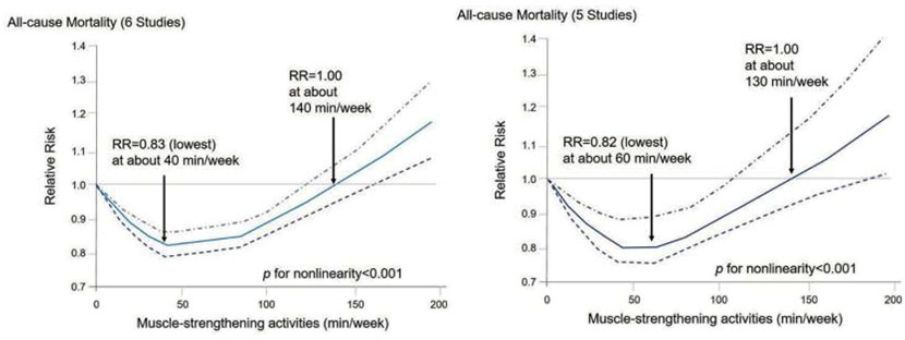
• How excessive exercise reduces your lifespan — Prolonged intense physical activity places chronic stress on the body, leading to issues like cardiac overuse injury and an increased risk of musculoskeletal injuries. Overtraining also impairs recovery, causing fatigue, reduced performance and a weakened immune system.
• Training over two hours weekly negates the advantage — When you’re doing strength training for a total of 130 to 140 minutes per week, the longevity benefits of exercise go down to the point as if you’re not exercising at all. In short, if you train for three to four hours a week, your long-term survival is worse than people who don’t do strength training at all.
• Excessive lifting leaves you worse off than being sedentary — Again, when you’re doing intense vigorous exercise in excess, you’re still better off than people who are sedentary. But for some (yet undetermined) reason, excessive strength training leaves you worse off than being sedentary.
• Aim for 20 minutes twice a week, not more — The lesson here is to keep strength training to 20 minutes twice a week on non-consecutive days, or 40 minutes once a week. Moreover, it’s just an add-on to your exercise regimen — don’t center your entire exercise sessions around it. Moderate-intensity exercise such as walking gives you far greater benefits.
• Even short weekly sessions protect against aging — Interestingly, this moderate amount of strength training aligns with findings from the Brigham Young University study,11 which showed that even small doses of resistance training — around 10 to 50 minutes weekly — result in measurable benefits to telomere length, slowing biological aging without the risks associated with overtraining.
To learn more about the benefits of weight training for older adults and how to incorporate it into your workout routine, read “Strength Training Turns Back the Clock on Your Biological Age.”
Frequently Asked Questions (FAQs) About Weight Training and Alzheimer’s
Q: Does weight training help prevent Alzheimer’s disease?
A: Yes. The study published in GeroScience showed that just six months of weight training twice a week preserved critical brain areas like the hippocampus and precuneus in older adults with mild cognitive impairment (MCI). These areas usually shrink early in Alzheimer’s, and the exercise group showed no volume loss, unlike the non-exercising control group.
Q: How does exercise protect the brain at a biological level?
A: Exercise activates several protective pathways:
•It boosts brain-derived neurotrophic factor (BDNF) and IGF-1, which help brain cells grow and repair.
•It reduces chronic inflammation, a major driver of cognitive decline.
•It improves blood flow and oxygen delivery to the brain.
•It regulates the HPA axis, lowering stress hormones like cortisol.
Q: What other brain benefits does strength training offer besides memory protection?
A: In addition to preserving brain volume, resistance training improves white matter integrity, supports nerve fiber health, and enhances verbal episodic memory. In the Brazilian study, five out of 22 participants in the training group improved so much they no longer met clinical criteria for MCI.
Q: How much strength training is enough to see brain and longevity benefits?
A: The sweet spot appears to be 20 to 40 minutes twice a week, or 40 to 60 minutes total per week. Research led by cardiologist Dr. James O’Keefe found that this level of training maximizes longevity benefits. Exceeding 130 to 140 minutes per week actually reverses these gains, increasing inflammation, injury risk, and stress on the body.
Q: Are lighter exercises like yoga or Tai chi also effective for brain health?
A: Yes. Mind-body exercises such as yoga and Tai chi lower cortisol and anxiety, improve mental clarity, and enhance executive function. One study showed that yoga not only reduced cortisol but also improved decision-making and memory test performance, demonstrating how gentle movement supports emotional and cognitive resilience.
- 1 Alzheimer’s Disease International, Dementia Statistics
- 2 Alzheimer.gov, What Is Mild Cognitive Impairment?
- 3, 4 GeroScience (2025), doi: 10.1007/s11357-024-01483-8
- 5, 6, 7, 8 News Medical, April 1, 2025
- 9 J Aging Res. 2020 May 14;2020:1407896
- 10 Mo Med. 2023 Mar-Apr;120(2):155–162
- 11 Biology 2024, 13(11), 883
Kefir Improves Memory by 66% in Alzheimer’s Patients
Reproduced from original article:
https://articles.mercola.com/sites/articles/archive/2025/05/19/kefir-improves-memory-alzheimers-patients.aspx
Analysis by Dr. Joseph Mercola May 19, 2025

Story at-a-glance
- Alzheimer’s disease affects memory and cognition, with the number of cases globally expected to reach more than 150 million by 2050. The disease is primarily driven by inflammation, oxidative stress, and gut-brain dysfunction
- Kefir consumption significantly improves memory and cognition in Alzheimer’s patients by reducing amyloid plaques, tau tangles, and inflammatory markers while protecting neurons
- C15:0, an essential fat found in grass fed dairy like kefir, protects against neurodegeneration by strengthening cell membranes, protecting mitochondria, and displacing harmful linoleic acid
- Iron accumulation in the brain accelerates Alzheimer’s progression through oxidative damage and ferroptosis; regular blood donation reduces iron overload
- Daily strategies such as optimizing beneficial gut bacteria, eliminating vegetable oils from your diet, and avoiding gluten offer protection against cognitive decline and neurodegeneration
Alzheimer’s disease (AD) is a progressive neurological disorder that gradually erodes memory, language, decision-making, and behavior. Over time, it strips away independence and identity. According to the World Health Organization (WHO), it accounts for 60% to 70% of all dementia cases.1 By 2050, the number of people living with Alzheimer’s is expected to hit more than 150 million globally.2
Alzheimer’s usually starts with simple forgetfulness like misplacing keys, repeating questions, or losing track of conversations. But as it worsens, it leads to disorientation, mood swings, personality changes, and eventually a total loss of autonomy.3 Inflammation, oxidative stress, damaged mitochondria, and gut-brain problems all play a role in its development.4
That understanding has led researchers to explore new strategies aimed at addressing the root causes of this condition. For instance, a recent systematic review published in the journal Brain Behavior and Immunity Integrative5 found that kefir, a probiotic-rich fermented milk drink, could be a promising tool for supporting Alzheimer’s management.
How Kefir Helps Prevent and Slow Alzheimer’s Disease
The featured study6 examined kefir’s potential as a complementary treatment for Alzheimer’s disease. The review included seven studies spanning invertebrates, rodents, and humans, and focused on kefir’s antioxidant, anti-inflammatory, and neuroprotective properties. Here’s what the findings revealed:
• Cognitive function improved across species — Improvements in memory and behavior were seen in rodents, while fruit fly models demonstrated increased survival and enhanced motor coordination. Kefir supplementation led to a 28% improvement in global cognition and a 66% increase in immediate memory performance, with delayed memory improving by 62%.
• Kefir lowered oxidative stress and preserved neurons — Markers of oxidative damage, like ROS, nitrotyrosine, and iNOS, were reduced after kefir treatment in rodents. Neurons in regions such as the hippocampus and cortex were also better preserved.
• Inflammation and neuronal damage decreased — Kefir reduced inflammatory markers such as NF-κB and caspase-3, which are linked to neuronal apoptosis. It also suppressed key inflammatory pathways (TLR4, MYD88, NLRP3) and reduced proinflammatory cytokines (TNF-α, IL-8, IL-12).
• Immune balance and gut health improved — Studies showed kefir modulated immune responses and restored intestinal mucosal integrity.
• Kefir enhanced insulin signaling in the brain — Some rodent models showed increased levels of insulin-degrading enzyme (IDE) in the hippocampus and better regulation of insulin receptors. These changes support neuron function and reduce amyloid buildup.
• Kefir’s unique bioactive compounds drive effects — Kefir contains peptides that block acetylcholinesterase (the same mechanism used by many AD drugs) and reduce oxidative protein damage. Kefiran, a complex carbohydrate found in kefir, also modulates inflammation and gut microbiota. These features make it a promising neuroprotective agent.
• Long-term use may sustain cognitive protection — Beyond short-term studies, kefir’s ability to regulate neurotransmitters (dopamine, serotonin, acetylcholine, and GABA) and promote brain-derived neurotrophic factor (BDNF) suggests ongoing benefits with continuous use.
Kefir Supplementation Improves Alzheimer’s Symptoms
One of the studies included in the featured review is a clinical trial published in Oxidative Medicine and Cellular Longevity,7 which evaluated the effects of 90 days of kefir-fermented milk on elderly patients with Alzheimer’s disease. This trial offered detailed insight into how kefir impacts the core drivers of neurodegeneration.
• Inflammation dropped sharply — Serum levels of proinflammatory cytokines decreased significantly following kefir supplementation. Cytokine ratios also improved, which indicates a shift from proinflammatory to more balanced immune response, which reduces amyloid buildup and protects against brain tissue loss.
• Oxidative stress was suppressed — Levels of superoxide, hydrogen peroxide, and peroxynitrite decreased by 30%, while nitric oxide bioavailability increased by 100%. This improves blood flow and protects neurons from oxidative damage.
• Apoptosis and DNA damage were reversed — DNA fragmentation dropped from 15% to 5%, and cleaved PARP-1 (a marker of programmed cell death) decreased by over 75%. The rate of apoptotic cells (cells that are naturally dying off) dropped by nearly half, while healthy cell populations rose, reflecting improved tissue stability. These changes suggest that kefir not only slows cellular damage but also promotes repair and survival at the tissue level.
• p53 signaling was activated — The expression of p53 tripled following supplementation. This master regulatory protein is essential for DNA repair, mitochondrial protection, and tumor suppression, and its activation helps explain kefir’s broad neuroprotective impact.
• A complex synbiotic effect — The kefir used in the study included both beneficial bacteria and yeast species. Its bioactive compounds, such as peptides, polysaccharides, and vitamins, worked together to modulate the gut-brain axis, enhance antioxidant capacity, and trigger neuroprotective pathways like GABA and BDNF.
• Safe and accessible — Kefir was well-tolerated, with no adverse effects reported. While this was an uncontrolled trial, the strength of the biological changes observed justifies future randomized studies and supports kefir’s use as a natural, low-risk intervention in early Alzheimer’s care.
Save This Article for Later – Get the PDF Now
Kefir Contains C15:0 — An Essential Fat That Protects Your Brain
Kefir isn’t just rich in probiotics and bioactive peptides. As a dairy product, it also contains pentadecanoic acid (C15:0), an essential saturated fat that has been recognized for its role in preventing cellular breakdown, reversing metabolic damage, and protecting the brain from neurodegeneration.
• C15:0 is essential for cellular resilience — This odd-chain saturated fat embeds into cell membranes, making them stronger and more resistant to oxidative stress. Unlike polyunsaturated fats (PUFs), which make membranes fragile, C15:0 stabilizes them. That’s important for brain cells, which are especially vulnerable to oxidation and mitochondrial decay in Alzheimer’s disease.
• C15:0 helps displace LA from your tissues — Research shows that individuals with Alzheimer’s have elevated levels of oxidized linoleic acid metabolites (OXLAMs) in their plasma.8 These toxic byproducts drive systemic inflammation, impair mitochondrial function, and contribute to ferroptosis, an iron-dependent form of cell death linked to neurodegeneration.9
C15:0 helps stop this process by displacing LA in cell membranes, reducing lipid peroxidation, and stabilizing cellular structures before damage takes hold. I have submitted a scientific paper detailing this mechanism and its importance for long-term LA detox — something I’ll be sharing more about in the near future.
• This fat is essential, but most people don’t get enough — C15:0 meets all the criteria of an essential fat. Your body doesn’t produce it in meaningful amounts, and deficiency is now linked to a cluster of problems collectively referred to as “cellular fragility syndrome.”10 These include fragile red blood cells, anemia, and dysmetabolic iron overload syndrome (DIOS), marked by excessive iron storage, especially in the liver.
DIOS increases the risk of ferroptosis, a destructive form of cell death triggered by iron and lipid peroxidation that targets mitochondrial membranes and shuts down energy production. This process may contribute to advanced fatty liver disease, steatohepatitis, and broader metabolic decline.
• C15:0 protects mitochondria and slows neurodegeneration — In the brain, C15:0 shields neurons from ferroptosis. This preserves energy production, supports cellular longevity, protects against aging and tissue damage, and helps maintain cognitive function as the brain ages.
• Grass fed kefir is a key dietary source — Modern agriculture has stripped dairy of much of its C15:0 by shifting to grain-fed cattle. Kefir made from grass fed cow’s milk retains more of this vital fat. Another good source is a serving of grass fed cheese or a tablespoon of butter, both of which provide 100 to 130 mg of C15:0, which is enough to support cellular repair and counteract LA damage over time.
For a deeper dive into C15:0 and why this fat may be essential, read “C15:0 — Found in Dairy — May Be an Essential Fat.”
Iron Overload — A Silent Accelerator of Alzheimer’s
Another factor that contributes to the development of Alzheimer’s disease is excess iron. While iron is essential for normal cellular function, too much of it leads to a dangerous cascade of oxidative stress, inflammation, and neural degeneration. A 2024 review published in Aging Medicine11 examined how iron buildup in a key brain area contributes to the development and progression of Alzheimer’s disease.
• The precuneus stores excess iron early in Alzheimer’s — The precuneus is the area of the brain responsible for memory, attention, and self-awareness, and is especially vulnerable to iron-induced oxidative damage. Multiple imaging studies show that individuals with mild cognitive impairment and Alzheimer’s disease have significantly higher iron levels in the precuneus.
• Iron overload drives amyloid and tau pathology — The review confirmed that elevated iron levels accelerate the aggregation of amyloid-beta plaques and tau protein tangles. Both of these are directly involved in killing neurons and causing cognitive decline.
• Iron disrupts mitochondria and raises oxidative stress — Iron accumulation leads to higher production of ROS, which damages cellular structures and impairs mitochondrial function. This weakens the brain’s energy supply and accelerates neuron death.
• Ferroptosis is the dominant cell death pathway — In the precuneus, iron overload initiates ferroptosis. Unlike apoptosis, this process causes catastrophic mitochondrial failure and is harder to reverse.
To address iron overload and reduce your risk of Alzheimer’s, I recommend donating blood regularly. Read How to Help Prevent and Treat Alzheimer’s Disease to learn more.
Additional Strategies to Prevent Alzheimer’s Disease
Preventing Alzheimer’s requires more than just one intervention. It takes a combination of daily strategies to reduce inflammation, protect neurons, and support metabolic and cognitive function. The following approaches offer a solid foundation:
• Optimize Akkermansia levels — Akkermansia muciniphila is a keystone gut microbe that supports gut barrier integrity, reduces inflammation, and produces short-chain fatty acids (SCFAs), which fuel the cells lining your colon. Alzheimer’s patients consistently show lower levels of Akkermansia.12
To optimize your levels, prioritize prebiotic-rich foods, fermented vegetables, and pharmaceutical-grade supplements that directly support Akkermansia growth. Tailor your carb intake to at least 250 grams daily, adjust it based on energy demands, and eliminate inflammatory vegetable oils to strengthen the gut-brain connection.
• Avoid gluten and casein — These proteins disrupt the blood-brain barrier and increase immune activation. Gluten, in particular, has been shown to weaken this barrier and allow bacteria to enter the bloodstream.13
That opens the door to cognitive decline, neuroinflammation, and diseases like Parkinson’s, anxiety, and depression. However, dairy fat, such as butter, is fine — it’s the proteins in pasteurized milk that are problematic.
• Include animal-based omega-3 fats, but don’t overdo it — Omega-3 fats DHA and EPA protect against the cell damage caused by Alzheimer’s disease, thereby lowering your risk of developing it or slowing its progression. However, keep in mind that they’re still PUFs, so add them into your diet in moderation. Focus on quality over quantity.
• Get vitamin D from sunlight — Low vitamin D correlates with faster cognitive decline and poor memory test scores. One study found a 40% drop in dementia risk with optimal levels.14 The best way to get vitamin D is through sensible sun exposure, aiming for a blood level of 60 to 80 ng/mL (150 to 200 nmol/L).
However, you need to purge vegetable oils from your body before going into the sun at solar noon. The LA in your skin oxidizes when exposed to sunlight, causing inflammation and skin damage. To protect your skin, avoid sun exposure during solar noon for four to six months as you work on removing LA from your body.
• Keep fasting insulin under 3 — Chronically high insulin promotes brain inflammation, insulin resistance in neurons, and accelerated aging. Reducing your insulin levels supports mitochondrial function and protects long-term cognition.15
• Eat folate-rich foods — Research shows that folate is a protective factor against Alzheimer’s disease.16 Vegetables, without question, are your best source of folate. Avoid supplements like folic acid, which is the inferior synthetic version of this compound.
• Eliminate mercury and aluminum — Mercury from dental fillings and aluminum from cookware, deodorants, and adjuvants disrupt brain chemistry. Both metals are linked to neurodegeneration.17 Use a biological dentist to safely remove amalgams and avoid products that contain aluminum.
• Exercise regularly — Physical activity increases blood flow to the brain, enhances neuronal plasticity, and supports the release of neurotrophic factors that help brain cells survive and thrive.18
• Eat antioxidant-rich foods — A good example is blueberries. Rich in anthocyanins and polyphenols, they help reduce oxidative stress and improve memory and brain function.19
• Challenge your brain daily — Lifelong learning strengthens neural networks and helps delay cognitive decline. Activities like learning an instrument, mastering a language, or solving complex problems all build brain resilience.20
• Avoid anticholinergics and statins — Anticholinergic drugs block acetylcholine, a key neurotransmitter for memory and attention. Statins interfere with cholesterol synthesis, deplete CoQ10, and inhibit the transport of fat-soluble nutrients into the brain. Both drug classes have been linked to increased dementia risk.21,22
Frequently Asked Questions (FAQs) About Kefir and Alzheimer’s Disease
Q: How does kefir support brain health in Alzheimer’s disease?
A: Kefir delivers a combination of probiotics, beneficial yeasts, peptides, and anti-inflammatory compounds that reduce amyloid buildup, protect neurons, and improve mitochondrial function. It also helps rebalance immune signaling and supports neurotransmitter production, making it a powerful dietary tool for cognitive protection.
Q: Can drinking kefir really improve memory in people with Alzheimer’s?
A: Yes. In one clinical trial, patients with Alzheimer’s who consumed kefir daily for 90 days showed a 66% improvement in immediate memory, a 62% improvement in delayed memory, and a 28% gain in overall cognitive function.
Q: What is C15:0, and why does it matter for Alzheimer’s?
A: C15:0 (pentadecanoic acid) is an essential fat found in grass fed dairy, including kefir. It strengthens cell membranes, protects mitochondria, and helps displace oxidized linoleic acid, which contributes to inflammation and neurodegeneration. Most people don’t get enough of it, yet it plays a key role in slowing cellular aging and protecting brain tissue.
Q: Why is excess iron a problem in Alzheimer’s?
A: Iron builds up in the precuneus region of the brain early in Alzheimer’s disease, where it fuels oxidative stress and triggers ferroptosis, a catastrophic form of cell death. This drives amyloid plaque formation, tau tangle development, and mitochondrial breakdown. Donating blood regularly helps reduce iron stores and lower your risk.
Q: What are the steps I can take to lower my Alzheimer’s risk?
A: There are a variety of daily strategies you can implement to reduce your risk, such as supporting your gut by optimizing Akkermansia muciniphila, eliminating gluten and vegetable oils from your diet, eating folate-rich foods, increasing your antioxidant intake, and getting sensible sun exposure, among others.
- 1 WHO, Dementia
- 2 Alzheimer’s Disease International, January 7, 2022
- 3 National Institute of Aging, Alzheimer’s Disease Fact Sheet
- 4 Int J Mol Sci. 2023 Sep 22;24(19):14450
- 5, 6 Brain Behavior and Immunity Integrative Volume 10, April 2025, 100115
- 7 Oxid Med Cell Longev. 2020 Jan 13;2020:2638703
- 8 Prostaglandins Leukot Essent Fatty Acids. 2012 Sep 5;87(4-5):135–141
- 9 Nutrients. 2023 Jul 13;15(14):3129
- 10 Metabolites 2024, 14(7), 355; doi: 10.3390/metabo14070355
- 11 Aging Med (Milton). 2024 Oct 22;7(5):649–667
- 12 Critical Reviews in Microbiology, 49(2), 151–176
- 13 Am J Lifestyle Med. 2022 Jan 11;16(1):32–35, Abstract
- 14 Alzheimers Dement (Amst). 2023 Mar 1;15(1):e12404, Highlights
- 15 Int J Mol Sci. 2021 Sep 15;22(18):9987, Abstract
- 16 Front Neurosci. 2021 Apr 14;15:661198, Abstract
- 17 Environ Res. 2020 Sep:188:109734
- 18 Front Aging Neurosci. 2023 Aug 4;15:1243869
- 19 Biomolecules. 2021 Jan 14;11(1):102
- 20 Dela J Public Health. 2021 Sep 27;7(4):124–127
- 21 Int. J. Mol. Sci. 2024, 25(23), 12850
- 22 JAMA Intern Med. 2019 Jun 24;179(8):1084–1093
Large-scale study reveals which beverage reduces dementia warning signs
Reproduced from original article:
https://www.naturalhealth365.com/large-scale-study-reveals-which-beverage-reduces-dementia-warning-signs.html
by: April 15, 2025
 (NaturalHealth365) Big Pharma spends billions developing dementia drugs while ignoring what might be right in your kitchen cupboard. A groundbreaking study involving thousands of older adults has just exposed an everyday beverage that significantly protects brain health in ways Western medicine has overlooked.
Published in the respected journal NPJ Science of Food, this extensive Japanese research finally provides solid evidence for what many natural health experts have claimed for years: regular consumption of green tea appears to shield the brain from age-related damage that leads to cognitive decline.
Brain scans reveal green tea’s remarkable protection against cognitive deterioration
This landmark Japanese research analyzed brain scans from nearly 9,000 older adults, comparing their beverage consumption habits with advanced brain imaging to uncover a striking pattern: individuals who consumed higher amounts of green tea showed dramatically fewer white matter lesions – the telltale brain abnormalities strongly linked to cognitive decline and dementia. The findings were unambiguous – participants who consumed three or more cups of green tea daily demonstrated significantly healthier brain structures compared to those who didn’t drink green tea regularly.
What makes this discovery particularly striking is that coffee consumption – despite its well-known ability to temporarily boost alertness – showed no protective effect whatsoever against these dangerous brain changes. The science clearly demonstrates that green tea contains unique compounds that coffee simply doesn’t possess.
The alarming dementia epidemic this natural solution could help combat
The timing couldn’t be more urgent. With almost 7 million Americans already battling Alzheimer’s and experts predicting this number could skyrocket to 13.8 million by 2060, we’re facing a cognitive health crisis of unprecedented scale.
While doctors continue prescribing medications with questionable results, this massive study points to a common household beverage that might help slow the dementia epidemic sweeping across our aging population.
The specific brain-saving mechanism revealed
The researchers identified a precise mechanism through which green tea appears to protect the brain: prevention of cerebral white matter lesions. These lesions represent actual damage to the brain’s communication pathways and are strongly associated with cognitive impairment, dementia, and increased stroke risk.
When researchers controlled for education, exercise habits, smoking, and dozens of other variables, the results remained stunningly clear: the more green tea participants drank, the healthier their brain scans appeared. People consuming the most green tea showed significantly fewer white matter lesions – the exact type of brain damage linked to cognitive decline.
The secret weapon hiding in your teacup? Scientists believe it’s the remarkable concentration of specialized plant compounds called catechins – particularly one known as EGCG – that give green tea its brain-protective power. These natural compounds possess something most antioxidants lack: the ability to actually cross the blood-brain barrier and directly shield delicate neural tissues from inflammation and oxidative damage.
While researchers have long known these compounds help fight heart disease, stroke and certain cancers, their ability to visibly protect brain structures on advanced imaging may prove to be their most valuable attribute as our population ages and dementia rates continue climbing.
How much green tea provides optimal protection?
The research identified a specific threshold for maximum benefit: three cups daily appears to be the optimal amount for brain protection. This aligns with a 2022 meta-analysis showing that each daily cup of green tea reduces dementia risk by approximately 6%.
Interestingly, some research suggests the benefits may not continue increasing with higher consumption – a study referenced by the researchers found that while 2-3 cups daily significantly reduced cognitive decline risk, the same effects weren’t observed beyond 4 cups.
Beyond the brain: Green tea’s whole-body health benefits
The cognitive protection provided by green tea represents just one aspect of its remarkable health-promoting properties. Regular green tea consumption has been linked to:
- Reduced cancer risk through multiple mechanisms
- Significantly lower cholesterol levels
- Decreased blood pressure
- Improved heart health
- Enhanced metabolic function
- Dramatic reduction in stroke risk (up to 24% with 2-4 cups daily)
Take a comprehensive approach to cognitive protection
While green tea appears to be an exceptionally powerful tool for brain health, researchers emphasize that maximum protection comes from a comprehensive approach that includes:
- Regular physical exercise, which increases oxygen-rich blood flow to the brain
- Consumption of antioxidant-rich fruits and nuts
- Including omega-3 sources like fatty fish in your diet
- Avoiding brain-damaging substances like alcohol
- Maintaining social connections and mental stimulation
Combining these protective habits with regular green tea consumption creates a powerful shield against cognitive decline that’s backed by hard science – without the laundry list of side effects found on prescription medication inserts.
Unlike experimental drugs that cost thousands and often disappoint in clinical trials, this research points to a simple, affordable solution that’s available everywhere. As millions face the prospect of cognitive decline, this evidence-based approach offers something precious: a practical strategy anyone can start today to help safeguard their mental sharpness for years to come.
Sources for this article include:
Mitochondrial Dysfunction in Neurodegenerative Disorders
Reproduced from original article:
https://articles.mercola.com/sites/articles/archive/2025/03/31/mitochondrial-dysfunction-neurodegeneration.aspx
Analysis by Dr. Joseph Mercola March 31, 2025

STORY AT-A-GLANCE
- Mitochondrial dysfunction is a key driver of neurodegeneration, with research showing that a single resting cortical neuron requires 4.7 billion ATP molecules every second for energy
- When mitochondria lose their efficient shape, electrons escape and form reactive oxygen species (ROS), triggering cellular damage and stress that particularly affects brain cells
- Research shows 42% of adults over 55 develop dementia by age 95, with projected new cases expected to double from 514,000 in 2020 to 1 million by 2060
- Mitochondria act as cellular calcium buffers — when this function fails, calcium floods cells and triggers the mitochondrial permeability transition pore, leading to widespread neuron death
- Key mitochondrial health strategies include eliminating seed oils, optimizing carbohydrate intake, reducing environmental toxin exposure, getting proper sun exposure and boosting NAD⁺ levels through supplements
You might be startled to learn that 42% of adults over 55 develop dementia by age 95.1 Dementia is characterized by memory loss, difficulties with language and reasoning, and an overall decline in the ability to perform everyday tasks. Left unmanaged, it spirals into more severe neurodegenerative disorders that undermine independence and quality of life.
A review published in Neurotherapeutics further highlights that a single resting cortical neuron consumes 4.7 billion adenosine triphosphate (ATP) molecules every second, underscoring how energy demands in your brain are immense and ongoing.2
I see this as direct evidence of why supporting cellular powerhouses — your mitochondria — is so central to preserving cognitive health. Chronic disruptions in those energy processes impose relentless stress on nerve cells, paving the way for memory problems and other neurological setbacks.
Mitochondrial Dysfunction Is at the Root of Neurodegeneration
The Neurotherapeutics review3 examined how different forms of disrupted energy processes in brain cells set the stage for progressive neurological disease. The researchers looked at various research findings that link faulty mitochondrial function to disorders affecting cognition, motor control and other higher-level tasks.
Their central goal was to pinpoint how malfunctioning mitochondria trigger the chain reactions seen in conditions such as Alzheimer’s disease, Parkinson’s disease and multiple other neurological syndromes.
• Evidence reveal how mitochondrial dysfunction drives brain changes — Investigators in this review did not limit their analysis to a specific group of patients. Instead, they consolidated evidence from a broad range of laboratory experiments and clinical observations targeting aging populations, individuals with rare mitochondrial disorders and those carrying known genetic mutations that alter mitochondrial function.
By weaving these areas together, the authors hoped to create a clearer picture of how compromised energy production leads to characteristic brain changes.
• Even small changes lead to significant damage — One of the paper’s most striking observations is how tiny structural shifts inside the mitochondria snowball into large-scale damage.4 When these organelles lose their efficient shape, electrons slip out of the normal energy pathway and team up with oxygen to form corrosive molecules called reactive oxygen species (ROS).
That surge in ROS sets off a cascade of biochemical stressors throughout brain cells, including direct attacks on important proteins and fats.
• The mitochondrion’s structure has a substantial role in neurofunction — As the authors state, “Excessive ROS production damages a variety of cellular components including proteins, lipids and DNA.”5 In short, the mitochondrion’s shape and integrity hold more power over neurofunction than many imagine.
Apart from the physical shape, researchers also honed in on the role of calcium balance. Healthy mitochondria function as a buffer by absorbing and releasing calcium ions to keep cells in balance.6
• The mitochondrial permeability transition pore leads to cell death — Once there’s a glitch, calcium floods the cell, and an emergency process called the mitochondrial permeability transition pore springs open.
The paper emphasizes that an uncontrolled opening of this pore triggers a wave of cell death, especially in your brain’s vulnerable neurons, where energy demands are already sky-high. It’s like watching a dam collapse because the main floodgate jammed.
Another intriguing angle involves how failing mitochondria disturb normal protein cleanup processes in the cell.7 The authors detail a scenario in which damaged mitochondria release proinflammatory signals, slowing down or outright blocking autophagy, the system cells use to clear out junk.
This slowdown contributes to the buildup of toxic plaques and misfolded proteins that characterize several neurodegenerative conditions. In practical terms, it means that your body’s usual housekeeping can’t keep up with the mess, and your brain is the unfortunate casualty.
Mitochondrial Dysfunction Tied to Parkinson’s, Alzheimer’s and Other Disorders
Throughout the review, there’s a clear focus on how each neurodegenerative disease taps into similar mitochondrial weaknesses.8 For instance, while Parkinson’s disease involves a breakdown in dopamine-producing neurons, and Alzheimer’s centers on beta-amyloid plaques, both conditions involve disruption of electron transport inside the mitochondria.
• One targeted intervention can influence many conditions — By comparing these processes side by side, the authors illustrate how one targeted intervention has benefits across multiple disorders. It’s a refreshing perspective that encourages looking beyond siloed research for cures or therapies. The researchers also address how the paper’s findings reflect on the broader population and our understanding of age-related memory and motor decline.9
• The connection between normal aging and mitochondrial collapse — They connect the dots between normal aging, which often features mild mitochondrial dysfunction, and more severe mitochondrial collapse seen in advanced disease states.
That means many people could be slipping down this slope long before typical symptoms even appear. According to the paper, identifying biomarkers of mitochondrial damage helps clinicians detect disease pathways early enough for effective interventions.
• ROS production modifies cellular signals — The authors also describe an intricate sequence of oxidative reactions that damage DNA, disrupt telomeres — the protective caps at the ends of chromosomes — and even alter the way genes are expressed.10
The review suggests that once ROS production speeds up, it doesn’t just drain energy — it also modifies cellular signals that keep neurons alive and functional. These modifications eventually tip cells into an energy crisis they cannot recover from, leading to unstoppable cell loss.
• Antioxidants help stabilize electron transport — Additionally, there’s discussion of how certain antioxidant strategies might stabilize electron transport by shielding the delicate proteins and lipids inside mitochondria.11
Some early-phase clinical trials, the paper notes, show promise in using compounds that block the worst of the oxidative assaults. While these lines of research are still evolving, they shine a bright light on the possibility of halting mitochondrial problems before serious neurological damage becomes entrenched.
Overall, this review underscores that protecting your cell’s power plants is a direct route to preserving brain function.12 By mapping the many crossroads where mitochondrial decline intersects with cognitive decline, researchers open doors to therapies that restore healthy energy production and help you maintain sharper memory, better coordination and greater resilience in the face of escalating demands on your brain.
Save This Article for Later – Get the PDF Now
The Growing Alzheimer’s Risk in Aging Populations
Understanding mitochondrial dysfunction’s role in neurodegeneration becomes urgent in the face of rising dementia cases. A study published in Nature Medicine13 tackled a massive data set on dementia, focusing on which groups are diagnosed most often, how early in life it occurs and how these trends shift over time.
Rather than exploring microscopic changes in the brain, this work looked closely at how social, genetic and age-related elements determine whether someone develops cognitive decline.
• Identifying the role of APOE ε4 in dementia development — Investigators pulled from a community-based study of thousands of participants, each free of dementia at the start, but varying in age, background and genetic traits.14 Their top priority was to measure how a person’s chances of developing dementia changed when factors such as sex, race and a specific genetic marker known as APOE ε4 came into play.
• Differences were seen between genders — By layering in long-term follow-up data and population statistics, they aimed to predict how many new dementia diagnoses would appear each year over the next several decades. A closer look revealed some dramatic differences between men and women.15
The paper found that women’s overall risk for dementia was higher than men’s when viewed across a lifetime, even though men often faced a greater likelihood of dying from other causes before cognitive problems fully manifested.
• Hormonal factors amplify the toll — In simpler language, men did not always reach the ages at which dementia most commonly appears. This gap sparked questions about how unique hormonal factors and longer lifespans amplify the toll on older women.
The same research found that Black adults were diagnosed with dementia at higher rates than White adults.16 This trend kicked in earlier, hinting that certain structural or social conditions accelerate the onset of memory loss.
• Certain factors affect dementia development — The paper noted that higher burdens of vascular complications, challenges with health care access and long-standing inequities could be part of the reason more Black adults developed dementia at younger ages. Investigators highlighted yet another twist: the APOE ε4 gene variant.17
This genetic factor often signals a heightened risk of Alzheimer’s disease, which falls under the broader dementia umbrella. Individuals carrying one copy showed a higher likelihood of facing cognitive problems, and those carrying two copies saw their odds jump even further.
• Dementia cases can rise to 1 million by 2060 — Looking ahead, the paper revealed a stark projection: around 514,000 new dementia cases occurred in 2020, but that total is expected to hit roughly 1 million by 2060.18 This doubling in newly diagnosed individuals points to significant population aging, where large segments of people are moving into the higher-risk age brackets at once.
Overall, the Nature Medicine paper19 suggests that the growing number of new dementia cases will not slow unless older adults gain more consistent access to early detection, better lifestyle options and interventions that protect their cognitive abilities — including optimizing your mitochondrial function.
How to Support Mitochondrial Health
You deserve straightforward ways to tackle an actual cause of neurodegeneration: a drop in cellular energy that wears down your nerve cells. I believe that if you support your mitochondria properly, you strengthen your brain and spare yourself from many issues that come with mitochondrial dysfunction. Below are five steps that focus on restoring mitochondrial health to boost your cellular power:
1. Eliminate processed foods and seed oils — I recommend shifting your diet away from seed oils like corn, soybean, safflower or canola. These oils contain linoleic acid (LA), a mitochondrial poison that compromises your cellular energy production. Aim to center your meals around wholesome foods such as fresh vegetables, grass fed butter or tallow, and clean collagen-rich proteins.
If you’re eating out, confirm what kind of oil they use in the kitchen — and opt out if it’s seed oil. This step helps protect your mitochondria from damage that accumulates over time, ultimately preserving your brain’s vitality.
2. Optimize your carbohydrate intake — Certain carbs are essential for steady energy output, especially keeping your neurons fueled. If you have a compromised gut, it’s important to start with easier-to-digest options, like white rice or slowly sipping dextrose water.
Over time, work in whole fruits and other nutrient-dense carbs. If you’re active, your needs are higher, so tailoring your intake ensures you’re not draining your mitochondria by consuming a low-carb diet.
3. Reduce exposure to environmental toxins — Your cells get bombarded by synthetic chemicals daily. Exposure to endocrine-disrupting chemicals (EDCs) in plastic, estrogen and pervasive electromagnetic fields (EMFs) impairs your cells’ ability to generate energy efficiently. As these pollutants build up, the mitochondria lose efficiency.
That’s why I recommend being proactive about reducing your exposure to environmental toxins. Consider household products made from natural materials and glass storage for leftovers. Sleeping in an EMF-free environment is also important, as it gives your cells a breather while your body recharges overnight. All of this lowers the stress your body must handle.
4. Get proper sun exposure — Daily sun exposure is important as it promotes cellular energy production by stimulating mitochondrial melatonin, offering powerful antioxidant protection. Avoid direct sunlight during peak hours (from 10 a.m. to 4 p.m. in most U.S. regions) until you’ve eliminated seed oils from your diet for at least six months, because accumulated LA in your tissues make you sunburn more easily.
5. Boost NAD+ Levels — Take niacinamide (50 milligrams three times daily) to increase NAD+ production, which helps your mitochondria generate more energy. NAD+ enables proper cell death signaling and supports your immune system’s ability to identify and remove damaged cells.
Frequently Asked Questions (FAQs) About Mitochondrial Dysfunction
Q: Why are mitochondria so important for brain health?
A: Mitochondria are the brain’s energy powerhouses, with a single resting cortical neuron using 4.7 billion ATP molecules every second. Mitochondrial dysfunction leads to energy deficits, oxidative stress, and neuron damage — all contributing to neurodegenerative diseases.
Q: How does mitochondrial dysfunction contribute to neurodegenerative disorders like Alzheimer’s and Parkinson’s?
A: Damaged mitochondria release reactive oxygen species (ROS) and disrupt calcium balance, triggering cell death and blocking cellular cleanup systems. This process causes toxic buildup and accelerates conditions such as Alzheimer’s and Parkinson’s.
Q: How widespread is dementia, and what are future projections?
A: Currently, 42% of adults over 55 develop dementia by age 95. New dementia cases are projected to double from 514,000 in 2020 to around 1 million by 2060 due to an aging population and genetic risk factors like the APOE ε4 gene variant.
Q: What lifestyle factors help protect mitochondrial function and reduce neurodegeneration risk?
A: Key strategies include eliminating seed oils, optimizing healthy carbohydrates, minimizing exposure to environmental toxins, getting proper sun exposure, and boosting NAD+ levels through niacinamide supplementation.
Q: What early signs and interventions are emphasized in preventing cognitive decline?
A: Detecting mitochondrial damage early through biomarkers, along with antioxidant therapies and targeted lifestyle changes, can help slow or prevent the onset of neurodegenerative diseases and age-related memory decline.
- 1, 13, 14, 15, 16, 17, 18, 19 Nature Medicine January 13, 2025
- 2, 3, 4, 5, 6, 7, 8, 9, 10, 11, 12 Neurotherapeutics December 19, 2023;21(1):e00292
Probiotics Offer New Hope for Alzheimer’s and Other Neurodegenerative Diseases
Reproduced from original article:
https://articles.mercola.com/sites/articles/archive/2025/03/20/probiotics-alzheimers-neurodegenerative-diseases.aspx
Analysis by Dr. Joseph Mercola March 20, 2025

STORY AT-A-GLANCE
- Probiotics may influence brain function through the gut-brain axis, offering benefits for neurodegenerative diseases like Alzheimer’s and Parkinson’s by improving gut health and reducing inflammation
- Studies suggest a link between gut health and cognitive function, indicating that an improved gut microbiome enhances mental health and may reduce inflammation associated with neurodegeneration
- Research indicates that probiotics could play a role in managing Alzheimer’s and Parkinson’s, offering a noninvasive treatment option that helps slow or reverse cognitive decline
- Biologically, the action of probiotics involves several interconnected pathways. By restoring a healthy balance of gut bacteria, probiotics reduce the production of harmful substances like lipopolysaccharides (LPS) that trigger inflammation in the brain
- Further studies are needed to confirm the long-term benefits of probiotics in neurodegenerative disease management, emphasizing the importance of continued research in this area
Neurodegenerative diseases (NDs) are disorders characterized by the progressive loss of structure or function of neurons, leading to their eventual death. This decline disrupts essential brain functions, resulting in symptoms that significantly impair daily living and overall quality of life.
Common neurodegenerative diseases include Alzheimer’s disease, Parkinson’s disease, and amyotrophic lateral sclerosis (ALS).1 Individuals affected by these conditions often experience memory loss, impaired movement, and difficulties with speech and coordination. As the diseases advance, patients may face severe cognitive and physical challenges, necessitating extensive care and support.
Neurodegenerative diseases now affect millions worldwide, with Alzheimer’s alone responsible for approximately 60% to 70% of dementia cases.2 On the other hand, the prevalence of Parkinson’s disease is expected to double by 2040, underscoring a growing public health concern.3 Additionally, ALS affects about 2 per 100,000 people in Europe and the U.S., with no known cure currently available.4
Risk Factors Associated with Neurodegenerative Diseases
Understanding the underlying causes of NDs is complex, as they often involve a combination of factors. For example, genetic mutations can predispose individuals to conditions like Alzheimer’s and Parkinson’s. According to a review published in the NPJ Parkinson’s Disease journal, genetic factors are linked to 56% to 79% of the risk for developing Alzheimer’s disease.5
• Environmental toxins — Exposure to heavy metals and pesticides, may contribute to neuronal damage. According to the aforementioned study:
“Several studies have suggested that lead (Pb), arsenic (As), and methyl mercury (MeHg) are also neurotoxins and can disrupt brain function, cause cognitive dysfunction, and increase the risk of AD and PD by disrupting mRNA splicing, the ubiquitin-proteasome system, the electron transport chain, and oxidative stress.”6
• Lifestyle factors — Poor diet, lack of exercise and chronic stress exacerbate NDs by affecting overall brain health and function. The progression from these underlying causes to full-blown NDs involves a cascade of biological events. Neurons, the brain’s communication cells, begin to lose their structure and function due to factors like oxidative stress and inflammation.
This deterioration disrupts the brain’s ability to process information, leading to symptoms such as memory loss and impaired motor skills. Over time, the cumulative effect of these disruptions results in the characteristic decline associated with NDs.
• Diagnosing NDs can be challenging — Often, symptoms overlap with other conditions, making it difficult to pinpoint the exact disease. For instance, memory loss could be attributed to normal aging, stress, or other medical issues, leading to potential misdiagnosis. Additionally, the lack of definitive biomarkers for many NDs means that diagnosis often relies on clinical assessments and patient history, which can be subjective and vary between practitioners.
• Limitations in diagnostic tools complicate the process — Imaging techniques like MRI and CT scans show brain changes, but these are not always specific to NDs. Blood tests and genetic screenings may offer clues but are not conclusive.
As a result, patients may undergo a lengthy diagnostic journey, experiencing uncertainty and anxiety while seeking answers. This underscores the need for more precise and reliable diagnostic methods to improve early detection and treatment outcomes.
Beyond the immediate symptoms, neurodegenerative diseases lead to profound emotional and financial burden for patients and their families. The progressive nature of these disorders often results in loss of independence, increased healthcare costs and emotional stress. Addressing these multifaceted challenges is crucial in improving patient outcomes and enhancing the quality of life for those affected.
Breakthrough Research Shows Probiotics’ Role in Alzheimer’s Disease Management
A recent study published in the journal Nutrients7 investigated the impact of probiotics on individuals diagnosed with Alzheimer’s disease. The research, conducted by Taiwanese researchers, aimed to determine whether introducing specific beneficial bacteria could influence cognitive function and reduce disease markers.
• Investigating the effects of probiotics — The study involved 60 elderly participants who were diagnosed with Alzheimer’s disease. They were assigned to either a probiotic treatment group or a placebo group over a 12-week period.
The probiotic group consumed a milk product containing strains such as Lactobacillus acidophilus, Lactobacillus casei, Bifidobacterium bifidum, and Lactobacillus fermentum. The primary goal was to assess any changes in cognitive function and Alzheimer’s disease markers as a result of the probiotic intervention.
• Probiotics improve cognitive function — Significant improvements were seen in the group that received probiotics compared to the placebo group. The probiotic group showed a notable increase in their Mini-Mental State Examination (MMSE) scores, indicating enhanced cognitive function. In contrast, those who took the placebo experienced a decline in their MMSE scores over the same period.
• Probiotics’ positive effects against inflammation — The probiotic group demonstrated a reduction in inflammation markers, which are often elevated in Alzheimer’s patients. “Considering these findings, specific probiotics demonstrate robust and effective antioxidant capabilities,” the study authors report.8
Save This Article for Later – Get the PDF Now
Probiotics Reduce Inflammation and Oxidative Stress
The mechanism behind these improvements is believed to involve the gut-brain axis, a communication network between the gastrointestinal tract and the brain.
• Probiotics balance your gut health — Probiotics help maintain a healthy balance of gut microbiota, which in turn influence brain function. By promoting a diverse and balanced microbiome, probiotics may reduce systemic inflammation and oxidative stress, both of which are implicated in the progression of Alzheimer’s disease. For more information on how gut health affects your mental health, read “Gut Health’s Impact on Mental Well-Being.”
• Benefits of SCFAs — Short-chain fatty acids (SCFAs) are metabolites produced by beneficial bacteria in the gut. SCFAs have been shown to support brain health by providing energy to neurons and reducing inflammation. The increased abundance of Lactobacillus species in the probiotic group likely contributed to higher SCFA levels, fostering an environment conducive to cognitive stability and improvement.
• Benefits are observed in a short amount of time — Cognitive improvements were observed after just 12 weeks of probiotic supplementation, suggesting that the benefits of probiotics can manifest relatively quickly. This rapid response underscores the potential of probiotics as a viable intervention for slowing or even reversing aspects of cognitive decline in Alzheimer’s patients.
Comparing the effects of probiotics to other variables in the study, probiotics consistently outperformed the placebo in all measured outcomes. While both groups participated in the same environment and received similar care apart from the probiotic intervention, only the probiotic group showed meaningful improvements in physiological markers related to brain health. This stark contrast emphasizes the specific role that probiotics play in enhancing brain health.
Probiotics’ Mechanisms of Action Against Neurodegenerative Disorders
Biologically, the action of probiotics involves several interconnected pathways. By restoring a healthy balance of gut bacteria, probiotics reduce the production of harmful substances like lipopolysaccharides (LPS) that trigger inflammation in the brain.9
• Enhancing the integrity of the gut barrier — They prevent the leakage of proinflammatory agents into the bloodstream, which could otherwise reach the brain and exacerbate neuroinflammation.10
• Affecting neurotransmitter production — Probiotics influence the production of neurotransmitters such as dopamine and gamma-aminobutyric acid (GABA).11 These chemicals are crucial for mood regulation and cognitive function. By increasing the levels of these neurotransmitters, probiotics contribute to improved mental health and cognitive resilience against the degenerative processes of Alzheimer’s disease.
Four Ways to Support Your Brain Through Gut Health
Your gut microbiome acts as a second brain, directly influencing your cognitive function and neurological health, and how you nourish it plays a significant role in your risk for neurodegenerative conditions. For example, a study published in Scientific Reports showed that certain bacterial strains in the gut, such as Collinsella, Lachnospira, and Veillonella, increased the risk of Alzheimer’s disease.12
However, they also identified strains that provide protective benefits against Alzheimer’s disease, such as Eubacterium nodatum and Eisenbergiella. These produce short-chain fatty acids (SCFAs), particularly butyrate, from dietary carbohydrates.
Butyrate nourishes your colonic epithelial cells, reinforcing the intestinal barrier. SCFAs also stimulate mucin production, creating a protective shield against harmful bacteria. Akkermansia muciniphila also produces SCFAs, and research has shown that Alzheimer’s patients tend to have very low levels of this important keystone species.13 Here are four proven strategies geared toward optimizing the Akkermansia in your gut to enhance your brain health:
1. Increase Akkermansia through targeted diet and supplementation — Prioritize foods and supplements that promote the growth of Akkermansia. Include well-cooked, prebiotic-rich vegetables and fiber sources that nourish this crucial bacterium.
Consider high-quality supplements specifically designed to enhance Akkermansia levels, supporting gut barrier integrity and reducing brain inflammation. Avoid processed foods and high-fat diets that can hinder Akkermansia growth.
2. Enhance beneficial short-chain fatty acids — Focus on increasing butyrate-producing bacteria alongside Akkermansia to protect against amyloid buildup in the brain. Incorporate fermented foods and resistant starches that feed these beneficial microbes.
Tailor your carbohydrate intake to support your unique microbiome, aiming for at least 250 grams of targeted carbs daily, and adjust based on your activity level to optimize Akkermansia and short-chain fatty acid production.
3. Support your gut-brain connection — Optimize your gut barrier integrity to prevent inflammatory compounds from reaching your brain. This includes removing inflammatory processed foods and supporting the growth of protective bacteria like Akkermansia, which strengthens your intestinal lining.
Maintain a balanced diet with sufficient carbohydrates tailored to your microbiome, and use pharmaceutical-grade supplements as needed to sustain Akkermansia levels.
4. Address systemic inflammation by promoting Akkermansia diversity — Lower inflammation throughout your body by enhancing microbiome diversity, with a specific emphasis on increasing Akkermansia.
Eliminate vegetable oils and other proinflammatory fats that damage gut bacteria, and incorporate foods that reduce inflammatory markers while supporting Akkermansia growth. Regularly monitor biomarkers such as C-reactive protein levels to track your progress in reducing systemic inflammation and maintaining a healthy gut microbiome.
Additional Strategies to Prevent Alzheimer’s Disease
Aside from optimizing your gut health and Akkermansia levels, there’s a plethora of helpful Alzheimer’s prevention strategies, several of which are outlined below:
• Avoid gluten and casein (primarily wheat and pasteurized dairy, but not dairy fat, such as butter) — As noted in a 2022 study,14 your blood-brain barrier is negatively affected by gluten. When bacteria enter your bloodstream, the risk of Alzheimer’s disease increases. Other cognitive disorders are linked to a weakened blood-brain barrier as well, such as Parkinson’s disease, anxiety and depression.
• Make sure you’re getting enough animal-based omega-3 fats — Omega-3 fats, namely EPA and DHA, help by preventing cell damage caused by Alzheimer’s disease, thereby slowing down its progression and lowering your risk of developing the disorder. That said, omega-3s are PUFAs, so don’t overdo it.
• Optimize your vitamin D level with safe sun exposure — Strong links between low levels of vitamin D in Alzheimer’s patients and poor outcomes on cognitive tests have been revealed. In a 2023 study,15 increasing vitamin D reduced dementia risk by 40%.
The best way to get vitamin D is through sensible sun exposure, aiming for a blood level between 60 and 80 ng/mL. However, you need to purge vegetable oils from your body before going into the sun at solar noon. The LA in your skin oxidizes when exposed to sunlight, causing inflammation and skin damage.
To protect your skin, avoid sun exposure during solar noon for four to six months as you work on removing LA from your body. Stick to sunlight during early morning and late afternoon in the meantime. For additional skin defense, you can take astaxanthin, a low-dose aspirin or molecular hydrogen. Niacinamide cream will also lower your risk of skin damage.
• Keep your fasting insulin levels below 3 — Insulin resistance is linked to accelerated brain aging, as well as neurodegeneration.16
• Eat a nutritious diet, rich in folate — Vegetables, without question, are your best form of folate. Avoid supplements like folic acid, which is the inferior synthetic version of folate. Research shows that folate is a protective factor against Alzheimer’s disease.17
• Avoid and eliminate mercury and aluminum from your body — Dental amalgam fillings, which are 50% mercury by weight, are one of the major sources of heavy metal toxicity. Make sure you use a biological dentist to have your amalgams removed. Sources of aluminum include antiperspirants, nonstick cookware and vaccine adjuvants.
• Make sure your iron isn’t elevated and donate blood if it is — A study18 published in Aging Medicine shows that excess iron increases your risk of Alzheimer’s disease by initiating the Fenton reaction, leading to increased oxidative stress.
• Exercise regularly — Exercise triggers beneficial changes that support cognitive function. Particularly, it’s been shown to improve blood flow to the brain, leading to an increase in biomarkers related to improved neuronal plasticity and better cell survival.19
• Eat blueberries and other antioxidant-rich foods — Wild blueberries, which have high anthocyanin and antioxidant content, are known to guard against neurological diseases.
• Challenge your mind daily — Mental stimulation, such as learning to play a musical instrument, is associated with a decreased risk of Alzheimer’s.20
• Avoid anticholinergics and statin drugs — Drugs that block acetylcholine, a nervous system neurotransmitter, have been shown to increase your risk of dementia. These drugs include certain nighttime pain relievers, antihistamines, sleep aids, certain antidepressants, medications to control incontinence, and certain narcotic pain relievers.
Statin drugs are particularly problematic because they suppress the synthesis of cholesterol, deplete your brain of CoQ10 and neurotransmitter precursors, and prevent adequate delivery of essential fatty acids and fat-soluble antioxidants to your brain by inhibiting the production of the indispensable carrier biomolecule known as low-density lipoprotein.
Frequently Asked Questions (FAQs) About Probiotics and Neurodegenerative Diseases
Q: How do probiotics impact neurodegenerative diseases like Alzheimer’s?
A: Probiotics influence brain function through the gut-brain axis by restoring gut microbiome balance, reducing inflammation, and improving cognitive function. Studies suggest that beneficial bacteria can slow or even reverse aspects of cognitive decline in conditions like Alzheimer’s and Parkinson’s.
Q: What research supports the benefits of probiotics for Alzheimer’s patients?
A: A study published in Nutrients found that Alzheimer’s patients who took probiotics for 12 weeks showed improved cognitive function and reduced inflammation compared to a placebo group. These improvements were measured using the Mini-Mental State Examination (MMSE) and inflammatory markers.
Q: What mechanisms make probiotics beneficial for brain health?
A: Probiotics help reduce inflammation by lowering harmful substances like lipopolysaccharides (LPS), enhancing gut barrier integrity, and influencing neurotransmitter production (e.g., dopamine and GABA). They also promote the production of short-chain fatty acids (SCFAs), which support brain health.
Q: Can improving gut health help prevent neurodegenerative diseases?
A: Yes. A diverse gut microbiome, particularly the presence of beneficial bacteria like Akkermansia muciniphila, plays a crucial role in reducing inflammation and protecting against neurodegeneration. Consuming prebiotic-rich foods, fermented foods, and maintaining a fiber-rich diet can support gut health.
Q: What other lifestyle changes can support brain health and reduce Alzheimer’s risk?
A: In addition to probiotics, strategies like avoiding processed foods, eliminating heavy metal exposure (e.g., mercury and aluminum), maintaining optimal vitamin D and omega-3 levels, exercising regularly, and engaging in mental stimulation activities can help prevent cognitive decline and support brain health.
- 1 Int J Mol Sci. 2020 Dec 14;21(24):9500
- 2, 7, 8 Nutrients. 2023 Dec 20;16(1):16
- 3 Clinical Medicine, Volume 20, Issue 4, July 2020, Pages 393-398
- 4 Amyotrophic Lateral Sclerosis and Frontotemporal Degeneration, 20(3–4), 264–274
- 5, 6 NPJ Parkinsons Dis. 2021 Aug 11;7:70
- 9 Front Immunol. 2021 Feb 26;12:578386
- 10 Front Immunol. 2023 Apr 24;14:1143548
- 11 Front Microbiol. 2023 Aug 17;14:1216674
- 12 Scientific Reports, Volume 13, Article number: 5258 (2023)
- 13 Critical Reviews in Microbiology, 49(2), 151–176
- 14 Am J Lifestyle Med. 2022 Jan 11;16(1):32–35, Abstract
- 15 Alzheimers Dement (Amst). 2023 Mar 1;15(1):e12404, Highlights
- 16 Int J Mol Sci. 2021 Sep 15;22(18):9987, Abstract
- 17 Front Neurosci. 2021 Apr 14;15:661198, Abstract
- 18 Aging Med (Milton). 2024 Oct 22;7(5):649–667, Iron Overload/Accumulation and Increase Risk of AD
- 19 Front Aging Neurosci. 2023 Aug 4;15:1243869
- 20 Aging Ment Health. 2021 Apr;25(4):593-601
Ozempic Linked to 19 Adverse Health Events
Reproduced from original article:
https://articles.mercola.com/sites/articles/archive/2025/02/22/ozempic-linked-to-19-adverse-health-events.aspx
Analysis by Dr. Joseph Mercola February 22, 2025
STORY AT-A-GLANCE
- GLP-1 receptor agonists like Ozempic and Wegovy, originally intended as diabetes medications, have gained popularity for weight loss, leading to global shortages despite having modest benefits
- Research shows these drugs reduce seizures and substance addiction risks, but they increase the likelihood of 19 other health conditions, including fainting, kidney problems and pancreatic issues
- Common side effects include nausea, diarrhea, vomiting and abdominal pain, with potential risks of acute pancreatitis and thyroid cancer, making the trade-off dangerous for users
- Ozempic’s manufacturer Novo Nordisk reported $40.6 billion in revenue, highlighting how the “magic pill” mentality and ultraprocessed food consumption create a profitable cycle for pharmaceutical companies and food manufacturers
- Instead of relying on weight loss drugs, focus on optimizing cellular energy production through dietary changes, avoiding vegetable oils and supporting your gut and mitochondrial health
By now, most people around the world have heard of Ozempic and Wegovy, which are GLP-1 receptor agonists. These drugs, originally made for treating Type 2 diabetes, cause rapid weight loss, thus attracting individuals who have been struggling to lose weight for a long time. In fact, the effectiveness of these drugs has led to a global shortage.1 However, as with many other drugs that promise immediate results, there’s a catch.
Ozempic Hailed as a ‘Miracle Drug,’ but the Downsides Are Sinister
A report from The Epoch Times2 covered a study published in Nature Medicine,3 showing the impact of certain GLP-1 receptor agonists, sold under brand names such as Ozempic and Wegovy. As noted by The Epoch Times, “The media, patients, and even some doctors have dubbed the medications ‘miracle drugs’ because of their profound weight-loss effects.”4
In addition to weight loss, the study also noted that these drugs also lower the risk of “seizures and addiction to substances such as alcohol, cannabis, stimulants and opioids.”5 It’s believed that these drugs affect the brain’s neurological pathways related to reward and impulse control, explaining how these changes in behavior occur.6
Despite these benefits, the researchers caution potential, as there’s a dark side to these drugs that mainstream media and Big Pharma do not want you to see. According to the report, Ozempic increases your risk of developing a slew of other serious health conditions:7
“Researchers warn that these benefits come with an increased risk of 19 health conditions, such as syncope (fainting), arthritic disorders, and kidney and pancreatic problems.”
Similarly, a 2022 study found that commonly reported problems include nausea, diarrhea, vomiting and abdominal pain. An increased risk for acute pancreatitis and thyroid cancer was also noted.8 Meanwhile, Nature Medicine claims the benefits are “modest” at best:9
“While GLP-1RA drugs display effectiveness against a wide array of health problems, the magnitude of associated benefits is modest — about a 10% to 20% reduction for most outcomes.”
A Closer Look at the Research
The Nature Medicine study analyzed 215,970 diabetics using GLP-1 receptor agonists and compared them to multiple control groups: 159,465 taking sulfonylureas, 117,989 using DPP-4 inhibitors, and 258,614 on SGLT2 inhibitors. An additional control group of 536,068 used all three medications, while a separate baseline group of 1,203,097 received only standard care.10
They then produced a master list of health outcomes related to GLP-1 agonist receptor usage. As noted by The Epoch Times, while these drugs produced benefits, there were also significant adverse health outcomes:11
“Compared to usual care, GLP-1RA use was associated with a reduced risk of substance use and psychotic disorders, seizures, neurocognitive disorders (including Alzheimer’s disease and dementia), coagulation disorders, cardiometabolic disorders, infectious illnesses and several respiratory conditions.
There was an increased risk of gastrointestinal disorders, hypotension, syncope, arthritic disorders, nephrolithiasis, interstitial nephritis and drug-induced pancreatitis associated with GLP-1RA use compared to usual care. The results provide insights into the benefits and risks of GLP-1RAs and may be useful for informing clinical care and guiding research agendas.”
In a report by GoodRx, Stacia Woodcock, PharmD., outlines the mechanisms that lead to weight loss and other supposed benefits related to taking these drugs:12
• They signal your pancreas to release more insulin — After a meal, your blood glucose levels go up. Usually, the pancreas releases insulin when this happens to lower blood glucose levels. But in Type 2 diabetics, the body doesn’t always release enough. Incretin mimetics work on the pancreas to help raise insulin levels after you eat, which then lowers your blood glucose level.
• They increase your body’s sensitivity to insulin — Your body may also not respond as well to insulin if you’re diabetic. Incretin mimetics help increase insulin sensitivity, so your body can respond better to insulin when it’s released.
• They tell your liver to stop making glucose — This helps stop the production of new glucose to keep blood levels down.
Save This Article for Later – Get the PDF Now
The Cycle of Ultraprocessed Food and Ozempic Wrecks Public Health
As the weight-loss effects of GLP-1 receptor agonists gained widespread attention, Big Pharma’s profits in this market skyrocketed. According to The New York Times, Novo Nordisk, the maker of Ozempic and Wegovy, reported $40.6 billion in revenue in 2024; this figure is expected to jump by 16% to 24% in 2025.13
In the Tucker Carlson Show interview featured above, Dr. Casey Means, a Stanford-educated surgeon now focusing on functional medicine, expounds on the idea that a “magic pill” that causes weight loss and other supposed benefits appeals to millions of users:14
“I think it’s very dark. It’s a stranglehold on the U.S. population. Almost like solidifying this idea that there is a magic pill — I mean, literally, the book by Johann Ari is called ‘Magic Pill’ — and convincing us that, you know, salvation from our chronic health issues is going to be found in a shot when we are living in a toxic stew that’s destroying our cellular biology.”
Elsewhere in the interview, Means shares how ultraprocessed food contributes to obesity, thus cycling back to taking drugs that cause weight loss. In the end, only Big Pharma and Big Food are the real winners here, as millions of adults struggle with the damage they cause:15
“We are the only species in the world that has an obesity and chronic disease epidemic, the only species in the world that has a chronic disease and obesity epidemic because of ultraprocessed food.
You think about every other animal in the wild — they’re eating real, natural foods except for domesticated animals, which are also getting chronic diseases just like humans because they’re eating our food. But every other animal, they’re able to regulate their satiety. They’re not eating themselves to death like we are. We’re literally eating ourselves to death …
This could be on track to be the most profitable medication ever in human history. It will be if the powers that be let it. And what the unfortunate part is that it doesn’t take our bodies out of the toxic stew that’s crushing our biology. Yes, we may melt some fat and muscle without changing any of the other levers that we just talked about that are crushing our biology. So, this is not the public health solution.”
Practical Ways to Address Underlying Metabolic Challenges
I believe the key to maintaining a healthy weight is optimizing your cellular energy production. This requires a multifaceted approach that takes effort to implement, but it leads to safer, healthier results — something GLP-1 receptor agonists don’t offer. With that in mind, here are my recommendations:
1. Avoid Ozempic and other GLP-1 agonist receptor drugs — The most obvious strategy is avoiding GLP-1 agonist receptors in the first place. If you’re tempted to start, you owe it to yourself to read the study to understand the dangers.
Pills and injections offer short-term relief, but they often mask deeper imbalances in how your cells produce and use energy. Real improvement happens when you remove the factors that strain your metabolism rather than rely on a drug to force quick weight loss.
2. Remove vegetable oils from your diet — If you regularly consume ultraprocessed foods, I recommend stopping right now and replacing them with real, whole foods. Processed foods contain linoleic acid-rich vegetable oils that disrupt your metabolic pathways and alter how your body stores fat. Instead, cook your meals using tallow, grass fed butter or coconut oil.
To protect your health from further damage, limit your intake of linoleic acid to less than 5 grams per day from all sources. I recommend using Cronometer, an online food tracker, so you don’t go over this recommended range.
3. Shift your carbohydrate sources gradually — Avoid making sudden dietary changes that can shock your system. If your gut is compromised, start by introducing easily digestible carbohydrates like whole fruit or white rice before incorporating more complex carbs.
For severe gut issues, sip dextrose water throughout the day as a temporary aid to support healing, but limit its use to about two weeks. The goal is to provide your cells with a steady source of easy-to-digest, healthy carbohydrates for energy.
4. Consider your protein and collagen intake — I suggest aiming for 0.8 grams of protein per pound of lean body mass and balancing that amount so that about one-third comes from collagen. Doing this supports muscle maintenance, tissue repair and hormone balance.
If you exercise frequently, you would probably do well with a higher intake, but take it slow and listen to how your body responds. Stable protein intake is foundational for regulating cravings and stabilizing energy.
5. Support your gut and mitochondrial health with other healthy habits — I recommend getting daily sunlight exposure while being mindful of timing. If your diet previously included vegetable oils, avoid intense midday sun for at least six months to reduce the risk of sunburn and skin damage. Additionally, grounding in low-EMF environments can be beneficial if available — oceans, particularly in North America, tend to be the safest option.
In addition, moderate-intensity movement, such as walking, supports cellular energy production and aids in weight management. Gradual, consistent effort in these areas strengthens your metabolism and leads to lasting health improvements. I believe this approach is far superior to relying on a so-called “magic” drug like Ozempic.
Boost Your GLP-1 Naturally with Akkermansia
The idea of a quick-fix pill may be tempting, but it comes at a long-term cost to your health. A better approach is to naturally boost your GLP-1 levels by supporting Akkermansia, a beneficial gut probiotic that produces a GLP-1-inducing protein. As noted in one study:16
“A. muciniphila increases thermogenesis and glucagon-like peptide-1 (GLP-1) secretion in high-fat-diet (HFD)-induced C57BL/6J mice by induction of uncoupling protein 1 in brown adipose tissue and systemic GLP-1 secretion.”
Many people lack Akkermansia due to various reasons, such as impaired mitochondrial function or an environment that is inhospitable to beneficial gut bacteria. Ideally, Akkermansia should comprise 3% to 5% of your total gut microbiome and it plays many different roles aside from promoting GLP-1.
For example, Akkermansia has the ability to produce mucin, which is a thick, gel-like substance that protects the gut lining from harmful pathogens, irritation from stomach acid and enzymes, as well as mechanical damage. Moreover, mucin helps nourish the gut bacteria already existing in your gut.
To boost Akkermansia naturally, eat plenty of berries and inulin-rich foods such as garlic, asparagus, bananas and leeks. Note, however, that if your gut health is currently out of whack, introducing high amounts of dietary fiber in your system all at once can worsen your gastrointestinal symptoms. So, work on healing your gut first, then gradually increase fiber intake.
- 1, 15 JAMA Netw Open. 2024 Aug 20;7(8):e2423385
- 2, 4, 5, 6, 7 The Epoch Times, January 28, 2025
- 3, 10, 11 Nat Med. 2025 Jan 20
- 8 Front Public Health. 2022 Oct 20;10:996179
- 9 WashU Medicine, January 20, 2025
- 12 GoodRX, October 2, 2023
- 13 The New York Times, February 5, 2025
- 14 YouTube, Tucker Carlson, August 17, 2024
- 16 Nature Microbiology volume 6, pages 563–573 (2021)
Proteins Linked to Alzheimer’s Disease Now Observable in Real Time
Reproduced from original article:
https://articles.mercola.com/sites/articles/archive/2024/12/31/proteins-linked-to-alzheimers-disease.aspx
Analysis by Dr. Joseph Mercola December 31, 2024

STORY AT-A-GLANCE
- Nearly 7 million Americans currently have Alzheimer’s disease. Projections show this number will increase to 13 million by 2050, according to the Alzheimer’s Association
- A study used liquid-based atomic force microscopy to visualize beta-amyloid protein development in real-time, identifying “superspreader” fibrils that accelerate disease progression across brain tissue
- Three major toxins that damage mitochondrial function are excessive linoleic acid (LA) intake, endocrine-disrupting chemicals from microplastics and electromagnetic field (EMF) exposure from electronic devices
- Certain pathogenic bacteria strains like Collinsella, Lachnospira, and Veillonella increase Alzheimer’s risk, while probiotic strains like Eubacterium nodatum produce protective benefits through short-chain fatty acids
- Prevention strategies include optimizing vitamin D levels, maintaining low fasting insulin, avoiding gluten and casein, exercising regularly and challenging your mind with new skills
According to the Alzheimer’s Association, nearly 7 million Americans are currently diagnosed with Alzheimer’s disease, the most common type of dementia. Furthermore, the situation is expected to worsen — by 2050, around 13 million Americans are projected to be diagnosed with this neurodegenerative disease.1
Alzheimer’s disease remains challenging for modern medicine to diagnose and treat. However, a recent study2 has made a breakthrough in understanding how the disease progresses, creating a foundation for future clinicians and researchers to build upon to create better treatment plans.
A Breakthrough in Observing Alzheimer’s Disease Progression
In a study3 published in Science Advances, researchers were able to visualize the development of amyloid-beta proteins to see how Alzheimer’s disease spreads. Beta-amyloid proteins have been observed using a variety of methods, several of which were reviewed in the study:
“Methodological advances have been able to resolve the ultrastructure of Aβ and tau protein aggregates formed along the primary nucleation pathway using cryo-electron microscopy (cryo-EM) and solid-state nuclear magnetic resonance spectroscopy.
Likewise, the aggregation kinetics and toxicity of these proteins have been investigated using ultrasensitive fluorescence-based assays, nanopore devices, and circular dichroism.
Details on the morphology, on-surface assembly, and chemical structure of Aβ and tau protein aggregates have also been made accessible using standard atomic force microscopy (AFM), video-rate AFM, and infrared spectroscopy.”
For context, beta-amyloid proteins aggregate in the brain, and they’re suspected of playing a key role in the development of Alzheimer’s. Hence, they’re what researchers usually focus on to find therapeutic solutions.4 Previous staining methods used for observational analysis distort the protein structures.
To find new ways to analyze the mechanisms of Alzheimer’s disease, the team used liquid-based atomic force microscopy (AFM). As explained by the authors:5
“Building on the observations from previous secondary nucleation studies, we postulated that it should be feasible with a high-resolution AFM to record elementary information such as oligomer size, shape, and adsorption site on individual fibrils with sub-10-nm diameter. Operating the AFM in an aqueous solution medium would allow the diffusion of oligomers on the fibril surface.”
Tracking the Progression of Alzheimer’s Disease in Real Time
Once the team settled on the methodology, they moved onto testing. To start, they agitated beta-amyloid samples for 30 minutes at room temperature. Then, they set up different experiments to observe the growth and progression protein aggregates from 30 seconds to 250 hours:6
“Nanoscale imaging supported by atomic-scale molecular simulations tracked the adsorption and proliferation of oligomeric assemblies at nonperiodically spaced catalytic sites on the fibril surface. Upon confirming that fibril edges are preferential binding sites for oligomers during embryonic stages, the secondary fibrillar size changes were quantified during the growth stages.”
From their observations of protein fibril development in real time, they were able to classify some of them as “superspreaders” based on their surface anatomy. Specifically, these superspreaders “feature highly active edges and surfaces where new protein molecules accumulate, eventually forming longer fibrils that may spread across brain tissue.”
As they spread, more protein aggregation occurs, effectively progressing the hallmarks of Alzheimer’s disease. Ultimately, the research team hopes that this breakthrough in fibril analysis will be able to help improve the diagnosis and monitoring of neurodegenerative diseases. “This work brings us another step closer to better understanding how these proteins spread in brain tissue of Alzheimer’s disease,” says Peter Nirmalraj, one of the authors of the study.7
Save This Article for Later – Get the PDF Now
Mitochondrial Dysfunction Is at the Heart of Alzheimer’s Disease
As researchers continue to come up with new tools to help diagnose and treat Alzheimer’s better, following a healthy lifestyle is important to reduce your risk. Prevention is by far your best option. I believe addressing mitochondrial dysfunction is key to protecting cognitive function, which is achieved by improving your cellular health.
The first thing to address is your diet. A study published in Neurology noted that eating an inflammatory diet raises your risk of dementia.8 In addition, I believe that there are three major toxins damaging your mitochondrial function by affecting intracellular calcium, which subsequently impacts cellular health.
Essentially, when calcium within your cells increase, your body produces superoxide and nitric oxide. These two combine to form peroxynitrite, a potent reactive oxygen species that contributes to poor health. Three pervasive toxins that raise intracellular calcium levels and drive peroxynitrite formation include:
1. Excessive linoleic acid (LA) consumption — LA is an omega-6 polyunsaturated fat (PUFA) found most abundantly in seed and vegetable oils and processed foods. I believe it is one of the most pernicious toxins of the Western diet, driving inflammation and chronic diseases among the populace.
Overconsumption negatively impacts your metabolic rate and gut microbiome, which are key factors for optimal health. To protect your health, I recommend keeping your LA intake below 5 grams a day.
Note that it’s virtually impossible to avoid LA, as most foods inherently have them, and your body does need some of it. The key is to avoid excess.
2. Endocrine-disrupting chemicals (EDCs) — Microplastics are so prevalent in our environment that humans ingest approximately a credit card’s worth of plastic every week.9 These microplastics contain endocrine-disrupting chemicals (EDCs), particularly phthalates and bisphenol A (BPA).
When these substances enter the body, they activate and overstimulate estrogen receptors. This increased estrogen activity raises intracellular calcium levels, which in turn leads to peroxynitrite formation.
To reduce your exposure, the first line of defense is minimizing your dependence on plastic products, such as bottled water and single-use food containers. Bringing a glass container and reusable tumbler for your water when going outside will significantly cut down your plastic usage. For additional strategies, read my article “How Microplastics Affect Your Reproductive Health.”
3. Electromagnetic field (EMF) exposure — The devices you use every day emit EMFs that activate voltage-gated calcium channel (VGCC) receptors within your cells. This triggers calcium influx, catalyzing peroxynitrite production.10
Major sources of EMFs include your cellphone, Bluetooth devices, wireless computer accessories and smart appliances that connect to your home’s Wi-Fi. For practical strategies on protecting yourself from EMFs, read my article “Study: Phone Radiation Kills Cheek Cells.”
Keep Your Gut Flora Healthy to Protect Cognitive Function
Your gut microbiome is one of the most crucial aspects for achieving optimal health. Research11 has shown that it plays a role in various functions, such as modulating the immune system, glucose metabolism and reducing insulin resistance. In addition to these findings, a study12 published in Scientific Reports showed that certain bacterial strains in the gut increased the risk of Alzheimer’s disease.
In the study, the researchers singled out strains such as Collinsella, Lachnospira and Veillonella as contributors to the risk of Alzheimer’s disease. According to the study authors:13
“In our analyses, Collinsella from the phylum Actinobacteria was identified as a risk factor for AD (Alzheimer’s disease) in both the discovery and replication samples … Importantly, an increased abundance of this genus has also been observed in AD transgenic mice and AD patients. Our findings provide evidence at the human genome-wide level of a connection between Collinsella and AD that supports previous observational studies …
In addition, we identified two Firmicutes genera as risk factors for AD (Lachnospira and Veillonella), with Veillonella being validated in the replication sample. Recently, it was reported that AD patients have an abundance of Veillonella in their oral microbiome. In the gut, it has been shown that an overabundance of species like V.parvula promotes intestinal inflammation …
The dual association of oral and gut abundance of Veillonella with disease points to this genus as a target for therapeutics and a potential bridge between conditions like gut inflammation and periodontitis with AD.”
The researchers also identified strains that provide protective benefits against Alzheimer’s disease, such as Eubacterium nodatum and Eisenbergiella. These produce short-chain fatty acids (SCFAs), particularly butyrate, from dietary carbohydrates.14
Butyrate nourishes your colonic epithelial cells, reinforcing the intestinal barrier. SCFAs also stimulate mucin production, creating a protective shield against harmful bacteria. Akkermansia also produces SCFAs, and research has shown that Alzheimer’s patients tend to have very low levels of this important keystone species.15
Carbohydrates Help Support Your Gut Health
Nurturing oxygen-intolerant beneficial bacteria, including Akkermansia, will strengthen your intestinal lining to create an environment that promotes overall wellness. On the other hand, low levels of oxygen-intolerant bacteria will lead to increased intestinal permeability, also known as leaky gut.
This causes openings in the intestinal barrier, allowing toxins, undigested food particles and harmful microbes to enter your bloodstream, resulting in systemic inflammation and chronic health issues.
To create an environment that allows your gut microbiome to thrive, increasing your intake of carbohydrates is a priority. Ashley Armstong provides some good recommendations for gut-friendly carbs, which include:
- Well-cooked white rice
- Sourdough bread
- Root vegetables like potatoes and sweet potatoes
- Fresh, ripe fruits
- Masa harina, or traditionally made tortillas
Also remember to include fermented foods. I’m a big believer of making your own fermented vegetables at home because they’re both simple and cost-effective to prepare. I don’t recommend buying commercially prepared fermented vegetables, as they often contain artificial sweeteners and additives, negating the benefits of fermented foods in the first place.
Additional Strategies to Prevent Alzheimer’s Disease
Aside from optimizing your gut health and mitochondrial function, there’s a plethora of helpful Alzheimer’s prevention strategies, several of which are outlined below:
| Avoid gluten and casein (primarily wheat and pasteurized dairy, but not dairy fat, such as butter) — As noted in a 2022 study,16 your blood-brain barrier is negatively affected by gluten. When bacteria enter your blood stream, the risk of Alzheimer’s disease increases. Other cognitive disorders are linked to a weakened blood-brain barrier as well, such as Parkinson’s disease, anxiety and depression. |
| Make sure you’re getting enough animal-based omega-3 fats — Omega-3 fats, namely EPA and DHA, help by preventing cell damage caused by Alzheimer’s disease, thereby slowing down its progression and lowering your risk of developing the disorder. That said, omega-3s are PUFAs, so don’t overdo it. |
| Optimize your vitamin D level with safe sun exposure — Strong links between low levels of vitamin D in Alzheimer’s patients and poor outcomes on cognitive tests have been revealed. In a 2023 study,17 increasing vitamin D reduced dementia risk by 40%.
The best way to get vitamin D is through sensible sun exposure, aiming for a blood level between 60 ng/mL and 80 ng/mL. However, you need to purge seed oils from your body before going into the sun at solar noon. The LA in your skin oxidizes when exposed to sunlight, causing inflammation and skin damage. To protect your skin, avoid sun exposure during solar noon for four to six months as you work on removing LA from your body. Stick to sunlight during early morning and late afternoon in the meantime. For additional skin defense, your can take astaxanthin, a low-dose aspirin or molecular hydrogen. Niacinamide cream will also lower your risk of skin damage. |
| Keep your fasting insulin levels below 3 — Insulin resistance is linked to accelerated brain aging, as well as neurodegeneration.18 |
| Eat a nutritious diet, rich in folate — Vegetables, without question, are your best form of folate. Avoid supplements like folic acid, which is the inferior synthetic version of folate. Research shows that folate is a protective factor against Alzheimer’s disease.19 |
| Avoid and eliminate mercury and aluminum from your body — Dental amalgam fillings, which are 50% mercury by weight, are one of the major sources of heavy metal toxicity. Make sure you use a biological dentist to have your amalgams removed. Sources of aluminum include antiperspirants, nonstick cookware and vaccine adjuvants. |
| Make sure your iron isn’t elevated and donate blood if it is — A study20 published in Aging Medicine shows that excess iron increases your risk of Alzheimer’s disease by initiating the Fenton reaction, leading to increased oxidative stress. |
| Exercise regularly — Exercise triggers beneficial changes that support cognitive function. Particularly, it’s been shown to improve blood flow to the brain, leading to an increase in biomarkers related to improved neuronal plasticity and better cell survival.21 |
| Eat blueberries and other antioxidant-rich foods — Wild blueberries, which have high anthocyanin and antioxidant content, are known to guard against neurological diseases. |
| Challenge your mind daily — Mental stimulation, such as learning to play a musical instrument, is associated with a decreased risk of Alzheimer’s.22 |
| Avoid anticholinergics and statin drugs — Drugs that block acetylcholine, a nervous system neurotransmitter, have been shown to increase your risk of dementia. These drugs include certain nighttime pain relievers, antihistamines, sleep aids, certain antidepressants, medications to control incontinence and certain narcotic pain relievers.
Statin drugs are particularly problematic because they suppress the synthesis of cholesterol, deplete your brain of CoQ10 and neurotransmitter precursors, and prevent adequate delivery of essential fatty acids and fat-soluble antioxidants to your brain by inhibiting the production of the indispensable carrier biomolecule known as low-density lipoprotein. |
- 1 Alzheimer’s Association, “Facts and Figures”
- 2 Science Advances, Vol 10, Issue 43, October 25, 2024
- 3 Science Advances, Vol 10, Issue 43, October 25, 2024, Introduction
- 4 News-Medical.net, October 28, 2024
- 5, 6 Science Advances, Vol 10, Issue 43, October 25, 2024, Abstract
- 7 Wiley Analytical Science, October 29, 2024
- 8 Neurology. 2021 Dec 14;97(24):e2381–e2391, Abstract
- 9 World Wildlife Fund, Assessing Plastic Ingestion from Nature to People
- 10 Open Journal of Veterinary Medicine, 11, 57-86, Abstract
- 11 Gut Microbes. 2024 Jan 4;16(1):229786, Diet with Probiotics and Their Impact on Gut Health
- 12, 13, 14 Scientific Reports volume 13, Article number: 5258 (2023), Discussion
- 15 Critical Reviews in Microbiology, 2023, 49(2), 151-176
- 16 Am J Lifestyle Med. 2022 Jan 11;16(1):32–35, Abstract
- 17 Alzheimers Dement (Amst). 2023 Mar 1;15(1):e12404, Highlights
- 18 Int J Mol Sci. 2021 Sep 15;22(18):9987, Abstract
- 19 Front Neurosci. 2021 Apr 14;15:661198, Abstract
- 20 Aging Med (Milton). 2024 Oct 22;7(5):649–667, Iron Overload/Accumulation and Increase Risk of AD
- 21 Front Aging Neurosci. 2023 Aug 4;15:1243869, Conclusion
- 22 Aging Ment Health. 2021 Apr;25(4):593-601, Abstract
Melatonin — A ‘Nighttime Guardian’ Against Alzheimer’s
Reproduced from original article:
https://articles.mercola.com/sites/articles/archive/2024/12/18/melatonin-alzheimers.aspx
Analysis by Dr. Joseph Mercola December 18, 2024

STORY AT-A-GLANCE
- Alzheimer’s disease affects over 50 million people worldwide, with poor sleep being a significant risk factor
- Disrupted circadian rhythms are common in Alzheimer’s patients and exacerbate cognitive decline; when your melatonin levels are balanced, you support the synchronization of your brain’s internal clocks, which enhances overall brain function and resilience
- Clinical studies show melatonin supplementation improves cognitive function in Alzheimer’s patients, with decreased melatonin levels serving as an early indicator of the disease
- Melatonin functions as a powerful antioxidant, supports immune system health, aids reproductive success, reduces post-operative pain and has anticancer properties; it also helps maintain the integrity of your blood-brain barrier, an important defense that keeps harmful substances out of your brain
- Natural melatonin production can be optimized through morning sunlight exposure, minimizing evening blue light, maintaining a dark bedroom and reducing stress before bedtime
Alzheimer’s disease is a formidable challenge, affecting over 50 million people worldwide and projected to surpass 150 million by 2050.1 If you or a loved one are concerned about cognitive decline, understanding Alzheimer’s roots is crucial. This condition is marked by the buildup of amyloid-beta (Aβ) plaques and neurofibrillary tangles (NFTs) in the brain, leading to neuronal loss and dementia.
While genetics play a role, lifestyle and environmental factors significantly influence your risk. Among these, sleep disorders like insomnia and sleep apnea stand out as modifiable risk factors linked to Alzheimer’s. Poor sleep disrupts essential brain processes, including the clearance of Aβ.
Not only does poor sleep contribute to Alzheimer’s, but the onset of Alzheimer’s also worsens sleep quality.2 By prioritizing healthy sleep habits, you support your brain’s natural defense mechanisms, reducing your risk of developing Alzheimer’s disease.
Melatonin — More Than Just a Sleep Aid
Melatonin, a hormone produced by your pineal gland, is widely recognized for regulating sleep cycles. But its benefits extend far beyond helping you fall asleep. Melatonin is secreted into your bloodstream at night, signaling to your body that it’s time to rest. This hormone not only influences peripheral organs but also plays a vital role in your central nervous system.
Further, if you’re looking to protect your brain health, melatonin might be your nighttime guardian. It interacts with specific receptors in your brain, initiating pathways that reduce the production of Aβ proteins and counteract their effects.3 Additionally, melatonin helps maintain the integrity of your blood-brain barrier, an important defense that keeps harmful substances out of your brain.4
As you age, melatonin production naturally declines, which may contribute to the increased risk of Alzheimer’s. By supplementing with melatonin, you restore some of its protective effects, supporting both your sleep quality and cognitive health. Incorporating melatonin into your nightly routine could be a simple yet powerful step toward safeguarding your brain against the ravages of Alzheimer’s.
Melatonin’s Anti-Amyloid and Tau-Reducing Effects
There’s debate over whether amyloid-beta is a symptom of Alzheimer’s, rather than a cause, and could have a protective role in the disease process.5 Nonetheless, when you take melatonin, it reduces the production of Aβ by promoting the non-amyloidogenic pathway, which prevents plaque formation.6
Moreover, melatonin doesn’t stop there — it also addresses tau protein abnormalities. Hyperphosphorylated tau proteins form neurofibrillary tangles, disrupting neuronal function and leading to cognitive decline. Melatonin has been shown to inhibit the enzymes responsible for tau hyperphosphorylation, thereby preventing the formation of these tangles.7
Additionally, melatonin enhances the clearance of Aβ from your brain by supporting astrocytes and improving the efficiency of the glymphatic system, your brain’s waste removal pathway.8 These dual actions make melatonin a powerful ally in reducing both Aβ and tau-related damage.
Save This Article for Later – Get the PDF Now
Melatonin’s Impact on Circadian Rhythms and Your Brain
Your body’s internal clock, or circadian rhythm, governs numerous physiological processes, including sleep, hormone release and cellular regeneration. Melatonin is a key regulator of these rhythms, ensuring that your body’s functions stay in sync with the day-night cycle. Disrupted circadian rhythms are common in Alzheimer’s patients and exacerbate cognitive decline.
When your melatonin levels are balanced, you support the synchronization of your brain’s internal clocks, which enhances overall brain function and resilience. By strengthening your blood-brain barrier (BBB), melatonin also helps protect your brain from harmful substances that could trigger inflammation and oxidative stress — both of which are implicated in Alzheimer’s disease.9
By maintaining robust circadian rhythms, melatonin helps ensure that your brain effectively removes toxins and supports neuronal health. If you struggle with irregular sleep patterns, supplementing with melatonin may help restore your circadian balance, providing a protective shield for your brain against the disarray that may lead to Alzheimer’s disease.
Clinical Evidence and Future Prospects of Melatonin in Alzheimer’s Prevention
Clinical studies are beginning to validate the promising effects of melatonin observed in animal models and cell lines. Research involving elderly individuals and patients with mild cognitive impairment has shown that melatonin supplementation can improve cognitive functions and sleep quality.10
For instance, a meta-analysis of 22 randomized controlled trials found that melatonin treatment over six to 12 months was associated with better cognitive performance in Alzheimer’s patients.
Additionally, higher physiological melatonin levels have been linked to larger hippocampal volumes, a brain region that’s important for memory and often compromised in Alzheimer’s. Pilot studies also suggest that melatonin, combined with bright light therapy, reduces agitated behaviors and improves sleep in dementia patients.11
Melatonin Levels as an Early Indicator of Alzheimer’s
Separate research underscores melatonin’s pivotal role not only in sleep regulation but also in the early detection of Alzheimer’s disease.12 A study published in the Journal of Pineal Research revealed that aged individuals exhibiting the initial neuropathological changes of Alzheimer’s already show significantly decreased levels of melatonin in their cerebrospinal fluid.
Their findings demonstrated a clear negative correlation between melatonin levels and the severity of Alzheimer’s neuropathology, specifically in the temporal cortex — the brain region where AD pathology typically begins.
This decline occurs even before the onset of clinical symptoms, suggesting that melatonin depletion is an early event in Alzheimer’s progression. By measuring cerebrospinal fluid melatonin levels, health care providers may be able to detect Alzheimer’s at a stage where interventions might be most effective.
The Pineal Gland — Your Brain’s Master Regulator
At the heart of melatonin production lies your pineal gland, a small but mighty organ nestled in the center of your brain. Often referred to as the “Seat of the Soul” by René Descartes, your pineal gland plays a key role in synchronizing your body’s internal clock with the external environment.13
It receives information about the light-dark cycle from your eyes and translates this into melatonin secretion. During darkness, your pinealocytes — specialized cells within your pineal gland — synthesize and release melatonin, signaling to your body that it’s time to wind down and prepare for sleep. This rhythmic production not only regulates your sleep patterns but also influences various physiological processes throughout your body.
As you age, your pineal gland may become calcified, reducing its efficiency and melatonin output, which may contribute to sleep disturbances and increased vulnerability to neurodegenerative diseases like Alzheimer’s.
Understanding the pivotal role of the pineal gland highlights the importance of maintaining its health through proper light exposure, minimizing nighttime blue light from screens and considering melatonin supplementation to support its natural functions and protect your cognitive well-being.
That being said, while melatonin is often described as a pineal hormone, only 5% of your body’s melatonin — which is also a potent anticancer agent — is produced in your pineal gland. The other 95% is produced inside your mitochondria — provided you get proper sun exposure, which is intricately involved in melatonin production.
Melatonin’s Multifaceted Benefits Beyond Sleep
Melatonin’s benefits extend far beyond helping you drift off at night. This versatile hormone acts as a powerful antioxidant and supports your immune system, reproductive health and even energy metabolism. For instance, melatonin influences insulin sensitivity and glucose homeostasis, playing a role in preventing Type 2 diabetes.14
A comprehensive umbrella review of meta-analyses published in Pharmacological Research also revealed that melatonin supplementation significantly improves pregnancy rates in women undergoing assisted reproductive technology (ART).15 This boost is attributed to melatonin’s role in enhancing the quality of embryos.
By acting as a powerful antioxidant, melatonin reduces oxidative stress in the reproductive environment, fostering healthier egg and sperm cells. Additionally, melatonin supports hormonal balance, which is necessary for successful implantation and pregnancy maintenance.
The review also revealed that perioperative patients who received melatonin required significantly fewer pain relievers.16 This pain-relief benefit is attributed to melatonin’s anti-inflammatory and analgesic properties, which help reduce the perception of pain and lower the need for additional pain medications.
Additionally, melatonin’s immune-boosting properties enhance your body’s ability to fight cancer while reducing inflammation and fatigue.17 Melatonin may also enhance the effectiveness of cancer therapies by inhibiting tumor growth and promoting apoptosis in cancer cells.
Melatonin Is a Key Player in Reducing Cellular Damage
As mentioned, melatonin serves as a formidable antioxidant, protecting your cells from the damaging effects of oxidative stress. A systematic review and meta-analysis explored melatonin’s impact on oxidative stress and found compelling evidence supporting its antioxidant properties.18
The analysis of 15 randomized controlled trials revealed that melatonin supplementation significantly boosted total antioxidant capacity while simultaneously reducing markers of oxidative damage, such as protein carbonyl (PCO) and malondialdehyde (MDA). These findings suggest that melatonin not only enhances your body’s ability to combat oxidative stress but also directly mitigates the harmful byproducts of free radical activity.
Lowering PCO levels means reducing the oxidation of proteins, which is vital for maintaining enzyme function and cellular integrity. Similarly, reducing MDA levels helps prevent lipid peroxidation, safeguarding your cell membranes from deterioration.19
How to Optimize Your Melatonin Production
While certain health conditions may benefit from melatonin supplementation, keep in mind that it also helps to optimize your body’s own production. Further, while melatonin has a high safety profile, using high-dose melatonin long term could be risky, as doses over 5 milligrams (mg) to 10 mg are likely to draw out heavy metals like mercury from your body.
Unless you’re following a good detoxification program and using a sauna regularly, these heavy metals could cause biological damage. Optimizing your melatonin production naturally starts with ensuring you receive sufficient bright sunlight during the day, as this helps regulate your circadian rhythm.
Try to spend at least 15 minutes in morning sunlight to help control melatonin levels, lowering them to appropriate daytime levels. This will help you stay alert during the day and improve your sleep quality at night.
As evening approaches and the sun sets, it’s important to minimize exposure to artificial lighting. Blue light from electronic devices and LED lights is particularly harmful because it suppresses melatonin production. If you need to use lights in the evening, choose incandescent bulbs, candles or salt lamps instead.
To counteract the blue light emitted by screens, consider using blue-blocking software or wearing blue-blocking glasses. Additionally, make sure your bedroom is completely dark when you sleep, as even light shining through closed eyelids reduces melatonin levels. If your bedroom isn’t entirely dark at bedtime, consider using a sleep mask or installing blackout curtains.
Engaging in stress-relieving activities like stretching or meditation before bed is also be beneficial. This is because melatonin release depends on the hormone norepinephrine. High stress levels, which increase cortisol production, inhibit the release of norepinephrine and, consequently, melatonin. Therefore, in addition to practicing stress-reduction techniques, try to avoid stressful activities — such as work-related projects — in the evening hours.
- 1, 2, 3, 4, 6, 7, 8, 9, 10, 11 Molecular Psychiatry August 11, 2024
- 5 J Alzheimers Dis. 2009 Oct; 18(2): 447–452
- 12 J Pineal Res. 2003 Sep;35(2):125-30
- 13, 14 Endotext, Physiology of the Pineal Gland and Melatonin October 30, 2022
- 15, 16, 17 Pharmacological Research February 2022, Volume 176, 106052
- 18, 19 Hormone Molecular Biology and Clinical Investigation November 13, 2020, Volume 41, Issue 4
Dimethyl Sulfoxide (DMSO): Benefits, Uses and Side Effects
Reproduced from original article:
https://articles.mercola.com/sites/articles/archive/2024/11/08/dimethyl-sulfoxide-dmso-benefits-side-effects.aspx
Analysis by Dr. Joseph Mercola November 08, 2024
STORY AT-A-GLANCE
- DMSO is an organosulfur compound used as a topical pain reliever and anti-inflammatory agent. It activates cellular stress responses at low to moderate levels, boosting resilience against future threats
- Research shows DMSO inhibits cancer cell growth and triggers apoptosis in various cancer cell lines, suggesting promise as a targeted therapy for slowing or stopping cancer progression
- In a mouse model of Alzheimer’s, low-dose DMSO improved visual function, likely due to its antioxidant properties. It may be useful as an early intervention in neurodegenerative diseases
- DMSO is related to MSM, which has shown benefits for joint health. It also exhibits antibacterial properties, inhibiting growth of various bacterial strains at relatively low concentrations
- While promising, DMSO has side effects including garlic-like odor and skin irritation. It increases absorption of other substances and may interact with certain medications, necessitating caution in its use
You may have heard of dimethyl sulfoxide (DMSO), an organosulfur compound, as a remedy for joint pain or inflammation. DMSO is produced both naturally and synthetically. It’s found in trace amounts in certain foods and widely used as a solvent in scientific research and industrial processes due to its ability to dissolve organic compounds.
In medicine, DMSO is used as a topical pain reliever and anti-inflammatory agent. It’s approved by the U.S. Food and Drug Administration for the treatment of interstitial cystitis, also known as bladder pain syndrome, a chronic condition that causes bladder pain, pressure and discomfort.1
DMSO is also used as a cryoprotectant to prevent cell damage during freezing.2 This compound shows promise for benefitting many health conditions, but it has a complex relationship with your cells. Recent research on yeast cells, for instance, reveals that DMSO’s effects depend heavily on concentration and exposure time, influencing whether the compound is healing or harmful.3
DMSO: A Cellular Stressor That Builds Resilience
At low and moderate levels, DMSO activates cellular stress responses that boost your resilience. However, at higher concentrations or with prolonged exposure, it impairs cell growth and may even prove toxic. Too little DMSO didn’t trigger the response, while too much overwhelmed the cells’ defenses.
The key lies in how DMSO interacts with cell membranes and energy production. Short-term exposure to moderate DMSO levels didn’t significantly damage yeast cell membranes or reduce their metabolic activity.4 But it did trigger stress response genes, priming cells to better handle future threats, explaining some of DMSO’s protective effects.
The study found that DMSO activated yeast cells’ environmental stress response (ESR) program at certain concentrations. This cellular alarm system, triggered by various mild stresses, prepares cells to better handle future, potentially more severe challenges.
It’s a bit like how exercise stresses your body in the short term but ultimately makes you stronger. This stress response activation helps explain why DMSO is helpful in cryopreservation or as a protective agent against radiation damage.
At higher doses though, DMSO began to interfere with cell division and survival. Your cells’ preferred energy source plays a surprisingly important role in how well they tolerate DMSO. The study found that yeast cells using primarily aerobic respiration, which relies heavily on oxygen, were most sensitive to DMSO’s toxic effects. In contrast, cells using fermentation or a mix of fermentation and respiration fared better.
This difference likely stems from how DMSO interacts with mitochondria, your cell’s power plants. Cells with highly active mitochondria are more vulnerable to factors that disrupt their delicate balance, including DMSO at higher doses. The compound may destabilize mitochondrial membranes, leading to increased production of harmful reactive oxygen species (ROS).
The researchers found that lowering oxygen levels in the yeast’s environment increased their DMSO tolerance across the board. This suggests that DMSO’s effects are closely tied to oxygen-dependent processes in your cells.
An Ally in the Fight Against Cancer
Studies reveal DMSO has another unexpected benefit: fighting cancer. Researchers investigated DMSO’s impact on both leukemia cells (representing blood cancers) and epithelial cancer cells (representing solid tumors).5 They found that DMSO significantly inhibited cell growth in all tested cancer cell lines, including MV4-11 and TF-1a leukemia cells, as well as Hep-G2 liver cancer cells and MCF7 breast cancer cells.
This inhibition was both dose and time-dependent, with noticeable effects starting at concentrations as low as 2% DMSO. At 10% DMSO, growth inhibition reached up to 69% in some cell lines after 72 hours of exposure. This suggests DMSO could be used as a targeted therapy to slow down or stop cancer growth.
The study didn’t just stop at observing growth inhibition — it explored how DMSO affects cancer cells. Using a trypan blue assay, which distinguishes between live and dead cells, the researchers found that 5% DMSO increased cell death rates from about 2% to 3% to 15% to 19% in leukemia cells after 48 hours. This indicates that DMSO isn’t just slowing cancer cell division; it’s actively killing these harmful cells.6
Under the microscope, DMSO-treated cancer cells showed significant changes in morphology. The cells shrank, their density decreased and, most importantly, their nuclei began to fragment. These nuclear fragments, visible as multiple dots within nondividing cells, are a hallmark of apoptosis, or programmed cell death.
The higher the DMSO concentration, the more pronounced these effects became. Even the adherent epithelial cancer cells, which normally form a smooth monolayer in culture, began to detach and cluster when exposed to DMSO. These observations suggest that DMSO could be triggering the cancer cells’ built-in self-destruct mechanisms.
Save This Article for Later – Get the PDF Now
DMSO Activates Cancer Cells’ Self-Destruct Sequence
Digging deeper into the molecular mechanisms, the researchers uncovered how DMSO sets off a cascade of events leading to cancer cell death. They found that DMSO significantly decreased levels of CDK2 and cyclin A — crucial proteins that regulate cell division. The CDK2-cyclin A complex, which plays a key role in driving cells from one phase of the cell cycle to the next, was particularly affected.7
By disrupting these regulators, DMSO effectively puts the brakes on cancer cell proliferation. But perhaps most tellingly, DMSO triggered DNA fragmentation in the treated cancer cells. This is considered a definitive sign of apoptosis, as it represents the cell systematically destroying its own genetic material. The researchers confirmed this by detecting increased levels of activated caspase 3, an enzyme central to the execution of apoptosis.
Interestingly, they didn’t see activation of caspase 9, suggesting that DMSO triggers apoptosis through the extrinsic pathway rather than the intrinsic mitochondrial pathway. This specific mechanism could be crucial in developing targeted cancer therapies that don’t harm healthy cells.
The study suggests that even at relatively low concentrations, DMSO has profound effects on cancer cell health and survival. Moreover, because DMSO is already widely used and well-tolerated in many medical contexts, it could potentially be fast-tracked for cancer treatment studies.
DMSO Improves Visual Function in Alzheimer’s Model
Early animal studies raised some concerns about DMSO causing eye problems, particularly affecting the lens. These findings were not found to translate to humans,8 however, and recent research offers a more reassuring perspective for those considering DMSO as part of their health regimen.
Research using a mouse model of Alzheimer’s revealed that DMSO, even at very low concentrations, significantly improves visual function.9 The study focused on 5xFAD mice, which are genetically engineered to develop Alzheimer’s-like symptoms. These mice typically experience a decline in contrast sensitivity, an important aspect of vision that’s also affected in human Alzheimer’s patients.
Remarkably, when treated with just .01% DMSO in their drinking water, the mice showed a marked improvement in their contrast sensitivity. This improvement was comparable to the effects seen with R-carvedilol, a drug specifically being investigated for Alzheimer’s treatment.
The fact that such a low dose of DMSO could produce these benefits is particularly intriguing, as it suggests DMSO’s therapeutic effects may have been underestimated in previous research where it was used merely as a vehicle for other drugs.
Early Intervention Potential with DMSO
The study’s findings point to DMSO’s antioxidant properties as a mechanism for its beneficial effects. DMSO is well-known as a powerful scavenger of hydroxyl free radicals, which contribute to oxidative stress, a factor in the development and progression of Alzheimer’s disease. The researchers observed that DMSO treatment corrected abnormalities in the retina that are associated with oxidative stress.
Specifically, they found that DMSO normalized the thickness of a particular layer in the retina (the ELM-RPE layer) that becomes contracted in the presence of oxidative stress. This correction suggests that DMSO is actively combating oxidative damage in the retinal tissue. Given that your retina is essentially an extension of your brain, these findings imply that DMSO could be providing similar protective effects throughout the central nervous system.
This is particularly exciting because oxidative stress is believed to be one of the earliest events in the development of Alzheimer’s, occurring even before the appearance of characteristic amyloid plaques and tau tangles.
Given these findings, there’s potential for DMSO as an early intervention in Alzheimer’s disease. The study focused on relatively young mice (4 months old) that were just beginning to show signs of visual impairment.10 The fact that DMSO was able to improve function at this early stage suggests it could be particularly valuable as a preventative measure or early treatment.
Early intervention is crucial in Alzheimer’s disease, as by the time cognitive symptoms become apparent, significant brain damage has often already occurred. If DMSO can help protect against oxidative stress and maintain neuronal health from the earliest stages of the disease process, it could slow or even prevent the progression to full-blown Alzheimer’s.
Moreover, the safety profile of DMSO at low doses is well-established, making it an attractive option for long-term use in at-risk individuals.
Beyond the Brain: DMSO’s Wider Implications
While this study focused on visual function and retinal health, its implications reach far beyond Alzheimer’s disease. The protective effects of DMSO observed in the retina extend to other tissues and organs throughout your body. Oxidative stress is implicated in a wide range of age-related conditions, from cardiovascular disease to arthritis.
For instance, DMSO is closely related to another compound that could benefit your joint health: methylsulfonylmethane (MSM). DMSO is a precursor to MSM, and both compounds share similar sulfur-based structures. Like DMSO, MSM has been shown to have significant anti-inflammatory effects, especially for joint pain. A randomized, double-blind, placebo-controlled trial in Japan explored the effects of MSM on mild knee joint pain in healthy individuals.11
Participants who took 2 grams of MSM daily for 12 weeks experienced significant improvements in their knee health compared to those taking a placebo. The study used a comprehensive measure of knee health (the Japanese Knee Osteoarthritis Measure) and found that MSM not only improved knee-specific symptoms but also enhanced overall health conditions.
This research suggests the sulfur-containing compounds in both DMSO and MSM may offer a range of health benefits, from neuroprotection to joint pain relief.
Given that DMSO can be converted to MSM in your body, using DMSO might provide some of the joint health benefits associated with MSM, in addition to its neuroprotective effects and anti-inflammatory and pain-relieving properties. These actions make DMSO an intriguing compound for overall health maintenance as you age.
DMSO’s ability to inhibit and kill a range of bacteria — even at low concentrations — has also been known for decades. Researchers tested DMSO against several bacterial strains, including Staphylococcus aureus, β-hemolytic streptococci, Corynebacterium acnes, Escherichia coli and Proteus species.12
They found that a 20% concentration of DMSO was enough to inhibit the growth of all these bacteria. So, even at relatively low concentrations, DMSO can effectively stop bacterial reproduction.
DMSO Risks, Dosing Suggestions and Other Considerations
While DMSO shows promising benefits, it’s important to approach its use with caution and awareness. As with any supplement or treatment, DMSO has side effects and contraindications that you need to be aware of. The most common side effect is a garlic-like taste or breath odor, which occurs because DMSO breaks down into dimethyl sulfide in your body. Some people may also experience skin irritation when DMSO is applied topically.
DMSO also increases the absorption of other substances through your skin, amplifying the effects of medications. This means you need to be careful about what comes into contact with your skin when using DMSO topically. It’s also important to note that DMSO interacts with certain medications, including blood thinners and steroids.
Additionally, high concentrations may cause liver damage if used improperly. Pregnant women and those with liver disease should use caution when considering DMSO. As always, I recommend consulting with a holistic health care practitioner to determine if DMSO is right for you.
Proper product selection and dosing are key to getting the best results. A Midwestern Doctor recommends looking for DMSO that has at least 99.9% purity and, if liquid, is stored in glass. For topical dosing, start with 70% and dilute it 50/50 with water. Gradually work your way up to the full-strength product as long as no skin irritation occurs. You can also progress to a 100% DMSO product if you have thick scars or are using it for specific health conditions.
For oral dosage, a typical starting dose is 0.5 to 1 teaspoon (of 70% or 100%). You may gradually increase the dose as long as you are tolerating it well. The maximum safe dosage is up for debate, but generally 3 teaspoons is considered the upper limit. Keep in mind that since DMSO has an unpleasant taste, you’ll likely want to mix it with milk or juice when consuming it orally. Be sure to mix it well so the DMSO doesn’t settle at the bottom.
Remember, while DMSO shows promise, it’s not a miracle cure. It should be considered as part of a holistic approach to health, including a nutrient-dense diet, regular exercise, stress management and other lifestyle factors that support your overall well-being. Used responsibly and under proper guidance, DMSO could be a valuable tool in your health arsenal, particularly for its antioxidant, neuroprotective, anticancer and anti-inflammatory properties.
- 1 Mayo Clinic, Dimethyl sulfoxide
- 2 National Library of Medicine, PubChem, Dimethyl Sulfoxide
- 3, 4 Scientific Reports volume 14, Article number: 21974 (2024)
- 5, 6, 7 Journal of Medical Discovery 2020; 5(4)
- 8 Arch Ophthalmol. 1968;79(4):423-427. doi: 10.1001/archopht.1968.03850040425011
- 9, 10 Acta Neuropathol Commun. 2024 May 31;12:85
- 11 Nutrients. 2023 Jun 30;15(13):2995. doi: 10.3390/nu15132995
- 12 JAMA. 1965;193(11):923-928. doi: 10.1001/jama.1965.03090110061015
Hallmarks of Alzheimer’s Are Stimulated by This Substance
Reproduced from original article:
https://articles.mercola.com/sites/articles/archive/2024/09/01/treating-mercury-toxicity-with-emeramide.aspx
Analysis by Dr. Joseph Mercola September 01, 2024
STORY AT-A-GLANCE
- Using nucleotide photoaffinity labeling technology, Boyd Haley, Ph.D., showed that mercury is the only heavy metal capable of causing a normal brain to develop the same biochemical abnormalities found in Alzheimer’s disease
- The enzyme creatine kinase is 98% inhibited in Alzheimer’s patients, and tubulin is inhibited by more than 80%
- Mercury causes the synaptic clefts to disappear and triggers the formation of neurofibrillary tangles, a major diagnostic hallmark of Alzheimer’s, by causing abnormal hyperphosphorylation of tau
- The chelating compound Haley developed, called emeramide or NBMI, tightly binds to mercury and expels it through your stool
- Phase I and Phase II drug trials has shown emeramide significantly lowers mercury burden in animals and humans; the drug is still going through the approval process; it is designated as an orphan drug for use as a mercury chelator in both the U.S. and the European Union
Boyd Haley, Ph.D., is a chemist specializing in the development of chemicals to chelate toxic metals, both from the environment and the human body. I had the opportunity to interview Haley (above) at the 2018 Academy of Comprehensive Integrative Medicine (ACIM) conference in Orlando.
Haley’s Ph.D. is in chemistry and biochemistry. He conducted research funded by the National Institutes of Health (NIH) for 25 years at the University of Wyoming and at the University of Kentucky. Early in his career, he developed a biochemical detection system called nucleotide photoaffinity labeling and has published studies on its usage.1 Haley explains:
“I took ATP and made it radioactive, which isn’t a big feat. But then I attached to that a molecule that would explode when it hit a photon of light. When it exploded, it made a very reactive intermediate that had a half-life of something like 10-12 or 10-13 seconds.
If ATP was bound to a protein, such as sodium potassium ATP [and] … you hit it with light, it would form a covalent bond at the binding site of ATP on the enzyme it was interacting with …
You could use these kinds of probes to see the difference between the ATP, guanosine diphosphate (GDP), cyclic adenosine monophosphate (AMP) and nicotinamide adenine dinucleotide (NADH) — all these binding proteins, to see how the energetics of the cell was changing.”
Haley’s Alzheimer’s Research
He later took a position with the Alzheimer’s Center, a research center for Alzheimer’s disease, where he collaborated with a former graduate student of his. The NIH funded their research for five years, which used Haley’s technology to assess the differences of ATP, GDP and cyclic AMP binding proteins in normal brains versus those with Alzheimer’s disease.
“There were dramatic differences,” he says. For example, the enzyme creatine kinase, which is a fundamental enzyme, is 98% inhibited in Alzheimer’s patients. They also discovered that tubulin — a major brain protein that holds an axon in its extended form and controls the growth direction of axons and dendrites — is inhibited by more than 80%.
In 1989, he published the paper2 “Aberrant Guanosine Triphosphate-Beta-Tubulin interaction in Alzheimer’s disease” in the Annals of Neurology, stating that “These results support the hypothesis that microtubule formation is abnormal in brains affected by Alzheimer’s disease.”
Haley goes on to recount the story of how he got into trouble with the NIH when he decided to investigate the influence of heavy metals on Alzheimer’s susceptibility. A popular theory at the time was that Alzheimer’s was caused by aluminum toxicity.
Using his technology, he was able to show that mercury was the only heavy metal capable of causing a normal brain to develop the same biochemical abnormalities — including abnormal tubulin — that you find in Alzheimer’s disease.
Haley claims his research has since been replicated and confirmed. According to Haley, mercury causes the synaptic clefts to disappear and triggers the formation of neurofibrillary tangles, a major diagnostic hallmark of Alzheimer’s, by causing abnormal hyperphosphorylation of tau.
He also published a paper3 in the respected medical journal Proceedings of the National Academy of Sciences in 1992, detailing how the presence of glutamine synthetase in the cerebrospinal fluid may be a potential diagnostic biochemical marker of Alzheimer’s disease, as well as more than 100 other studies,4 including a review of the relationship between mercury and autism,5 and research showing how the chelating agent he developed, emeramide (NBMI), protects against the cytotoxicity of mercury.6
Biochemical Abnormalities Are Stimulated by Mercury
Beta-amyloid, which many associate with Alzheimer’s, is not the actual cause of the disease. It’s just a marker; it’s a result of the disease. However, you can cause beta-amyloid buildup in the brain by treating neurons with mercury.
“What happens is mercury inhibits the expression of neprilysin, which is the main protease in the brain used to chew up beta-amyloid. Mercury doesn’t affect beta-amyloid, but what it does do is it keeps the protease, the cleanup enzyme, from being expressed,” he explains.
“If you give mercury at low levels, very low levels, to tissues that are going to live for a while, you’ll see a buildup of beta-amyloid protein. The bottom line is: 6 out of 6 of the major biochemical abnormalities and pathological hallmarks of Alzheimer’s disease can be stimulated by adding mercury.
I can tell you that was something that NIH, or the people who run NIH at the very top, did not want to hear … They said beta-amyloid is the cause of Alzheimer’s disease. That made them heroes — they found the cause, so now they would find the cure …
But they don’t want to look at it being something simple. There’s no money to be made if you tell people, ‘If you don’t want to get Alzheimer’s disease, don’t expose yourself to mercury.’
Mercury is not the only cause. I would never say that, and I never did say that. I said, ‘Mercury is the major exacerbating factor7 because we put dental amalgams in our mouth, and the major exposure, the source of mercury in our body, comes from them [sic] amalgams, according to the World Health Organization (WHO).'”
Save This Article for Later – Get the PDF Now
The Transformation of a Skeptic
It’s interesting to note that Haley was in fact highly skeptical of the idea that dental amalgam released mercury before he started studying the matter. Like so many others, he assumed the U.S. Food and Drug Administration and the American Dental Association would never allow something truly toxic to be placed in people’s mouths.
His scientific investigations eventually convinced him that amalgams are a major source of mercury exposure that can indeed exacerbate and trigger chronic illness — something he details in his 2014 paper,8 “Evidence Supporting a Link Between Dental Amalgams and Chronic Illness, Fatigue, Depression, Anxiety and Suicide.”
Haley also recounts the twists and turns in his life that brought him to investigate the links between mercury toxicity and autism, and how vaccines can be a source of toxic mercury exposure. While thimerosal (mercury-based preservative) has been removed from many childhood vaccines, it’s still used in some.
One tipoff that thimerosal was bad news came from a 1977 report from Toronto Hospital, where 10 of 13 infants died after having their umbilical region treated with merthiolate (thimerosal) to kill bacterial infection. Merthiolate is no longer in use, as it was discovered that these infants died from mercury toxicity.
This report revealed that thimerosal turned into ethyl mercury, which the infant body cannot eliminate. Despite that, a mere decade later, in 1988, the U.S. Centers for Disease Control and Prevention decided thimerosal was an appropriate preservative for use in vaccines given to newborn babies and infants.
How Genetics Influence Your Mercury-Elimination Capacity
Haley completed his Alzheimer’s research in 1988, just over 30 years ago, yet he’s never been invited to an Alzheimer’s conference to present his work. He has also published a book in which he proposed a mechanism for why having two copies of the ApoE2 gene renders you more or less immune to Alzheimer’s.
The ApoE2 gene has two cysteine molecules on the surface, whereas ApoE4 — which is a major risk factor for Alzheimer’s — has two tyrosine molecules. These are amino acids on the structure. The cysteine amino acid on E2 binds effectively to mercury, whereas the tyrosine on E4 cannot bind to mercury at all.
As a result, having two copies of the ApoE4 gene places you at a significant disadvantage, as your brain cannot eliminate mercury naturally, whereas having two copies of ApoE2 is highly protective because your brain has the ability to clear out mercury.
It is also helpful to note that Dr. Dale Bredesen who wrote the book “The End of Alzheimer’s,” believes the ApoE4 allele may actually protect against Alzheimer’s if you are metabolically flexible and regularly engage in intermittent or partial fasting.
Therapeutic Interventions to Address Mercury Toxicity
Alzheimer’s disease is associated with oxidative stress. While mercury is not a redox metal, meaning it cannot create hydroxyl free radicals, mercury does displace iron and kaempferol, and when mercury displaces iron, it stops ATP production in that electron transport system.
By displacing iron from the iron sulfur centers mercury also blocks the cytochromes, as cytochromes require iron to work. “There are publications now showing that mercury exposure totally screws up the metabolism of iron in the body,” Haley says.
The chelating compound he developed, called emeramide or NBMI,9 tightly binds to both mercury and free iron, which is also highly toxic. As such, emeramide can also be used in the treatment of hemochromatosis, a genetic disease that causes chronic iron overload.
Drawbacks of Most Popular Chelating Agents for Mercury
Haley also discusses the drawbacks of using dimercaptosuccinic acid (DMSA) or 2,3-dimercapto-1-propanesulfonic acid (DMPS) to detoxify mercury — the two most commonly used chemical chelators. According to Haley, they are in fact not true chelators. Rather they form a “sandwich complex” where each molecule of mercury will have two DMSA molecules attached to it, opposed to just one.
A significant problem is their ability to translocate mercury from the blood and other organs and concentrating it in the kidneys, thereby causing renal failure. What’s more, most of the mercury is not in your blood but rather in your cells, and neither DMPS nor DMSA can enter the cell, Haley claims. They only remove mercury from your blood.
“I initially developed the idea that I had to have a hydrophobic chelator that would get into the mitochondria, into the DNA … Mercury is hydrophobic. It’s uncharged. It’s a gas. It goes through the biomembrane. You have to have a chelator that does the same thing.
[The mercury] starts out as a gas. It goes in as Hg0 when you breathe mercury vapor [from your mercury dental fillings], and then it goes wherever it wants. [If you’re eating fish], then it will be methyl mercury, but it’s the same thing. Methyl mercury is also membrane-permeable.
It goes right through membranes because it binds. It’s CH3Hg+. But if it’s in the blood, there’s a high level of chloride, and chloride binds that negative charge, so you end up with some of the Hg methyl mercury in the chloride form that can go right through the membrane because it’s uncharged. That’s the reason it gets through the brain so effectively,” Haley says.
“[T]hen, in the brain or in any tissue, it gets converted into Hg2+ by an enzyme called catalase … and then it becomes very toxic; it’s charged, and then it won’t go out [of the cell].”
Haley’s Decision to Develop a Better Chelator
Haley’s decision to develop a better chelating agent for mercury was the result of failed attempts to alert health authorities to the very real dangers of thimerosal in vaccines.
“One night I was sitting at home. I have a daughter who has a Ph.D. in molecular biology and toxicology. She called me up when she was writing her Ph.D. thesis. She said she found a website that mentioned me, and it wasn’t very complimentary, to say the least.
She was kind of sad and teary. It made me angry that I just let those people go and say those things … I remember that night [in 2002] very vividly. I sat down with a glass of red wine … and said, ‘How do I win? I can’t out-PR these guys. You cannot out-PR the CDC’ … That was the night I decided, ‘I’m a chemist. I make things. I’m going to make a better chelator.’ That’s the way I went.
I wrote a grant. I got some funding to try and make the chelators that would enter the cells … If you’re going to use a chelator, the first thing it has to be is nontoxic itself … It had to be hydrophobic [to] pass the blood-brain barrier and get in the cells …”
Haley recounts the history of how emeramide was developed — and describes the differences between it and DMPS and DMSA. Importantly, emeramide is nontoxic and binds very tightly to mercury. It’s also a very potent antioxidant, with two glutathione “arms.” (Glutathione is a powerful antioxidant produced in your body that is instrumental in detoxifying mercury and other toxins.)
Haley believes its antioxidant power comes from the glutathione components, which scavenge hydroxyl free radicals. Other testing showed each molecule of emeramide scavenges three hydroxyl free radicals. While it stops the toxicity, it does not repair any of the damage already done, which will need to be addressed through other means.
Why Was Haley’s Initial Product Shut Down by the FDA?
Haley’s first product, developed in 2006 and sold between 2008 and 2010 under the name Oxidative Stress Relief (OSR), was shut down by the FDA in 2010 after a complaint was filed. Haley explains the circumstances:
“When they shut me down, [my attorney, an FDA expert] told me, ‘Dr. Haley, this is silly. The compound has in-structure, dicarboxyl benzoate, which is found in cranberries and cystamine, which is on the terminal end of coenzyme A. It’s just cysteine without the carboxylic acid group. It’s a natural product.
Two natural products [combined], just like slow-release niacin and n-acetyl cysteine … It can be [sold as] a natural product and a supplement if it contains any one of a natural product or any combination of two’ …
That’s what [the FDA rules] said. And then they changed that. We call it the Boyd Haley rule now. [The FDA] said, ‘Not if you put [two natural products] together chemically’ …”
In essence, Haley was targeted, and the FDA changed the rules to make their targeting stick. In the end, Haley chose not to fight the FDA in court. “I don’t have that kind of money,” he says. He closed down his business and no penalties or formal legal action were ever taken by the FDA. Haley’s attorney told him he would need to develop a chemical chelator that doesn’t exist naturally, and go through drug approval. This is the route he took with emeramide.
Emeramide Phase I Studies
Emeramide is the active pharmaceutical ingredient (API). The drug itself is called Irminix, and it is designated as an orphan drug for use as a mercury chelator in both the U.S. and the European Union, because neither the FDA nor the European Medicines Agency (EMA) have an official treatment for mercury toxicity.
“We got it started and we took it through all animal trials that they requested we do. You have to get incredibly high levels to have an effect on an animal — 100 times more than you would ever give a human being. We use 4 to 5 milligrams (mg) per kilogram of body weight to treat a person for mercury toxicity.
We were giving these animals a minimum of 290 mg per kilogram of body weight to make them sick. Some animals don’t get sick at all. Humans don’t. I mean it’s different. But there’s nothing in there that’s not reversible. It’s something that you stop and it goes away. It’s totally safe …
We did all that, and then we got permission to do a Phase I study in Sweden. That’s when you give the drug in single doses for a period of time and go up higher and higher, and then you give multiple doses for a week …
We got up to 600 mg a day for two weeks in a Phase I study in humans with no adverse effects at all. I mean nothing happened; 600 mg is way more than you and I may ever need to take; 300 mg would be a good amount …
It peaks in your blood within two hours. About 60% to 80% … is absorbed … In test animals, we showed that it did the same thing and that it concentrates and it peaks in all tissues of the body at the same time. It gets in the brain. You get more of it in the kidney and the liver than you will get in the brain, but it does get into the brain. It crosses the blood-brain barrier and is effective in eliminating the iron out of the brain [as well] …”
Phase II Studies
Phase II studies were done on Ecuadorian gold miners, who use mercury during the refining and purification process, showing it decreased the mercury level in 10 of 11 miners. “Their urinary mercury levels dropped dramatically. Their blood levels went down also,” Haley says.
“It was the people at the EMA advisory group who told us to go to South America or Africa or someplace where mining gold is thrust on those people. The adverse effects [of the mercury exposure] — stomachaches, headaches and diarrhea — were [also] dramatically improved in those who took the drug.
Mercury does all of this and some other toxic side effects. The problem with mercury is there’s no endpoint that you can point at that the FDA will say they’ll accept as a proof that you’ve done it.”
Once you’ve taken the emeramide, the mercury is excreted through your stool. And, contrary to most other chelators, you are not required to use a binder to get it safely out. Haley adds:
“We’ve looked at the cytochrome P450 (CYP) enzymes or the P450 system and the mercury NBMI complex, which when it binds to it, it never lets go … The toxicity is eliminated very quickly when you take NBMI … [In] about a month, most of it is out … [after] just one dose.”
The applications, of course, apply not only to those with mercury dental fillings, but also autistic children who have mercury toxicity, and people with neurodegenerative diseases such as Alzheimer’s, ALS, Parkinson’s, Huntington’s and others. To hear anecdotal reports of improvements and recovery for some these conditions, please listen to the interview in its entirety.
Haley has himself been taking about 200 to 300 mg of emeramide daily since 2006, as a preventive measure. He’s now 83 years old, and claims the compound has helped him maintain his cognition. He also has the blood glutathione level of a teenager.
It may also help people who struggle with chronic obstructive pulmonary disease (COPD) due to smoking, which exposes you to high amounts of toxic metals. A Phase II study has also been performed on COPD patients to make sure it’s not toxic for this groups of patients.
More Information
Emeramide is still under drug development but can be obtained via expanded access, named patient use, compassionate use or special use, depending on the country you’re in. An early access application and prescription, required by the EMA, is available on the company’s website, EmeraMed.com, along with more details by country.
If you have questions about the company itself, which is based in Ireland, you may request an information packet via email at Info@EmeraMed.com. While the product is given away for free to those who qualify for early access, a two-week treatment package costs about $600 for Irish medical board fees, insurance and mailing.
OSR used to sell for $30 for a month’s worth of treatment and was sold as a dietary antioxidant. “When you make it a drug, it’s a lot more expensive,” Haley says. It’s still unclear exactly how much Irminix (also now called emeramide and OSR#1)10 will sell for.
“I mean it’s definitely not going to be anything like ($600),” Haley says. “The real slow-down here is that if you’re going to get it drug-approved, you have to show it’s nontoxic. You have to do the Phase I study. And then you have to do the Phase II study and the Phase III study. Those are efficacy (tests) to show your drug works.
How do you show that your drug is binding mercury in a group of Americans in which none of them — according to the FDA or to science or the NIH — are mercury-toxic? Because you have to be [at a certain level] in your urine level to be [considered] mercury-toxic.
That is scientifically incorrect because the people who don’t excrete mercury have very low urinary and blood levels. They build it up in their cells, and that’s what goes down [when using Irminix] …
We now have found a [test] group in Colombia, South America — A young boy found a jar of liquid mercury. He took it to his school, shared it with his friends. The process of all that made about 125 people very mercury-toxic, and they’re not gold miners, so they’re not being [continuously] exposed. We initiated a study in Colombia on those people, because they … do have very high levels. That’ll be able to show [that emeramide works].”
- 1 PNAS September 1, 1974;71(9):3367-3371
- 2 Annals of Neurology 1989 Aug;26(2):210-5
- 3 Proceedings of the National Academy of Sciences December 15, 1992;89(24):11949-11953
- 4 Academictree.org Boyd E. Haley Scientific Publications
- 5 Journal of Trace Elements in Medicine and Biology September 2016;37:8-24
- 6 International Journal of Toxicology October 12, 2011
- 7 The Relationship of the Toxic Effects of Mercury to Exacerbation of the Medical Condition Classified as Alzheimer’s Disease, by Boyd Haley (PDF)
- 8 Neuro Endocrinol Lett. 2014;35(7):537-52
- 9 Emeramed.com
- 10 FandaChem