Oral

now browsing by category


‘Ozempic Teeth’ — How GLP-1 Injections Can Ruin Your Oral Health


Reproduced from original article:
https://articles.mercola.com/sites/articles/archive/2025/08/11/ozempic-teeth.aspx


Analysis by Dr. Joseph Mercola     
August 11, 2025

Story at-a-glance

  • More than 13.5 million people are now using GLP-1 injections like Ozempic, up from 450,000 in 2018 — a sharp rise linked to an equally sharp increase in side effects
  • Patients using Ozempic are reporting rapid tooth decay, bleeding gums, and dental infections — even in people with no history of oral disease or poor hygiene habits
  • Reduced saliva and increased stomach acid — two known side effects of GLP-1 drugs — set the stage for bacterial overgrowth, acid erosion, bad breath, and irreversible enamel loss
  • Weight loss from GLP-1 drugs often includes bone and muscle mass, not just fat — putting users at higher risk for fractures, osteoporosis, and long-term structural damage
  • The oral bacteria triggered by Ozempic use, such as P. gingivalis, are linked not just to gum disease but to heart disease, pneumonia, and even Alzheimer’s

Over 13.5 million people today are now taking GLP-1 drugs — an astounding surge from nearly 450,000 prescriptions in 2018.1 Once prescribed as a drug for managing Type 2 diabetes, these injections are now being used for weight loss, and sold under brand names like Ozempic, Wegovy, and Mounjaro.

It seems like there’s no stopping their popularity. Unfortunately, the same can be said for the side effects being linked to these drugs. Social media is now abuzz with the latest concern being linked to GLP-1 medications. Apparently, people who are using these drugs are now coming down with sudden dental issues.

This Shortcut to a Slimmer Waist Could Ruin Your Smile

A news report from Fox News highlights yet another rising concern about GLP-1 medications, particularly how these drugs are triggering tooth- and gum-related issues. According to the report, patients and doctors are now seeing a consistent pattern of dental problems linked to these medications.2

Beware of “Ozempic teeth” — Much like the infamous “Ozempic face,” this refers to oral health symptoms, such as tooth decay, gum disease, oral infections, bad breath, and dental collapse, that are showing up in people who are taking these medications. Even those with no prior history of dental disease are experiencing these side effects.

Dental professionals are reporting these effects — While there is still no scientific evidence linking GLP-1 use with dental issues, medical experts are now seeing a pattern.3 Dr. Ricky Marshall, an oral health practitioner based in Arizona, has noticed the increasing number of patients who are reporting similar symptoms. “Patients have reported dry mouth while on Ozempic in both dental practices I work in,” he said.

Why does Ozempic cause dry mouth? Marshall believes that the dry mouth side effect is one of the main reasons for the dental problems linked to these weight loss drugs. But why does this occur in the first place? Apparently, semaglutide, the active ingredient in these injections, affects your salivary glands and reduces saliva production. According to an article in The Conversation:

“It isn’t exactly clear why semaglutide has this effect on the salivary glands. But in animal studies of the drug, it appears the drug makes saliva stickier. This means there’s less fluid to moisten the mouth, causing it to dry out.”4

Your Digestive and Dental Health Are Connected

Unfortunately, those who use these weight loss drugs are often unaware of the side effects, as many of them aren’t indicated on the drugs’ information leaflet. But, according to The Hearty Soul, even if there are no direct warnings about these side effects, there are signs. “For example, one of the common side effects that appears on the label is acid reflux. This increase in acid can start to erode your teeth over time.”5

Ozempic slows down digestion — This is what causes your stomach to produce more acid. Having higher amounts of stomach acid then results in acid reflux (heartburn), and when the acid goes back up and reaches your teeth, it “will absolutely contribute” to tooth decay, Marshall said.6

Another one of Ozempic’s major side effects is vomiting — Semaglutide delays digestion, which can cause bloating, nausea, and vomiting. When you repeatedly vomit, the hydrochloric acid in your vomit erodes your teeth’s enamel, the protective outer surface. In particular, the back surface of the teeth is more prone to being damaged.7

Bad breath is another concern — The reduced saliva production in your mouth allows bacteria like Streptococcus mutans, Lactobacillus strains, and Porphyromonas gingivalis to thrive, which leads to foul odor. Lack of saliva means your tongue is not being cleaned properly, which further contributes to halitosis.8

Ozempic alters how you taste food — Dr. Daniel Rosen, a New York-based obesity specialist, said that people who take the injection also change the way they view food — a phenomenon dubbed as “Ozempic tongue.” While this is mainly due to the different taste receptors on the tongue, the effect could be related to your brain, too.

“You have to remember that taste is not just in your tongue it is also in your brain. We know GLP-1s lessen the dopamine hit from food, making the experience less enjoyable, which could be why tastes start to shift. Think of seeing a photo of a juicy burger on a menu and that first bite with the explosion of flavor in your mouth and juices dripping down to your chin,” Rosen said.

“That is the pleasure system in the brain responding to food. If all of that is dampened or turned off in the brain because of the GLP-1 medications you can see why someone would say food tastes different or that things don’t taste the same. Patients might say everything tastes the same, or food tastes so bland.”9

Your oral health is not an isolated concern — It reflects the state of your systemic well-being. For example, the bacteria P. gingivalis produced as a side effect of Ozempic use not only leads to periodontal disease, but also impairs your innate immune response and raises your risk of Type 2 diabetes, heart disease, pneumonia, autoimmune disease, and mental health issues.

These recent alarms about Ozempic are a clear indication of how it’s setting off a silent breakdown, and if you’re not paying attention, the damage could become irreversible. Read “Ozempic and Other Weight Loss Drugs Linked to 162 US Deaths” to learn more about the damaging effects of these drugs.

Save This Article for Later – Get the PDF Now

Download PDF

You Lose More Than Just Body Fat — You Lose Bone and Muscle, Too

Another striking yet underreported side effect of GLP-1 drugs? Rapid loss of bone and muscle mass. With every injection, you’re just not shedding body fat; you’re also shrinking your bones and lean muscles, which has severe health consequences. According to an article in LADBible, “Losing this muscle can impair someone’s strength and joint stability, while reduced bone density also raises the risk of osteoporosis, a condition which can cause brittle bones.”10

Nearly half of your weight loss could be coming from your bones and muscles — In one clinical trial on semaglutide, 140 participants were tracked over 68 weeks to observe its effects on obese individuals. While the participants lost 23 pounds of fat, they also lost 15 pounds of lean muscle mass. That’s nearly 40% of their total weight loss coming from tissue the body actually needs to survive and thrive.11

The elderly are more severely affected — According to a Daily Mail article, individuals already dealing with age-related degeneration — postmenopausal women, older adults, and those with low baseline muscle mass — are suffering the worst outcomes from these weight-loss injections. Professor Carl Heneghan, director of Oxford University’s Centre for Evidence-Based Medicine, said:12

“Any drug that reduces muscle mass and bone density is a bad idea for people who are frail and those vulnerable to fractures with osteoporosis. The evidence is clear — these drugs carry significant risks and the longer a patient stays on them, the greater the risk.”

Alarmingly, young people are also affected — Ozempic’s effects on bone and muscle loss aren’t just seen in the elderly. The LADBible article highlights how Avery, a 30-year-old singer, developed full-blown osteoporosis and osteopenia after just one year on Ozempic, taking the weight loss drug without a prescription.13

Osteoporosis typically affects postmenopausal women or elderly adults. So seeing this disease emerge in a young adult with no prior history is a clear warning sign that something is very wrong. On her social media, the singer warned people not to make the same mistake she did. “It’s become very normalized, but it’s really dangerous. I just can’t believe I did that damage to myself,” Avery said.14

These findings show how these “miracle drugs” alter your physiology at a foundational level. And if you keep using these shortcuts, you could find yourself paying a steep price for that slimmer waistline.

Your Oral Health Affects Your Overall Health Far More Than You Think

As I’ve mentioned in my previous articles, your mouth serves as a window to your physical and mental health — it’s the first indicator that something is not functioning properly inside your body. What’s more, the damage that Ozempic causes to your oral health could pave the way for worse health issues, such as:

Cardiovascular disease — A review published in the Journal of Endodontics analyzed data from 12 research papers and found that people who have already lost all their natural teeth or only have fewer than 10 natural teeth remaining were 1.66 times more likely to die from heart-related issues compared to those with more teeth.15

Infections — Having gum disease means your gums are inflamed; when this happens, pathogenic bacteria and viruses can enter your body through the leaky nature of your blood vessels. Once inside, they can spread to various areas via the bloodstream. When these harmful microbes reach your organs, they lead to inflammation and trigger disease.

Alzheimer’s disease — A 2023 study published in the Neurology journal found that periodontitis (gum infection) causes your hippocampus to shrink — a marker of this neurodegenerative disease.16

When multiple systems in your body are struggling, your gums are often the ones that break down first. This is because although the gum tissues regenerate quickly, they are also exposed to constant bacterial challenges. That makes them both vulnerable and valuable as an early warning system. For this reason, I recommend implementing strategies to protect your oral health.

In addition, if you’re using Ozempic and are experiencing the telltale oral symptoms, it’s a sign that something more sinister is going on inside your body. The best course of action is to simply stop using these drugs.

Strategies to Manage Your Weight Naturally

You don’t need to rely on GLP-1 drugs to bring your weight back to a healthy state. Instead, I recommend optimizing your cellular energy production through a multifaceted approach that involves various diet and lifestyle changes.

While it takes effort to implement, the results are safer, healthier, and long-term — something Ozempic and other weight loss drugs just can’t offer. Here are strategies that I recommend for healthy weight management:

1. Avoid Ozempic and other GLP-1 drugs — Don’t fall for the hype. Although these medications initially seem efficient in eliminating excess fat, their benefits are usually short-lived and the side effects are damaging.

2. Eliminate vegetable oils from your diet — If you regularly consume ultraprocessed foods, I recommend stopping right now and replacing them with real, whole foods. Processed foods contain linoleic acid (LA)-rich vegetable oils that disrupt your metabolic pathways and alter how your body stores fat. Instead, cook your meals using tallow, grass fed butter, or coconut oil.

I also recommend downloading my new Health Coach app, which will come out soon. It has Seed Oil Sleuth™, a unique feature that will help identify every hidden source of seed oils in your meals. It also calculates your daily intake to the nearest tenth of a gram. Just scan the QR code below to join the early-access list to the app.

3. Shift your carbohydrate sources gradually — Avoid making sudden dietary changes that will shock your system. If your gut is compromised, start by introducing easily digestible carbohydrates like whole fruit or white rice before incorporating more complex carbs. If you have severe gut issues, sip dextrose water to provide your cells with a steady source of easy-to-digest, healthy carbohydrates for energy. After a week or two, begin to transition over to whole fruit or white rice.

4. Consider your protein and collagen intake — I suggest aiming for 0.8 grams of protein per pound of your ideal body weight and balancing that amount so that about one-third comes from collagen. This will help support muscle maintenance, tissue repair, and hormone balance.

If you exercise frequently, you might need to slightly increase your intake. My suggestion is to take it slow and listen to how your body responds. Stable protein intake is foundational for regulating cravings and stabilizing energy.

5. Support your mitochondrial health with other healthy habits — Getting daily sun exposure is one example; however, if you’re still consuming vegetable oils, make sure to avoid intense midday sun for at least four to six months. The LA in these oils migrate to your skin and oxidize under sun exposure, causing skin damage. Read “The Role of Sun Exposure in Optimizing Your Cellular Health” for more information.

Get enough sleep at night, minimize your stress, and incorporate moderate-intensity movement, such as walking, in your lifestyle. These strategies will support your cellular energy production and aid in weight management.

Remember, the shortcut to optimal health — and healthy weight loss — does NOT exist. If you’re still tempted to start using weight loss medications, use caution; make sure to read the studies, familiarize yourself with the side effects, and make sure you understand the dangers of these drugs.

Frequently Asked Questions (FAQs) About Ozempic and Oral Health

Q: What are “Ozempic teeth,” and why are people talking about them?

A: “Ozempic teeth” refers to a cluster of dental issues — such as tooth decay, gum infections, dry mouth, and enamel erosion — reported by users of GLP-1 weight-loss drugs like Ozempic, Wegovy, and Mounjaro. These side effects are not listed on drug labels, but dentists and patients are seeing a consistent pattern of sudden, severe oral deterioration.

Q: How do GLP-1 drugs like Ozempic damage your teeth and gums?

A: These drugs reduce saliva production, which normally helps clean the mouth and protect teeth. They also slow digestion, increasing acid reflux and vomiting — both of which bathe the teeth in acid. This combination erodes enamel, inflames gums, and creates the perfect environment for bacteria to thrive.

Q: Who is most at risk for these oral and bone-related side effects?

A: People with already compromised dental health, dry mouth, or poor digestion are especially vulnerable. Older adults, postmenopausal women, and those with low muscle mass are also at higher risk for severe bone loss and fractures caused by the rapid loss of lean tissue on these drugs.

Q: What’s causing the bad breath and strange taste changes reported with Ozempic use?

A: Reduced saliva allows harmful bacteria to thrive, leading to bad breath. Some users also report “Ozempic tongue,” where food tastes bland or unappealing. This shift is likely due to how GLP-1 drugs affect dopamine and taste perception, altering how the brain processes food-related pleasure.

Q: What are safer, long-term alternatives to GLP-1 weight loss drugs like Ozempic?

A: Instead of relying on injections, focus on restoring your body’s ability to produce energy efficiently. Start by removing vegetable oils from your diet, gradually reintroduce healthy carbohydrates like fruit or white rice, especially if your gut is sensitive, and prioritize protein and collagen (0.8 grams per pound of ideal body weight, with one-third from collagen). Support your mitochondria with sun exposure, quality sleep, stress reduction, and consistent movement like walking.

New study exposes the hidden cause of migraines and chronic pain

Reproduced from original article:
https://www.naturalhealth365.com/new-study-exposes-the-hidden-cause-of-migraines-and-chronic-pain.html


by: |

poor-oral-health-linked-to-chronic-pain(NaturalHealth365)  A recent study has uncovered what might be the most overlooked cause of chronic pain that affects millions of women worldwide: the bacteria living in your mouth.  Scientists from the University of Sydney have discovered a disturbing connection between poor oral health, specific oral bacteria, and chronic pain conditions, including migraines, fibromyalgia, and functional abdominal pain.

This research challenges everything we thought we knew about pain disorders and suggests your dentist might be as important to pain management as your neurologist.

The shocking link between your mouth and chronic pain

The comprehensive clinical study, published in April 2025 in Frontiers in Pain Research, examined the relationship between oral health scores, oral microbiota, and various pain presentations in women with central sensitization disorders.

Researchers found that participants with lower oral health scores suffered more migraines and higher pain levels throughout their bodies.  Even more concerning, four specific oral pathogenic species were significantly associated with bodily pain:

  • Solobacterium moorei: An anaerobic pathogen associated with bad breath that helps harmful biofilms thrive
  • Dialister pneumosintes: A periodontal pathogen linked to cerebral and neck abscesses
  • Fusobacterium nucleatum: A known oral pathogen implicated in numerous health conditions
  • Parvimonas micra: An opportunistic pathogen recently identified as a biomarker for colorectal cancer

Migraines and your microbiome

Women with migraines had significantly lower oral health scores than those without.  Participants in the lowest oral health quintiles were much more likely to suffer from frequent and chronic migraines.

A specific genus of bacteria called Mycoplasma was implicated in migraine at every taxonomical level studied.  One species in particular, Mycoplasma salivarium, was significantly more abundant in migraine sufferers.  This same bacterium has been isolated from patients with temporomandibular joint (TMJ) pain.

The study showed that each one-point increase in oral health score was associated with a 5-8% reduction in migraine risk!

The gut connection

The researchers also found a significant difference in oral health scores according to the severity of functional bowel disorder symptoms.  Those with the worst oral health scores had the most severe functional abdominal pain.

This finding supports growing evidence for what scientists are now calling the “oral-gut-brain axis,” where problems in the mouth can cascade through the digestive system and ultimately affect brain function and pain perception.

How mouth bacteria trigger system-wide pain

The human microbiota produces and induces substances that influence pain signaling throughout the body:

  • Bacterial activity can induce substance P, calcitonin gene-regulated peptide (CGRP), and vascular endothelial growth factor (VEGF)
  • Bacteria can alter DNA methylation and affect neurotransmitter balance
  • Lipopolysaccharide (LPS) from gram-negative bacterial cell walls can induce pro-inflammatory cytokines

Dr. Sharon Erdrich, lead researcher, explained: “We hypothesize that poor oral health sets the stage for translocation of bacteria and microbial metabolites to the systemic circulation.  These metabolites and bacteria trigger heightened pain signaling and defects in pain mechanisms, contributing to the pathogenesis of idiopathic nociplastic pain.”

What’s particularly concerning is that Western medicine rarely considers oral health when treating chronic pain conditions.  Patients often endure years of ineffective treatments without anyone examining what’s happening in their mouths.

What can you do now?

While more research is needed to determine whether improving oral health can directly reduce chronic pain, there are several steps you can take:

  1. Prioritize your oral hygiene: Brush thoroughly twice daily, floss daily, and consider using an oral irrigator
  2. See a biological dentist: Find a dentist who understands the connection between oral health and overall wellness
  3. Address existing dental issues: Don’t ignore signs of gum disease or infections
  4. Consider oral probiotics: These may help restore a healthier balance of bacteria
  5. Reduce sugar consumption: Sugar feeds harmful oral bacteria
  6. Stay hydrated: Proper hydration supports healthy saliva production

The future of pain management starts in your mouth

This research suggests that for many chronic pain sufferers, the road to relief might begin with dental care rather than another pain medication.

For millions suffering from migraine, fibromyalgia, and other unexplained pain conditions, this research offers new hope – and a compelling reason to take a closer look at what’s happening inside their mouths.

If you are ready to discover the hidden secrets to optimal oral health and how it can transform your overall wellness, get access to Jonathan Landsman’s life-changing Holistic Oral Health Summit featuring 33 of the world’s top experts revealing breakthrough strategies to eliminate infections, prevent disease, and enhance your wellbeing through proper oral care.

Sources for this article include:

Frontiersin.org
EMJreviews.com

No Proof MMR Vaccine Is ‘Safer’ than Measles, Mumps or Rubella Infection, Physician Group Says

© January 14th 2025 Children’s Health Defense, Inc. This work is reproduced and distributed with the permission of Children’s Health Defense, Inc.
Want to learn more from Children’s Health Defense?
Sign up for free news and updates from Robert F. Kennedy, Jr. and the Children’s Health Defense.
Your donation will help to support us in our efforts.
Reproduced from original article:
https://childrenshealthdefense.org/defender/measles-mumps-rubella-infection-mmr-vaccine-risk

The risk of permanent disability or death from the MMR vaccine may be greater than the risk from a measles, mumps or rubella infection because large enough vaccine safety studies haven’t been done, according to a collection of new documents released by Physicians for Informed Consent.

word "risk" and mmr vaccine

The risk of permanent disability or death from the MMR vaccine may be greater than the risk posed by measles, mumps or rubella infection because large enough vaccine safety studies haven’t been done, according to a collection of new documents released by Physicians for Informed Consent (PIC).

The collection includes disease information statements for measles, mumps and rubella, and a vaccine risk statement for the MMR vaccine.

According to the Mayo Clinic, measles is a viral infection typically accompanied by a skin rash, fever, cough, runny nose, sore throat, inflamed eyes and tiny white spots on the inner cheek.

Mumps and rubella also are viral infections. According to PIC, all three viral infections typically resolve on their own with proper rest and hydration in almost all cases.

Dr. Shira Miller, PIC’s founder and president, told The Defender, “The main takeaway is that the MMR vaccine has not been proven safer than measles, mumps and rubella.”

PIC is a nonprofit that delivers data to doctors and the public so they can “evaluate the data on infectious diseases and vaccines objectively, and voluntarily engage in informed decision-making about vaccination.”

Miller explained that the MMR vaccine clinical trials didn’t include enough subjects to be able to prove that the risk of permanent disability or death from the vaccine is less than the risk of permanent disability or death from measles, mumps or rubella.

The number of measles, mumps or rubella infections that result in permanent disability or death is so low that researchers would need to have at least 50,000 subjects in a clinical trial to be able to show that the vaccine is safer than the disease.

The MMR vaccine’s clinical trials fall very short of that benchmark, according to PIC’s statement on MMR vaccine risk.

Prelicensure clinical trials for vaccines, including the MMR shot, are “relatively small and usually last no longer than a few years,” according to the Centers for Disease Control and Prevention’s (CDC) 2024 “Manual for the Surveillance of Vaccine-Preventable Diseases.”

The 2024 edition of the CDC manual doesn’t specify exactly how many subjects are in these “relatively small” trials. However, the 2011 edition stated that “relatively small” meant that such trials are “usually limited to a few thousand subjects.”

The rate of disability or death among healthy children from any of those three diseases is incredibly rare. PIC wrote:

“For children under age 10 at normal risk (i.e., with normal levels of vitamin A and infected after birth), the pre-vaccine annual risk of death or permanent disability from measles, mumps, and rubella respectively was 1 in 1 million, 1 in 1.6 million, and 1 in 2.1 million. …

“Therefore, the cumulative annual risk of a fatal or permanently disabling case of any of those diseases was about 1 in 500,000, and the risk over a 10-year span was 1 in 50,000.”

In other words, clinical trials would need at least 50,000 subjects to detect one case of death or disability from a measles, mumps or rubella infection.

Meanwhile, no safety studies on the MMR vaccine have been done that looked for possible genetic mutations, impaired fertility or cancer, according to the product’s package insert.

Also, seizures from the MMR vaccine occur five times more often than measles-related seizures.

Dr. Liz Mumper, a pediatrician, praised PIC for releasing the collection of data on measles, mumps and rubella, and on the MMR vaccine.

“Most parents have not had access to the information contained in the thoughtful analysis done by Physicians for Informed Consent. Parents should recognize that the risk of bad outcomes from a measles infection — if their child lives in a developed country with clean water and is not immune-deficient — is extraordinarily rare, as PIC reports.”

Unfortunately, she added, recent U.S. media reports “sensationalized” the risks of measles.

What’s typically missing from measles media reports

PIC’s statement on measles cited numerous facts commonly overlooked in many media reports on measles outbreaks, including:

  • The U.S. measles mortality rate dropped dramatically before a measles vaccine was introduced in 1963.
  • Immunity from the MMR vaccine wanes so that by age 15, roughly 60% of vaccinated children are susceptible to subclinical measles virus infections.
  • Studies have suggested a link between a naturally acquired measles infection and a reduced risk of Hodgkin’s and non-Hodgkin’s lymphomas.
  • Studies also suggested a link between a naturally acquired measles infection and a lower risk of asthma, eczema and hay fever.
  • Malnutrition — particularly vitamin A deficiency — is a primary cause of over 100,000 measles deaths in underdeveloped countries.

Mumper said that the risk of bad outcomes from a measles infection drastically declined with improved public health and better nutrition long before MMR vaccines were available.

“The risk of bad outcomes has always been more for children in developing countries who are more likely to have nutritional deficiencies including vitamin A and lack access to clean water,” Mumper added.

The Trump Administration Must Bring Moderna to Heel

Reproduced from original article:
https://brownstone.org/articles/the-trump-administration-must-bring-moderna-to-heel

The Trump Administration Must Bring Moderna to Heel

The Trump Administration Must Bring Moderna to Heel

Last week, independent journalist Alex Berenson reported that a preschool-aged child died of “cardio-respiratory arrest” after taking a dose of Moderna’s Covid mRNA vaccine during its clinical trials. Despite federal requirements to report all trial information, the company withheld the truth for years as it raked in billions from its Covid shots.

The extent of the cover-up remains unknown, but Moderna, headed by CEO Stéphane Bancel, disregarded federal law requiring companies to report “summary results information, including adverse event information, for specified clinical trials of drug products” to clinicaltrials.gov. The company, not the government, is responsible for posting all results, and failure to report the death of a child constitutes a clear breach of US law, which threatens civil action against any party that “falsifies, conceals, or covers up by any trick, scheme, or device a material fact.”

To this point, pharmaceutical companies have remained largely immune for their role in perpetrating globally-scaled deception resulting in thousands of vaccine injuries and billions in profits. They have enjoyed a liability shield courtesy of the PREP Act, which offers protections for injuries resulting from vaccines; that indemnity, however, does not extend to non-compliance with federal regulations, material misstatements or omissions of fact, or other offenses.

The death of the child only became known because of an obscure European report released last year, which revealed that Moderna has known about the death for over two years while it continues to advertise Covid shots to children as young as six months old.

Moderna’s European filing also revealed that the company withheld trial results demonstrating that children under 12 who received the vaccine were ten times more likely than those who received the placebo to suffer “serious side effects.” Without any evidence, Moderna claimed that the side effects, including the death of a child, were unrelated to the shots.

The incoming Trump administration offers a rare opportunity to hold pharmaceutical companies accountable and to investigate the depth of the cover-up.

The FDA is responsible for enforcing the reporting of vaccine trial results, but recent heads of the agency such as Scott Gottlieb and Robert Califf have been fanatical supporters of Big Pharma. Trump’s choice for FDA, Dr. Marty Makary, presents a stark contrast to his predecessors. Makary has criticized the US Government’s reluctance to acknowledge the role of natural immunity in preventing Covid infection, and he opposed the widespread vaccination of children. He testified to Congress, “In the U.S. we gave thousands of healthy kids myocarditis for no good reason, they were already immune. This was avoidable.”

President-elect Trump has tapped Robert F. Kennedy, Jr., perhaps the most well-known critic of the Covid vaccines, to lead the Department of Health and Human Services, which oversees the FDA. He has named Dr. Jay Bhattacharya, an author of the Great Barrington Declaration, as his choice to head the National Institutes of Health. Further, Senator Ron Johnson (R-WI) told Berenson that he plans to subpoena the FDA once Republicans become the majority party in the Senate this month.

President Trump’s first term was ultimately defined by his failure to fulfill his pledge to “drain the swamp.” A corrupt bureaucracy, personified in many ways by Dr. Anthony Fauci, aided and abetted by advisors like his son-in-law, Jared Kushner, hijacked the president’s agenda. Now, the Trump administration has an unlikely yet monumental opportunity for health reform, which can start on January 20 with an investigation into Moderna’s cover-up.

The Covid response doomed Trump 1.0. Whether one regards this as a monumental error, the betrayal of a president by his advisors, an event beyond the president’s control, or a deeper and more complex plot involving everything and everyone associated with the government, both in the US and around the world, there is no question of the scale of the calamity for the public. The shots are part of that, the capstone failure of a long line of foreshadowing with lockdowns and all that was associated with pre-pharmaceutical interventions. The antidote came not as a cure but, for many, the disease itself.

There must be truth if not justice.


Published under a Creative Commons Attribution 4.0 International License
For reprints, please set the canonical link back to the original Brownstone Institute Article and Author.

Author

  • Brownstone Institute

    Articles by Brownstone Institute, a nonprofit organization founded in May of 2021 in support of a society that minimizes the role of violence in public life.


Why Does the COVID Vaccine Persist In The Body?

Reproduced from original article:
https://www.midwesterndoctor.com/p/why-does-the-covid-vaccine-persist

The consequences of the questionable decisions made to manufacture the mRNA vaccines

DEC 24, 2024

Recently, new data emerged showing that the COVID vaccines persist for up to 700 days within patients (and likely longer). As this is quite concerning to many, I was required to write an article explaining how this happens, and how it relates to the egregious production process that characterized the COVID-19 vaccines.

Upsides and Downsides

A lot of things in life are trade-offs, and as I’ve gotten older, more and more I’ve come to appreciate how many things in our society boil down to the fact that the options for addressing them all have significant downsides, so in many cases no solution exists which is satisfactory to all parties involved.

As such, this dilemma is typically managed by some combination of the following:

• Having a biased focus which emphasizes the benefits of an approach a side supports and downplays its downsides (or conversely disproportionately focuses on the downsides of an opposing position). To this point, I’ve had countless issues I’ve debated both sides of and been able to effectively persuade audiences of each one—which highlights how subjective many of the entrenched beliefs we hold actually are (and, in turn, is why I put so much work here into fairly presenting both sides of each controversial topic I cover).

• Sweeping the downsides under the rug and gaslighting the populace into believing they don’t exist.

• Blitzing the public into supporting a questionable policy before they have time to recognize its downsides, and if that fails, overtly forcing them to go along with it.

Note: I believe one of the reasons why governments frequently do horrible things to their people is because they are put in the position of having to “solve” a problem (but with no truly satisfactory way to do it), so they become habituated to using the three previous strategies to push their chosen policies along and simultaneously develop a collective mentality that those questionable approaches are necessary for the “greater good.”

There are many different manifestations of this dilemma, many of which I believe are essentially reflective of a foundational concept in medicine—sensitivity and specificity.

An ideal diagnostic test would catch every instance of a disease (100% sensitivity) and simultaneously never have a false positive (100% specificity). Unfortunately, in almost all cases, this is impossible to do, and instead a trade-off exists where you can either prioritize sensitivity (which leads to a significant number of false positives) or prioritize specificity (which leads to a significant number of false negatives). During COVID for example, a decision was made to prioritize sensitivity with the PCR tests (by having a high replication cycle thresholds) so no cases of COVID would be missed, but this resulted in such poor specificity that the PCR tests effectively became worthless (except for drumming up fear) since they produced so many false positives.

As such, when tests are designed, attempts are made to ensure there is a good balance between sensitivity and specificity. In some cases this is successful (e.g., there are many lab results we will take at face value), but in many other cases, given the technology involved, it’s not really possible to do so (or it is, but lobbying led to overdiagnosis so a medical product could be sold).

Similarly:

• Many policies in the justice system aims to enact fall into this same situation. For example, an ideal death penalty is robust enough to deter murder (and keep violent criminals away from the public), but simultaneously lenient enough that it doesn’t accidentally execute innocent individuals. Since there is no way to have both perfect sensitivity and specificity on this, different states take radically different approaches to how they enforce the death penalty (lying all along the spectrum between sensitivity and specificity). Likewise, our judicial system was founded under the principle “innocent until proven guilty” whereas many other countries have judicial systems that are the exact opposite.

• In medicine, one of the greatest challenges is finding the appropriate dose, as people differ, so what might be a safe and therapeutic dose for one person could be toxic for another. As such, standardized doses are typically chosen by finding the best overall balance between efficacy (a sufficient dose) and safety (avoiding a toxic dose), but for many drugs, the standardized dose leads to many more sensitive patients becoming severely injured by the drugs (which is then commonly “addressed” by gaslighting those injured patients).
Note: a much more detailed discussion on the art of dosing can be found here.

• Every medical intervention has its risks and benefits, and ideally, the job of a physician should be to accurately weigh those to determine the best treatment for a patient (while simultaneously conveying what they are to the patient). Unfortunately, in many cases, they don’t (which is a large part of why patients are so dissatisfied with the medical system).

Overall, there are three key takeaways from this paradigm I wish to focus on in this article:

1. It is often incredibly difficult to find an acceptable balance between sensitivity and specificity, and many of the conventions our society now follows were the result of years of debate and protest from both sides to find a palatable middle ground between the two.

2. In almost any sphere I frequently find rushed attempts to find an acceptable balance between two conflicting positions to be immensely flawed and prone to creating significant issues in the future.

3. Many of the issues with the vaccine program are encapsulated by this framework.

The Forgotten Side of Medicine is a reader-supported publication. To receive new posts and support my work, consider becoming a free or paid subscriber. To see how others have benefitted from this newsletter, click here!

How Vaccines “Work”

Note: many believe the immune system is one of the least understood parts of physiology, something I would argue is a result of immunological research being focused on making profitable pharmaceuticals (e.g., vaccines) rather than understanding how it works.

In the classical conception of immunity, there are two types, innate immunity and adaptive immunity, with the innate response being relatively nonspecific (so it can work against a wide variety of infectious threats, including those the body has never seen before) and the adaptive one, which is uniquely suited to eliminating the specific invading organism.

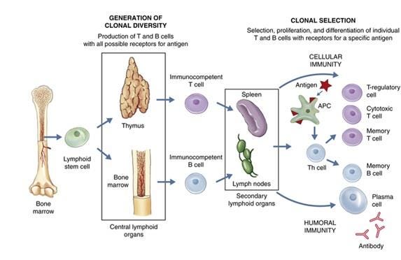
The most widely accepted explanation for how the adaptive immune system works is “clonal selection theory” which states that the immune system:
1. Uses a random generation process to create a vast pool of potential antigen matching sequences.
2. Has vast number of different immune cells that each have those sequences attached to them circulate the blood stream.
3. Waits for one of those immune cells to contact an invading pathogen that the sequence it carries matches.
4. Have each of the immune cells be programmed to start rapidly reproducing once they get a sequence match.
5. Through the previous 4 steps, make it possible to produce a large number of immune cells which are specific to an invading organism (because they can bind to their antigens and alert the rest of the immune system to the organism’s presence), and thus effectively neutralize the infection.
6. Once the process is complete, to leave behind memory B cells, which match the invading pathogen and are able to stimulate the immune response in a much more rapid fashion (thereby shortening the time that steps 3 and 4 take).

The theory behind vaccination is that if the specific immune response and memory B cells can be created before the body encounters a dangerous microbe, this can:

• Allow the body to mount a robust immune response before a harmful invading organism has time to multiply within the body and cause significant damage.

• Cause individuals to rapidly clear infections (rather than needing to wait for the adaptive response to kick in), thereby reducing or eliminating the amount of time they can spread the disease into the population.

• Cause individuals to develop an immune response at the site of infection (e.g., the membranes of the nose and throat), thereby preventing the organism from being able to colonize those areas and thus preventing its transmission.

Being able to do this is hence immensely appealing to governments, as it allows a single intervention (the vaccine) which can easily be distributed to everyone in a top-down manner (which is what governments are good at doing) to address a longstanding problem (infectious disease outbreaks) and more importantly, to allow the government to present the appearance of working in earnest to safeguard the public’s health. Because of this appeal, throughout history, governments will get deeply invested in vaccine programs, and then once issues arise with those programs, double-down on the vaccine (e.g., with mandates) rather than reconsider the wisdom of the vaccine program.
Note: in a previous article, I showed how this misguided and tyrannical conduct has existed ever since the first vaccine (smallpox).

Vaccine Production

To “work” vaccines aim to mass produce a dangerous organism’s antigen without the organism itself being present and then administer that antigen into the body. By doing so, the intermediate stage of an infection (where the organism has already reproduced enough inside its host to have a large number of antigens be available to match a circulating immune cell) can be achieved without the individual being in danger of being damaged or overwhelmed by the infection.

Unfortunately, unlike chemicals which can be rapidly synthesized, antigens are complex enough that they can only be produced by biological systems. As such, to produce the antigen, one of the following is typically done.

• Mass produce the infectious organism, then “kill” it so that its antigens can be collected, but the organism itself is not able to cause infections.

• Genetically modify another organism to mass produce a desired antigen, then kill it and extract the antigen (e.g. the HPV vaccine does this with modified yeasts).

• Modify the live pathogen (typically a virus) so that it can still cause the infection and reproduce inside the recipient but simultaneously is “weakened” so that it is less likely to cause illness.

• Genetically modify a “benign” virus to contain the antigen but be unable to replicate in the human body, then mass produce it outside the body, and have the body develop an immune response to the virus and the antigen on it once it is injected.

• Introduce mRNA into cells so human cells can produce large amounts of the desired antigen, which the immune system then sees (e.g., on the surface of the cells) and develops an immune response to.

The basic problem is that none of these approaches are perfect, and each has both its ups and downsides. For example:

• Most can create autoimmunity.

• In those where only a single antigen is used (and the virus spreads from human to human), if the vaccine actually works, it rapidly stop working because the pathogen quickly evolves a new antigen that no longer matches the vaccine.

• It contrast, the multi-antigen ones (which don’t have that issue) are typically live attenuated vaccines, which then can cause those with weakened immune systems to develop infections from the vaccine itself (e.g., this happens with the polio vaccine—which is why the primary cause of polio now is from the vaccines rather than natural infections, but also can happen with others like the shingles and measles vaccine).

Furthermore, some infectious diseases respond fairly well to vaccination, but the majority do not, so at this point, the vaccine industry has already picked all the “low-hanging fruit” and hence faces an existential struggle to develop new proprietary (patentable) vaccines it can bring to market. For example, had it not been for COVID-19 (SARS-CoV-2), a SARS vaccine would have never been brought to market as it was well recognized the SARS virus was poorly suited for vaccination (which what we then saw throughout the pandemic).

Finally. even if a vaccine “works” it still has to be manufactured, and there are numerous cases of the tradeoffs being made resulting in a disaster. For example:

• To make the inactivated polio vaccine, the live polio virus had to be exposed to formaldehyde. However, the challenge with this was that if too much formaldehyde was used, it would damage the antigens on polio to the point they no longer matched those on the poliovirus, whereas if too little was used, some of the polioviruses would remain active and could then give the vaccine recipient polio. The creator of the vaccine (Salk) opted to prioritize efficacy over safety, which the government in turn supported, even when one of their own scientists (Bernice Eddy) warned them against releasing the vaccine (as it caused polio in her lab). That 1955 vaccine then infected at least 220,000 people with live polio virus in Cutter’s vaccine, of whom 70,000 developed muscle weakness, 164 were severely paralyzed, and 10 died.

Note: an identical issue had happened on a smaller scale (9000 infections, 12 severe cases, 6 deaths) in 1935 with an earlier version of the inactivated polio vaccine. Likewise, (as I showed here) there have been dozens of incidents where an insufficiently inactivated or attenuated diphtheria, rabies or yellow fever vaccines severely injured hundreds of people (as the attenuated vaccines faced a similar issue with it being easy to over or under attenuate).

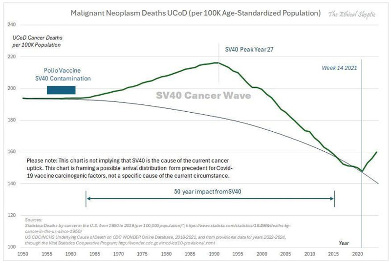
• Growing viruses for vaccines requires having a cell culture to grow them in. Monkey kidney cells were chosen because they worked well for doing this, but unfortunately were contaminated with the cancer causing SV40 virus. In 1962Eddy again warned the government about the vaccine, but they still chose to give it to the public (and retaliated against her for speaking out), which in turn led to a wave of cancer sweeping through America, which until the COVID-19 vaccines was unprecedented:

Note: many other viral vaccines (particularly the live ones) also have had harmful viral contaminants identified within them, but unlike SV40, that contamination has not been acknowledged. Most noteably, a strong case can be made that HIV emerged from virally contaminated vaccines (that had been grown in monkey tissues).

• After a potentially dangerous strain of influenza (due to it having similarities to the 1918 influenza) was identified, a rush began to make an emergency vaccine for it (despite the FDA’s chief influenza expert Morris accurately warning that strain posed no risk to America). Since it took a while to cultivate the virus for a live attenuated vaccine, in order to make the vaccine be produced fast enough to hit the market before the influenza strain disappeared, a decision was made to hybridize it with the PR8 strain, a fast growing influenza strain directly descended from the 1918 influenza. Morris warned against doing this, but was ignored (and fired). That 1976 vaccine subsequently injured a large number of people (including some of our patients) and was a publicity disaster for the US government.

The anthrax vaccine used during (and after) the Gulf War required growing large amounts of the bacteria, killing them, and then filtering out the most toxic components from the final vaccine preparation. The issue the manufacturer ran into was that because of how dirty the vaccine was, its contaminants clogged the filters the manufacturer used, so “solve” the problem and be able to manufacture the vaccine at scale for the military, the manufacturer opted to use larger filters which did not clog, but also didn’t filter many of the toxic components out of the final products—which resulted in one of the most harmful vaccines in history being unleashed upon our military.

• Due to the challenges in producing it, the DTP vaccine (another killed bacterial vaccine) was notorious for having hot lots that killed their recipients (or left them with brain damage). In fact, one FDA pertussis specialist in 1976 stated:

Pertussis vaccine is one of the more troublesome products to produce and assay. As an example of this, pertussis vaccine has one of the highest failure rates of all products submitted to the Bureau of Biologies for testing and release. Approximately 15-20 percent of all lots which pass the manufacturer’s tests fail to pass the Bureau’s tests.

Eventually, the injuries that vaccine created led to so many lawsuits that the manufactures could not afford to continue producing the vaccine, at which point, the 1986 Vaccine Injury Act was passed. This shielded the manufactures from all future liability from it (hence allowing them to stay in business), and eventually incentivized the production of a safer but more costly pertussis vaccine.

• Frequently when an antigen is produced, it cannot solicit a sufficient immune response (unless a lot of it is used—which frequently makes the vaccine too costly to produce). To solve this problem, cheap (and toxic) adjuvants which enhance the immune response to the antigen are used, thereby allowing an affordable amount of antigen to be required for the final product. When the HPV vaccine was developed, it was discovered that its antigen (along with standard adjuvants) could not mount a sufficient immune response to get FDA approval, so a decision was made to use an experimental (but much stronger adjuvant) which worked—but also gave a large number of recipients autoimmune disorders (at least 2.3%). Nonetheless, that trade-off was also accepted to get it to market.

In short, if you look at all these cases, a consistent pattern should be clear. Whenever there is a choice between getting a dangerous vaccine to market or holding off because there isn’t a way to do it safely, the vaccine industry will always do the risky approach (especially in “emergency” situations) as they know they can unconditionally rely upon the US government to promote the product as “safe and effective” and then legally shield them from the disaster which inevitably follows.

COVID-19 Vaccine Hurdles

When COVID-19 began, the industry faced three major issues:

• Whoever was the first to develop a successful vaccine would make a lot of money, but those whose products took longer to reach the market would like miss out on the bonanza.

• There was a finite amount of time the lockdowns could be sustained (which made people want to vaccinate so they could be “free”) and it was very likely the population would rapidly develop herd immunity to COVID-19—so they was a limited window to get a vaccine to market.

• It was extraordinarily difficult to make a safe and effective vaccine for SARS (e.g., decades of work had not yielded a viable product).

Fortunately for the industry, the WHO (and Bill Gates), in 2010, had enacted their “Decade of Vaccine” plan, and with the World Economic Forum (between 2014-2016) had developed a framework for pushing through emergency vaccines that could bypass the regulatory process in the event of a health “emergency.” This framework gave lavish lavish fiscal incentives for vaccine manufacturers and positioned unaccountable organizations like the WHO, Gates foundation, or the World Economic Forum as the directors of a future pandemic response.

Shortly after that framework was finished, the FDA on January 13, 2017 released extremely detailed regulations for obtaining emergency use authorizations, and five days later, Gates publicly announced his plan to the world. This framework was endorsed by pharmaceutical companies, including Pfizer Moderna and J&J, and when Operation Warp Speed was finally conducted in 2020, it mirrored the framework Gates had previously developed.

Note: Event 201, a Gates funded “simulation” exercise modeling the release of a dangerous SARS virus from China was conducted on October 18, 2019. Reading through it in December of 2019 allowed me to accurately predict how COVID-19 would play out. Likewise, on 9/4/2019, Gates invested 55 million in the company that produced Pfizer’s mRNA vaccine—which in two years was worth 550 million.

So as you might expect, the industry chose to adopt the fastest possible production pathway, and was quickly granted the legal immunity (and lavish funding) necessary to accomplish that.

mRNA Vaccine Challenges

Note: one of the major advantages to the mRNA platform was that its production turn around time was much faster than existing alternatives (e.g., growing a virus in chicken eggs). This was a key reason why Fauci’s agency made decades of investments to develop the platform (as with the existing options, seasonal flu vaccines had to start being produced long before the circulating strain was known—which frequently led to the annual flu vaccine being for the wrong strain).

For the mRNA vaccines to “work,” the following needed to occur:

1. An antigen needed to be chosen that was highly likely to elicit a robust immune response that suggested SARS-CoV-2 immunity (and hence could win approval).

2. mRNA matching that antigen needed to be produced at scale.

3. The mRNA needed to be able to get into cells.

4. Once it got into the cells, the mRNA needed to be produce sufficient protein to create an immune response.

Many who have examined Pfizer and Moderna’s vaccines were perplexed by the design that was chosen, as many of the immensely harmful decisions that were chosen strongly imply the vaccine was deliberately designed to harm as many people as possible. While this very well might be the case, many of those issues can instead be explained by the fact each of those 4 challenges was addressed in a way that prioritized getting a vaccine to market rather than producing a safe one.

For example, the spike protein was the most reactive (immunogenic) part of the virus and necessary for SARS to infect cells, so it was an ideal vaccine target. However, it was also a terrible antigen to chose as:

• The spike protein had a significant overlap with human tissues so it was likely to trigger autoimmunity if produced throughout the body.

• The spike protein was highly toxic, so if it was mass produced within the body, it likely would injure the recipient.

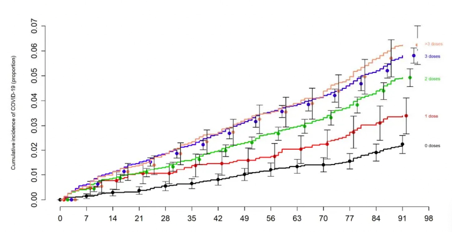
• It was a rapidly mutating part of the virus, which guaranteed the circulating spike protein would quick evolve into a variant the vaccine did not work against it—leading to the remarkable situation where we mandated a vaccine for an extinct virus (which, in turn, caused those vaccinated to become more likely to catch COVID-19 as their immune systems were continually primed to respond to a different virus from that they were exposed to)—something best demonstrated by the Cleveland Clinic’s study of 51,011 people):

Similarly, when I was trying to understand the acute toxicity of the vaccines, I suspected the lipid nanoparticles used to produce the vaccines had to be responsible for their acute toxicity (e.g., which could be seen within seconds of it being put into blood) as those effects onset far faster than it seemed possible for significant numbers of spike proteins to be produced.

After I looked into, I realized that for decades it had been deemed impossible to produce mRNA carrying lipid nanoparticles that had sufficient safety and efficacy to produce viable vaccines (to the point pioneers in the technology like Robert Malone who had invested decades of work into it abandoned it as they felt it was impossible to make a lipid nanoparticle which were not cytotoxic). In turn, when I reviewed leaked Pfizer regulatory documents, I noticed that their lipid nanoparticle had been chosen on the basis of it being the only one that had efficacy—again suggesting safety wasn’t taken into consideration.

Note: there were many other safety issues with mRNA techonolgy (e.g., synthetic mRNA induced immune suppression and antigenic spike protein coating cells and causing the immune system to destroy those cells) that are beyond the scope of this article.

mRNA manufacturing

In my eyes, the two biggest production issues with the mRNA vaccines were producing them at scale and then having them produce spike protein in the body at scale.

Note: these were easy to predict as one of the greatest challenges the pharmaceutical industry, and particularly biotech faces is producing their products at scale—a process which typically takes years to work out, but for Operation Warp Speed needed to be done in a few months.

mRNA design

A major challenge with synthetic mRNA was that the immune system would rapidly break it down, which resulted in not enough of the spike protein being produced to trigger a sufficient immune response. This in turn was “solved” through pseudouridylation, a process where pseudouridine replaces uridine in mRNA molecules as pseudouridylated mRNA resists immune system degradation.

Unfortunately:

• This process occurs in a very limited and tightly regulated manner inside the body. At the time the mRNA vaccines were developed, there was limited understanding on the biological significance of pseudouridylation, however many (e.g., Robert Malone) felt the preliminary data showed this approach had serious risks (e.g., immune suppression).

• These existing technology for pseudouridylation causes it to occur in a random and haphazard way. As such, rather than being able to determine a “safe” dose of pseudouridylation, an all or nothing approach had to be adopted, which in turn led to a significant number of vaccines having “excessive” pseudouridylation (either due to how much of it happened to the mRNA or where in the mRNA the pseudouridylation occurred). In short, this approach was extremely reckless, and akin to playing Russian roulette but hoping everything worked out.

In turn, the pseudouridylation did “solve” the mRNA degradation problem (to the point it won an unscrupulous Noble Prize), but it also created a new issue—the mRNA (and spike protein production) persisting in the body for a prolonged period, and likely due to the haphazard nature of the vaccine’s production, it persisting much longer in some cases that others. Lastly, while this invention “won” the Noble Prize, there were also viable mRNA vaccines that did not utilize it (and hence did not have the risks it entailed).

Note: Codon optimization (which increases protein production from mRNA) may have also caused the vaccine to excessively produce spike protein in the cells.

mRNA preservation

One of the major issues with synthetic mRNA is that its immensely fragile, so even if it could be protected from immune degradation within the cells (e.g., via pseudouridylation) it still was highly susceptible to common environmental factors. As such, when the lots of finished vaccines were examined by regulators, they determined there was both significant degradation of the mRNA and significant variability in what mRNA was preserved.

This is one of the many tables from the leaked EMA documents (which shows how quickly mRNA is destroyed at normal temperatures).

As this degradation and the resulting “truncated mRNA” was one of the primary concerns from the drug regulators Pfizer “solved” this problem by having all of their vaccines be ultra frozen (under the belief this would prevent mRNA degradation). However, this was largely for show, and before long the practice was abandoned (e.g., many vaccine sites had vials sitting outside throughout the day).

Note: at the time I learned about it, I thought that the primary issues would be broken mRNA sequences producing unintended and potentially harmful protein sequences. We now believe it’s actually the opposite, and that this breakdown process was a blessing in disguise, as evidence gradually accumulated that the older vaccines were and the further from their production site they were injected into the body, the less toxic they were—suggesting that the vaccine’s toxicity was mitigated by portions of its mRNA breaking down and hence preventing it from producing as much spike protein.

Process 1 vs. Process 2

The original process used to produce the mRNA vaccine had two ways it could be done. In the cleaner process (which was used to produce the vaccines for the trials), mRNA was produced through controlled replications with minimal contaminants present. Unfortunately, this process could not be scaled.

As such, an alternative approach was done:

• E. coli bacteria were genetically modified to have DNA that contained the spike protein, antibiotic resistance, and the SV-40 promoter.

• Those bacteria were mass produced, then doused with an antibiotic (so those without the resistance gene and thus the spike protein gene) died and did not contaminate the final product.

• The remaining bacteria are killed and their DNA is extracted.

• An mRNA polymerase is used turn that bacterial DNA into vaccine mRNA.

• Everything besides the mRNA is eliminated.

• The mRNA is packaged into lipid nanoparticles.

The essential problem with this process was that it was not feasible to remove many of the contaminants from each stage of production and there was significant room for variability at each stage (and thus in the final product). While many contaminants could be an issue, the vaccine safety community primarily focused on the plasmids as:

• They’ve repeatedly proved plasmids were present in the vaccine.

• The SV40 promoter (a key part of the cancer causing SV40 virus) was grafted onto the plasmids as this was an effective way to increase mRNA production.

• Plasmids had the potential to alter the human microbiome and genome (especially since the SV40 promoter will bring DNA into the nucleus)—which is particularly concerning since vaccine plasmids have now been shown to get into cell nuclei.

• If there was a genomic integration, it was likely creating indefinite spike protein production in the cells.

• When vaccine lots were analyzed for their plasmid levels, it was found the lots which more frequently injured their recipients had higher levels of DNA plasmids (suggesting either harmful genomic integration was occurring, that the plasmids were toxic absent genomic integration, or that they were a proxy for other harmful contaminants).

Inconsistent Manufacturing

Each of the previous points illustrates that the vaccine industry was not ready to bring the mRNA vaccines to market and that the manufacturing of them was rife with errors. Many other points also support this such as:

Japan pulled 1.63M vials of Moderna’s vaccine after visible metal particles were found in them and when examining vaccine vials, Ryan Cole found glass shards in the vaccines. Both of these suggest the production of the vaccines was rushed to the point basic quality control steps were not taken.

• When Ryan Cole did a recorded examination of the vaccines with Del Bigtree they found:

We looked at all the different vaccines and I think one of the conclusions we came away with is that it’s just a hodgepodge..there were vaccines that seems as though there were no particles within them, almost nothing there it was almost like a saline shot and then there were Pfizer’s that were just packed with them and you just get the sense that the manufacturing of this is totally inconsistent…some were more concentrated and some were less.

Note: this suggests that there was either very poor mixing when the vaccines were packaged (leading to some having lots of the lipid nanoparticles and others none) or that the vaccine manufacturers were unable to produce enough vaccine to meet the existing orders and switched to packing placebo vials to meet their contracted orders.

In one mass spectrometry examination of 4 vaccine vials, it was determined that the lipid nanoparticles, but not the mRNA, were present in each of the vials. This suggests that the vaccine was not prepared in a consistent manner, or that the manufacturers ran out of mRNA to fill the vaccines with.

• A large body of evidence from the adverse event reporting databases (compiled here) demonstrated that the toxicity of the vaccine lots greatly varied, which again was likely explained by inconsistencies in their manufacturing.

Note: in a previous article about the mRNA lipid nanoparticles, I showed how inconsistencies in the manufacturing of the vaccines likely explained why the vaccines tended to affect different organs in the body (as their charge was affected by how much mRNA they contained and the area they deposited in the body was influenced by their charge) and why some individuals had acute reactions to them.

Blot Gate

One method of analyzing which proteins are present is with an approach known as “Western Blot.” Periodically, individuals will fake Western Blots (which can be detected because its very easy to identify computer generated ones).

With the COVID-19 vaccines, Western Blots were meant to serve as a quality control measure which ensured the mRNA vaccine were producing their intended protein. However, when we examined the available Western Blots we discovered:

• Some of them were computer generated (and hence likely fake).

• Others showed proteins besides the intended vaccine antigen were there (possibly due to broken RNA fragments being turned into proteins).

All of this again suggested that there were serious manufacturing issues with the COVID vaccines, but a decision was made to sweep all of that under the rug to protect the vaccine manufacturers (particularly since the drug regulators willfully ignored this fraud).

Note: this is similar to how there was extensive fraud throughout the COVID-19 clinical trials to exaggerate vaccine efficacy and safety (which essentially invalidated all the data was gathered), yet even after the trial participants and trial supervisors repeatedly notified the FDA, nothing was done. Likewise, shortly before the vaccine rollout, Vanity Fair published an article highlighting the serious issues in Americas’s vaccine manufacturing plants and that the FDA essentially was unable to monitor them—hence arguing things would get even worse during Operation Warp Speed.

mRNA persistence

Once the COVID-19 vaccine hit the market and the injuries began to mount, we noticed a three curious patterns.

1. The susceptibility to vaccine injuries greatly differed, but in many cases seemed to cluster (e.g., I knew a husband and wife who got vaccinated at the same time and both have near fatal complications from the vaccine).

2. A significant number of individuals appeared to be sensitive to vaccine shedding, something that is supposed to be “impossible” but nonetheless was occurring in a fairly repeatable manner to a large number of people, with symptoms similar to COVID vaccine injuries but typically less severe.
Note: everything we know about shedding is discussed here.

3. Many individuals with vaccine injuries appeared to be suffering toxic reactions to spike proteins in their blood steam months if not years after vaccination. For example:

• Autopsy studies (conducted on individuals who died suddenly up to 6 months after the vaccine) have shown that their tissues were flooded with spike protein. Beyond the inflammatory and necrotic responses to the spike protein being their likely cause of death, it also suggested those individuals had had large amounts of spike protein being perpetually produced within their bodies.

• Both myself and Pierre Kory have come across numerous cases of individuals with vaccine injuries who, months, if not years after vaccination, respond to spike protein binders (suggesting unbound spike protein was causing their issues). Additionally, those individuals either remained recovered, significantly regress, or partially regress once the binder is stopped (implying there is a sustained but possibly diminishing production of spike protein in the body in many vaccine injured patients).

• The only commercially available test for the spike protein (offered by Quest) measures existing antibodies to the spike protein receptor binding domain, and provides values anywhere from 0-25,000 (or higher than 25,000). Clinically, we’ve seen that long COVID rarely get levels above 4,000, whereas in those with vaccine injuries, it can be anywhere from 0-25,000 with many being over 25,000. In turn, a rough (but not precise) correlation exists between the antibody levels (which do not directly measure spike protein levels) and a patient’s illness (along with its levels improving or worsening generally correlating to a patient’s symptoms improving or worsening). Curiously, many patients do not have their antibody levels decline with time (which is what you typically see after a COVID infection), which again suggests spike protein is being produced within the body that then stimulates an immune response. Conversely however, some of these cases may instead have been from people who had antibody levels far above 25,000 who then had their antibody levels decline (but this cannot be detected as they are still above the 25,000 cut off).
Note: Alex Bereneson has also shared reports of vaccinated individuals who’ve had spike protein antibody levels over 25,000 for months after vaccination.

Pivotal mRNA Studies

When the COVID vaccines were pitched to the public, two of the greatest concerns were that the experimental gene therapies could change our DNA and that they would persist in the body for a prolonged period. To address these sales barriers, the media continually platformed experts like Paul Offit and Anthony Fauci) who dismissed us by continually saying things like:

  1. The vaccines cannot enter the nucleus of the cell
  2. mRNA from the vaccines breaks down rapidly in the cell, so it does not have time to enter the nucleus and change your DNA.
  3. mRNA is not DNA, so believing mRNA can change DNA represents a fundamental lack of knowledge of biology.

These points raised red flags to me, as beyond their being issues with each of them, no data was ever provided to disprove genomic integration of the vaccine (which would have been quite easy to do). As such, I assumed the vaccines did integrate into the genome, and evidence would eventually emerge that they did (and likewise that at least some of the vaccines would continually produce spike protein in the recipients).

Throughout this process, two very important, but largely forgotten studies came out.

Note: due to the extreme scientific embargo on anything which challenged the COVID narrative, it is somewhat of a miracle either of these were published.

The first was a March 2022 study from Stanford which showed that both vaccine mRNA and spike protein persisted at high levels at least 2 months after vaccination. This was highly unusual for mRNA, and suggested something (e.g., the pseudouridylation) was preventing it from breaking down. Most importantly, the study did not go beyond 8 weeks, so it was likely the mRNA was persisting for a much longer time.

The second was a January 2023 study (with many Harvard authors) which found in adolescents and young adults who developed myocarditis within a few days of vaccination, that compared to controls, they had significantly higher levels of free spike protein circulating in their blood (due to them not forming antibodies which bound it). This in turn suggested that those who were reacting the worst to the vaccines lacked the ability to form antibodies which could counteract its effects in the body, which both explained why some individuals were so sensitive to the vaccine (e.g., shedding) but also that those likely to “benefit” from vaccination also would have mounted a robust response against a natural infection (hence invalidating the justification for getting vaccinated).

Note: with the smallpox vaccines, vaccination was deemed successful if there was a strong inflammatory reaction at the vaccination site, whereas if no reaction occurred, the vaccination was deemed unsuccessful and the individual was repeatedly revaccinated (as when it didn’t “take” the individuals could still get smallpox). Many early medical dissidents observed that severe reactions to the smallpox vaccine typically followed the vaccine not “taking.” They hence came to believe that the smallpox vaccine working was due to the recipient having a functional immune system that could both already fight off smallpox and create the superficial inflammatory reaction to the vaccine and that the vaccine was simply taking credit for what their immune system could already do. This study made me wonder if something similar has occurred with the COVID-19 vaccines.

Most recently, Yale’s immunology team (which had previously strongly endorsed using the vaccine to prevent COVID and to treat long COVID—which is often disastrous) conducted a longterm study of the effects of vaccination on immune function. Recently, they shared their preliminary results with the participants and disclosed that they were having difficultly publishing the study due to the political pushback they were receiving.

Note: it is extremely common for studies which challenge a medical dogma to never be published. For example, the study demonstrating COVID vaccination causes shedding was stonewalled for well over a year, as I show here, many studies demonstrating harm from the childhood vaccines have been blocked from publication (or even being conducted), and in one well known incident, the CDC deleted data that incriminated the vaccine program. Likewise, I recently highlighted how once the ultrasound industry took off, research into its dangers abruptly ended (due to funding being withdrawn and journals refusing to publish it), which eventually created a collective amnesia that this data ever existed.

As the study was being conducted, a few of us were contacted by participants in the trial who kept us appraised of what was going on, but asked us not to disclose any of it so that we would not interfere with the trial’s publication (as Yale putting their name behind a study demonstrating longstanding immunological injury would make “long-vax” become a medically accepted condition—but the scientific community tends to react quite badly to study results being prematurely leaked).

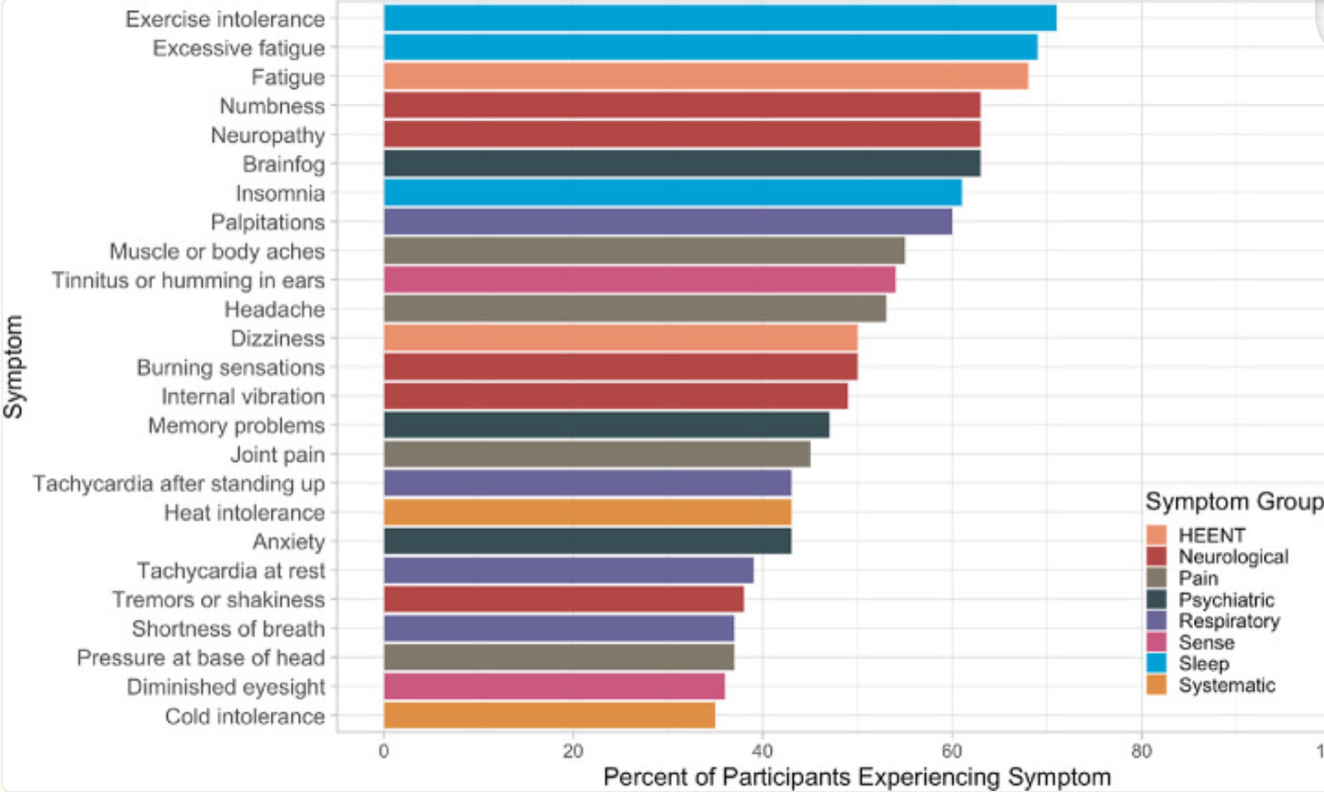
Prior to that meeting, preliminary data had been published a 2023 pre-print (which has still not been published) which detailed the common symptoms seen in the 241 participants with post vaccination syndrome (PVS), which match what we’ve seen in clinical practice:

To quote the study:

In conclusion, people reporting PVS after covid-19 vaccination in this study are highly symptomatic, have poor health status, and have tried many treatment strategies without success. As PVS is associated with considerable suffering, there is an urgent need to understand its mechanism to provide prevention, diagnosis, and treatment strategies.

Note: these results were discussed in more detail in this October 2023 online conference (e.g., the mast cell component of the illness). From watching this conference, my impression was that the investigators sincerely want to help the trial participants, but due to the unpleasant implications of their findings, are in a very challenging position (hence why their 2023 pre-print has still not been published).

Recently, Alex Berenson decided to use his platform to publicize what was disclosed at the recent meeting, at which point, I felt it was appropriate to share some of what we’d learned, and shortly after it became a trending topic on 𝕏. There I highlighted that:

• There was a sustained drop in CD4 levels which the Yale group suspected could account for the sustained immune suppression following vaccination (e.g., AIDS is characterized by severe CD4 suppression). I do not know the average CD4 drop they saw, but one participant in the trial (who has extensive post vaccine symptoms) shared with me her labs which included:

• Study participants were found to have sustained vaccine spike protein in their blood (e.g., 700 days after vaccination), which led the researchers to suspect the vaccine was integrating into the genome.

Note: while there are no commercially available spike protein tests (instead we have to test the antibody levels), research institutions like Yale (and the studies I cited above) have access to tests for free spike protein. Fortunately, after years of work, a commercially available (and affordable) blood spike protein test appears to be just around the corner.

All of this briefly means:

• There is objective proof that long vax is a real syndrome. Unfortunately, given the pace at which science works, it will likely be a few more years before it is formally acknowledged (which will likely dovetail with new pharmaceuticals to treat it entering the market and all remaining interest in the COVID vaccines disappearing). However, given that Trump promoted these vaccines (and has been unwilling to distance himself from them), I could see left-wing institutions like Yale accelerating their publication timeline so that this becomes widely publicized throughout his presidency.

• The persistence of spike protein in the body indicates that the 2022 Stanford study would have found positive results if it had tested patients more than two months post vaccination and that the spike protein antibody titers are indicative of spike protein persisting within the body.

Note: I recently discussed this topic with Dr. Malone (who I consider to be the most knowledgeable people in this area). We are both of the opinion that while genomic integration may play a role in spike protein persistence, the more probable explanation is simply that the body cannot break down the mRNA (and possibly the spike proteins) due to how it was modified. Presently, the data does not exist to quantify the scale of spike protein genomic integration, but with what is currently known (which could change as more data becomes available) cellular production of vaccine mRNA is most likely not responsible for the majority of the free spike protein found in the vaccine injured individuals.

Conclusion

Throughout my life, as I’ve come to feel that because of the bad trade-offs inherent to many policies or technologies, those behind them (particularly the government) will take an approach akin to trying to pound a square peg through a round hole (as government always defaults to utilizing the force at its disposal to solve the problems it encounters). In contrast, whenever I encounter situations where there really does not seem to be a good way to balance the trade-offs, I take that as a sign I need to consider a completely different approach rather than forcing the one I’ve committed to into working.

With COVID for example, I realized near the start that it would be an exercise in futility to address it with a vaccine—a truth much of the world has now had its eyes opened to. Instead, it was my assessment from the start that the best option would be to quickly develop viable treatments for the illness that could prevent severe complications from it and then allow infected individuals to recover with a strong immunity to the disease (and as we’ve now seen, natural immunity is vastly superior to vaccine immunity for COVID-19).

Unfortunately, rather than heed that approach, our medical apparatus decided to do everything it possibly could to push the vaccine upon us, while simultaneously doing all they could to bury the myriad of effective off-patent treatments developed for COVID-19.

Since the pathway Bill Gates put into place for lucrative accelerated approvals is still in place, I believe this highlights how important it is for us to actually understand how these technologies work and the trade-offs involved with them (which are never disclosed). In turn, it is my sincere belief that if the public had known part of what I presented here, they likely would have never taken the COVID vaccines. Similarly, as I’ve tried to illustrate here, contamination and poor production is a systemic problem with vaccines, and were robust independent testing to be conducted (so people actually knew what was in the vaccines they were taking), the demand for them would likely disappear until the industry was forced to clean up its act.

Overall, it is my belief that the most effective way to stop these unsafe products is to simply have enough people boycott them that they become financially unsustainable (and due to the new era of information diffusion we are walking into thanks to platforms like 𝕏, it’s actually possible). The FDA and CDC have lost an immense amount of trust because of how flagrantly they lied to the public, and have thus far refused to take any accountability for their actions—something that will likely change once financial pressures (e.g., people no longer buying the drugs rubber-stamped by the FDA) force the agency to make genuine amends for its conduct throughout the pandemic.

While COVID-19 was a profound tragedy, because of it we now have an extraordinary opportunity to fix this continually proliferating corruption, and I sincerely thank each of you who has helped to make this moment possible.

An updated index of all the articles published in the Forgotten Side of Medicine (including the DMSO ones) can be viewed here. Additionally, to learn how other readers have benefitted from this publication and the community it has created, their feedback can be viewed here.

Who Knew Humans Have a Third Set of Teeth?


Reproduced from original article:
https://articles.mercola.com/sites/articles/archive/2024/03/16/third-set-of-teeth.aspx
The original Mercola article may not remain on the original site, but I will endeavor to keep it on this site as long as I deem it to be appropriate.


Analysis by Dr. Joseph Mercola     
March 16, 2024

third set of teeth

STORY AT-A-GLANCE

  • Dental experts are exploring a medication to regenerate teeth, initially targeting anodontia, which could revolutionize dental care
  • Wisdom teeth, often called “third molars,” aren’t a separate set, but the final teeth to emerge in adulthood, evolving from ancestral needs
  • Gingivitis, if left untreated, can progress to periodontitis, highlighting the importance of proactive oral hygiene to prevent tooth loss
  • Gum disease not only affects oral health, but it also correlates with brain health, indicating a broader impact on overall wellness
  • Simple practices like oil pulling with coconut oil and maintaining a diet rich in whole foods complement oral hygiene routines, promoting healthier teeth and gums

Most people are born with 20 baby teeth and 32 permanent teeth. But around 1% of people have a condition called hyperdontia, which means they have extra teeth, known as supernumerary teeth.1

These extra teeth can be either baby or permanent, and there might be one or several of them. However, according to Katsu Takahashi, who heads the dentistry and oral surgery department at the Medical Research Institute Kitano Hospital in Osaka, Japan, about 1 in 3 cases of hyperdontia lead to the growth of a third set of teeth.

What’s interesting is that Takahashi and his colleagues believe that all humans might have once had the ability to grow a third set of teeth, but over time, this ability was lost.2 There’s even evidence suggesting that the “buds” for a third set of teeth may still exist.

Pioneering a Tooth Regenerative Drug

Sharks and certain reptiles have a unique ability: Their teeth continuously regenerate throughout their lives, sometimes as frequently as every two weeks.3 Could humans also tap into this regenerative power to grow new teeth once they’re lost? Takahashi and his team firmly believe so, and they’re actively developing a medication to try to make this a reality.

Their groundbreaking work, published in 2021, unveiled a protein produced by the uterine sensitization-associated gene-1 (USAG-1 gene)4 that hampers tooth growth in mice. By using a neutralizing antibody medicine to block USAG-1,5 the mice successfully grew new teeth.6 Now, the team is focused on translating these findings into a medication for humans, aimed at addressing anodontia — the complete absence of teeth — in children aged 2 to 6.

The clinical trials are slated to commence in July 2024, with the potential for the product to reach dentists’ offices by 2030.7 Takahashi told The Mainichi:8

“The idea of growing new teeth is every dentist’s dream. I’ve been working on this since I was a graduate student. I was confident I’d be able to make it happen … In any case, we’re hoping to see a time when tooth-regrowth medicine is a third choice alongside dentures and implants.”

Does a Third Set of Teeth Naturally Exist in Humans?

In a 2023 review published in Regenerative Therapy, Takahashi and colleagues detailed the progress made in tooth regeneration. They wrote, “Anti-USAG-1 antibody treatment in mice is effective in tooth regeneration and can be a breakthrough in treating tooth anomalies in humans.”9 They continued:

“With approximately 0.1% of the population suffering from congenital tooth agenesis and 10% of children worldwide suffering from partial tooth loss, early diagnosis will improve outcomes and the quality of life of patients. Understanding the role of pathogenic USAG-1 variants, their interacting gene partners, and their protein functions will help develop critical biomarkers.

Advances in next-generation sequencing, mass spectrometry, and imaging technologies will assist in developing companion and predictive biomarkers to help identify patients who will benefit from tooth regeneration.”

In humans, the potential for tooth regeneration hinges on what experts like Takahashi refer to as a “third dentition,” an additional set of teeth believed to naturally occur in humans.

“In addition to the permanent dentition in humans, a ‘third dentition’ with one or more teeth can occur. In some cases, this third dentition is thought to develop as a partial dentition following permanent dentition,” they explain. “In humans, a rudimentary epithelial form of the third dentition has been identified … Detection of the third dentition during early childhood facilitates the visualization and characterization of hyperdontia in the mouth of infant and some fetuses.”10

Apart from studies on mice, researchers also experimented on ferrets and found that using antibodies targeting USAG-1 resulted in tooth regeneration, akin to the concept of a third dentition. “This result is encouraging given that ferrets share dental patterns similar to those of humans,” they noted in Regenerative Therapy.11

While further testing for safety and effectiveness is crucial, the team remains optimistic about the potential of this treatment to stimulate tooth growth in humans.12

“Compared to dental implants and dentures, antibody-based treatment is more cost-effective and uses a naturally existing third dentition in humans at certain ages. Anti-USAG-1 antibody treatment in mice is effective for tooth regeneration and can be a breakthrough in treating tooth anomalies in humans.”

Save This Article for Later – Get the PDF Now

Download PDF

Understanding Wisdom Teeth: Decoding the Role of ‘Third Molars’

Wisdom teeth, often known as “third molars,” are not a separate set of teeth; rather, they are the final set of teeth to emerge in adulthood.13 It’s believed that these molars, which typically appear between the ages of 18 to 24, might have served a purpose for our ancestors, who had larger jaws and more teeth.14

However, as human jaws have evolved to be smaller, wisdom teeth sometimes fail to erupt fully through the gums. When they do, they can pose issues if there isn’t adequate space. Oral health specialists commonly advise removing wisdom teeth if they’re growing in at odd angles, causing discomfort, experiencing tooth decay or impacting adjacent teeth, leading to inflammation.

Despite these recommendations, many parents choose to have their teenagers’ wisdom teeth extracted preventatively, even in the absence of apparent issues. Statistics suggest that around 5 million individuals undergo wisdom teeth removal annually, with many of these procedures potentially being unnecessary.15 A 2005 Cochrane Review also indicated that a significant number of wisdom teeth extractions could be avoided.16

The review highlighted the importance of judicious decision-making based on specific indicators for extraction, which could potentially reduce the need for surgical procedures by 60% or more. Furthermore, the authors suggested that closely monitoring asymptomatic wisdom teeth might be a reasonable approach. A subsequent 2020 Cochrane Review reiterated the ongoing debate surrounding the removal of asymptomatic and disease-free wisdom teeth:17

“Impacted wisdom teeth can cause swelling and ulceration of the gums around the wisdom teeth, damage to the roots of second molars, decay in second molars, gum and bone disease around second molars and development of cysts or tumors.

It is generally agreed that removing wisdom teeth is appropriate if signs or symptoms of disease related to the wisdom teeth are present, but there is less agreement about how asymptomatic disease-free impacted wisdom teeth should be managed.”

The Link Between Oral Health and Your Body’s Health

The medication researchers are testing will initially be designed to promote tooth regrowth in those with anodontia, but its success could pave the way for broader applications, including addressing tooth loss. However, it’s crucial to recognize that declining oral health not only affects your teeth but also impacts your overall well-being.

Without preventive oral hygiene, you can develop gingivitis, an inflammatory condition triggered by the buildup of plaque or bacteria on teeth. One of the symptoms of gingivitis is red, bleeding gums. Left untreated, gingivitis can progress to periodontitis, a severe infection that may lead to tooth loss.

Furthermore, dental health significantly influences brain health, as evidenced by studies linking gum disease to hippocampal atrophy, the shrinking of a brain region associated with Alzheimer’s disease. In one study18 involving 172 individuals aged 55 and older,19 both gum disease and tooth count were associated with changes in brain structure. Participants with mild gum disease and fewer teeth exhibited accelerated shrinkage in the left hippocampus.

In this cohort, the researchers found that people with one less tooth experienced more brain shrinkage at a rate that was equivalent to nearly one year of brain aging.

A systematic review and meta-analysis of 13 studies revealed a marked increase in the risk of Alzheimer’s disease and mild cognitive impairment among individuals with periodontal disease compared to those without.20 This risk was particularly pronounced in individuals with severe periodontal disease. Moreover, beyond cognitive decline, periodontitis has been linked to various systemic diseases, including:21

Diabetes Heart disease Respiratory disease
Adverse pregnancy outcomes Cancer Nervous system diseases

Recognizing Warning Signs of Oral Health Issues

Nearly half of adults aged 30 or older — about 46% — exhibit signs of gum disease, while approximately 9% have severe gum disease.22 However, the tricky part is that many individuals are unaware of their condition because gum disease often remains “silent,” showing no signs or symptoms until it reaches more advanced stages.23

In the early phase of gingivitis, you might notice that your gums bleed during brushing, flossing or when eating hard foods. Additionally, your gums may appear red or swollen. As the disease progresses, your gums might recede, making your teeth look longer. You may also experience loose teeth, mouth sores, bad breath and pus between your gums and teeth.24

Unlike anodontia, a rare genetic disorder resulting in tooth loss, the loss of teeth later in life can often be prevented by taking proactive oral health measures. Consistent oral hygiene practices, such as regular brushing, flossing and tongue scraping, coupled with routine cleanings by a mercury-free biological dentist, play a crucial role in maintaining healthy teeth and gums.

Adopting a lifestyle that prioritizes a diet rich in fresh, whole foods is also vital for promoting a naturally clean mouth and good oral health. Another beneficial practice is oil pulling, which involves swishing a small amount of oil, like coconut oil, around your mouth for approximately 20 minutes before spitting it out into the garbage.

Oil pulling, when combined with regular brushing and flossing, can help reduce gingivitis and plaque, as well as bacterial colony counts in saliva.25 Only a small amount of oil is needed to achieve good results — 1 tablespoon for adults and 1 teaspoon for a child.

Coconut oil, known for its antibacterial and antiviral properties, is particularly suitable for oil pulling. Research has shown that coconut oil pulling is as effective as chemical mouthwash (chlorhexidine) in reducing plaque, gingival index score, bleeding-on-probing and gingivitis,26 highlighting its potential benefits for oral health.

While the concept of growing a third set of teeth holds promise, the potential side effects of using medication to achieve this result are uncertain. For now, staying vigilant about your oral health can help ensure that your teeth remain firmly in place — where they belong — at any stage of life.

Here’s Why You Should Consider Not Chewing Gum


Reproduced from original article:
https://articles.mercola.com/sites/articles/archive/2024/01/10/chewing-gum-downsides.aspx
The original Mercola article may not remain on the original site, but I will endeavor to keep it on this site as long as I deem it to be appropriate.


Analysis by Dr. Joseph Mercola     
January 10, 2024

chewing gum downsides

STORY AT-A-GLANCE

  • Global chewing gum sales are expected to reach $48.68 billion in 2025, but there are downsides to gum chewing to be aware of
  • If you chew gum excessively, it signals to your body that it’s time to digest food when it’s really not, which could have health implications
  • When you chew gum, it may increase the amount of air and saliva that you swallow, leading to bloating, pressure on the stomach and possibly worsened acid reflux symptoms
  • Gum is essentially made of plastic and often contains titanium dioxide (TiO2) nanoparticles, artificial colors and other harmful additives
  • Chewing gum is also linked to headaches and temporomandibular disorders, and poses a significant threat to the environment due to plastic pollution

Chewing gum is a common pastime worldwide, with global sales expected to reach $48.68 billion in 2025. In North America alone, the chewing gum market may reach $3.5 billion by 2024.1 Some people chew gum for the flavor while others use it more as a tool to reduce food cravings or help quit smoking.

Still others chew gum as a form of stress relief, as chewing, also known as mastication, may reduce anxiety.2 But before you get hooked on this seemingly innocent habit, there are several downsides to chewing gum that you should be aware of.

Chewing Triggers Biological Processes in Your Body

Chewing, and especially chewing slowly, helps with the mastication-to-digestion process, starting in your mouth. Chewing helps break down your food faster, and stimulates the production of saliva, which contains an enzyme called lingual lipase to help break down fats, and which helps (quite a bit) when you swallow. The longer you chew, the more time those enzymes have to start breaking down your food.

The process makes digestion easier on your stomach and small intestine, because digestion takes a lot of energy. Chewing food thoroughly makes it easier for your intestines to absorb the nutrients in the foods you eat.

Chewing also increases glucagon-like peptide 1 (GLP-1). As a peptide hormone, GLP-1 is, among other things, part of a group of incretin hormones, which are released when you eat to regulate insulin, along with many other functions.3 Along with affecting insulin, GLP-1 may influence the nervous system, leading to an appetite-reducing response.

A link may also exist between histaminic neurons in the brain and the periodontal ligament and the masseter muscle — one of four muscles involved in chewing — to influence blood sugar levels.4 These processes are beneficial when you’re about to eat, but if you chew gum excessively, it signals to your body that it’s time to digest food, when it’s not.

Chewing Gum May Cause Digestive Issues, Jaw Troubles

When you chew gum, it may increase the amount of air and saliva that you swallow.5 This can lead to bloating, pressure on the stomach and possibly worsened acid reflux symptoms.6 Titanium dioxide (TiO2) nanoparticles are also common in gum. They’re added as a whitening agent to increase brightness and resistance to discoloration.

Research shows, however, that chronic exposure to TiO2 nanoparticles significantly decreases intestinal barrier function while increasing reactive oxygen species generation, proinflammatory signaling and intestinal alkaline phosphatase activity, which plays a role in intestinal health.7

Iron, zinc, and fatty acid transport also significantly decreased following exposure to TiO2 nanoparticles. In a Binghamton University news release, it’s explained:8

“Acute exposures did not have much effect, but chronic exposure [three meal’s worth over five days] diminished the absorptive projections on the surface of intestinal cells called microvilli.

With fewer microvilli, the intestinal barrier was weakened, metabolism slowed and some nutrients — iron, zinc, and fatty acids, specifically — were more difficult to absorb. Enzyme functions were negatively affected, while inflammation signals increased.”

The European Union banned titanium dioxide for food use in 2021 after the European Food Safety Authority Panel on Food Additives and Flavourings found it “can no longer be considered as safe when used as a food additive.” The panel concluded that TiO2 particles “have the potential to induce DNA strand breaks and chromosomal damage” and a “concern for genotoxicity could not be ruled out.”9

Temporomandibular disorders, which include those relating to the temporomandibular joint, or TMJ, are also associated with gum chewing.10 Incidence of TMD symptoms, including clicking and pain, were higher in gum chewers than non-chewers.11 If you chew gum on one side of your mouth more often than the other, it can also cause jaw muscle imbalance.

Anytime you overuse a certain set of muscles, it can lead to contracted muscles and related pain, including headaches, earaches, and toothaches over time.

Download this Article Before it Disappears

Download PDF

Gum Was Once Natural, Now Contains Plastic

Gum is a processed food that contains questionable ingredients. These include “gum base,” which is what makes gum chewy. The exact ingredients that make up gum base are often kept quiet as a trade secret, but they may include:12

  • Fillers — These provide texture and bulk to the gum and are usually calcium carbonate or magnesium silicate (talc).
  • Elastomers — Long synthetic polymer molecules such as polyvinyl acetate.
  • Emulsifiers — These chemicals help keep flavors and colors mixed.
  • Softeners — Vegetable oil and lecithin are used to keep the product soft and chewy. Once these have washed away and been swallowed, the gum gets stiff.

It wasn’t always this way. People have been chewing gum for thousands of years. One 5,700-year-old piece of chewed gum from Denmark was found to be made of birch pitch, a substance obtained by heating birch bark.

Interestingly, birch pitch contains betulin, a compound with antiseptic properties, so it’s possible the “gum” was chewed for medicinal purposes.13 Other ancient gum was made from chicle, which comes from the sap of the Sapodilla tree.14

Modern scientists sought to find a recipe that provided the same characteristics of these natural substances using synthetic chemicals that were easier to source. The U.S. Food and Drug Administration’s list of additives15 permitted for direct addition to food for consumption includes Section 172.615 that covers the chemicals allowed in chewing gum.

These are the additives they say “may be safely used in the manufacture of chewing gum”16 and bear the name “chewing gum base,” which:17

“… means the manufactured or partially manufactured nonnutritive masticatory substance comprised of one or more of the ingredients named and so defined in paragraph (a) of this section.”

The list of ingredients the FDA allows in gum base includes the following, which are plastics, rubbers and waxes:18

  • Butadiene-styrene rubber
  • Isobutylene-isoprene copolymer (butyl rubber)
  • Petroleum wax, Petroleum wax synthetic
  • Polyethylene, one of the most widely used plastics, included in plastic wrap, grocery bags, drainage pipes and bulletproof vests
  • Polyvinyl acetate, one of the ingredients found in PVA glue, which you may know as school glue and wood glue19

As author David Jones wrote in Just One Ocean, “You probably had no idea that you were chewing on what is essentially a lump of malleable plastic and that’s not surprising, because the manufacturers don’t actually tell you as much — they kind of dodge around the detail.”20

Toxic Food Chemicals Are Common in Chewing Gum

In addition to titanium dioxide nanoparticles, synthetic food dyes, including Red No. 40, Yellow No. 5, Yellow No. 6 and Blue No. 1, are often added to chewing gum. Such dyes are linked to hyperactivity and other neurobehavioral problems in children.21 As noted by the Environmental Working Group:22

“The California health agency [Office of Environmental Health Hazard Assessment] also found that current federal safe intake levels of these dyes might not protect children’s brain health. Current legal levels were set by the Food and Drug Administration decades ago and do not take recent research into account.

Human studies have also linked synthetics dyes to learning difficulties and restlessness in sensitive children. In the EU, products containing Red No. 40, Yellow No. 5, and Yellow No. 6 must contain the warning ‘May have an adverse effect on activity and attention in children.’”

Artificial sweeteners are also common in chewing gum. While sugar-sweetened gum isn’t recommended, as it can promote tooth decay,23 artificial sweeteners like aspartame are toxic. A systematic review and meta-analysis conducted by the World Health Organization revealed “potential undesirable effects from long-term use of NSS [non-sugar sweeteners], such as an increased risk of Type 2 diabetes, cardiovascular diseases and mortality in adults.”24

Further, even sugar-free gum can be bad for your teeth if it contains acidic additives. Acid-containing sugar-free gum, which may be fruit-flavored, may increase the risk of demineralizing enamel on your teeth, causing dental erosion.25

Chewing Gum May Trigger Headaches and Release Mercury From Fillings

Excessive gum-chewing may be an underrecognized trigger for headaches, particularly in children and adolescents. One study involved 30 children with a median age of 16 years who suffered from chronic migraine or tension headaches. After quitting gum-chewing for one month, headaches went away completely in 19 of them while another seven had a reduction in headache frequency and severity.26

When the children then started chewing gum again, their headaches returned within days. It’s possible the headaches may be linked to chewing-gum-induced TMJ, which may cause headaches. A separate systematic electronic search of the literature concluded:27

“Despite the limited evidence, it seems reasonable to suggest that headache attacks may be triggered by gum-chewing in migraineurs and in patients with tension-type headache …

Although larger randomized studies will be necessary to definitely establish the relationship between gum-chewing and headache across different populations, it seems cautionary to suggest that subjects with migraine or tension-type headache should avoid or limit gum-chewing in their lifestyle.”

While it’s often stated that chewing gum may reduce appetite, one study found gum chewers’ meals end up being less nutritious than those eaten by non-gum-chewers.28 Chewing gum was also linked to increased meal size and reduced nutrient adequacy. Mint gum chewers were also less likely to eat fruit but their intake of snack foods was not affected, possibly because the minty flavor in the gum makes fruits — but not junk foods — taste bitter.

If you have mercury fillings, you should also know that chewing gum may cause this known neurotoxin to release from the fillings into your body. According to one study:29

“… chewing gum has been shown to increase the release rate of mercury vapor from dental amalgam fillings … The impact of excessive chewing on mercury levels was considerable.”

Every time you chew, mercury vapor is released and quickly finds its way into your bloodstream, where it causes oxidative processes in your tissues. If you chew gum, you’re going to be chewing often, which is why it’s particularly problematic for those with mercury fillings.

Gum Is ‘One of the Biggest Threats to Our Ecology’

Another often overlooked reason to give up gum? It poses a significant risk to the environment because it’s made of “nonbiodegradable hydrophobic polymers.”30 Because many people toss their gum on sidewalks and streets, gum pollution is a major pollution problem.

According to a review in Current World Environment, “Each year, chewing gum generates more than 105 [100,000] tonnes of “plastic” garbage. Thus, the discarded nonbiodegradable residue of the gum produces plastic pollution. Every year, enormous sums of money are spent to clean up the abandoned gum from the streets.”31

So, not only may chewing gum have adverse effects on your own health, collectively it’s contributing to considerable environmental damage as well. If you’re chewing gum for stress relief, consider other options like meditation, yoga and the Emotional Freedom Technique (EFT). If it’s the flavor you’re after, try adding fresh mint leaves or cinnamon to water for a healthy alternative.

– Sources and References

Root Canals Cause Breast Cancer-Frequently

Reproduced from original OMNS article (OrthoMolecular News Service):
http://orthomolecular.org/


Subscribe to the free Orthomolecular Newsletter: http://orthomolecular.org/subscribe.html
Go to the OMNS Archive: http://orthomolecular.org/resources/omns/index.shtml
Orthomolecular Medicine News Service, December 17, 2023

Commentary by Thomas E. Levy, MD, JD

OMNS (December 17, 2023) Breast cancer causes enormous morbidity and mortality around the world, and its traditional treatment, along with the relentless progression of the disease, greatly impacts the quality of life for both patients and their families. This cancer basically targets just women, as only a relatively minimal number of cases occur in men (roughly 1%). [1] Yet, despite its predilection for women, breast cancer is still the most common of malignancies (aside from non-melanoma skin cancers) statistically in the overall population. It continues to be the leading cause of cancer deaths across the planet. [2]

Redox Biology and Toxins

Reduction and oxidation basically refer to the movement of electrons between molecules. When a normal biomolecule with a normal electron content is depleted of one or more electrons, it becomes oxidized. And when that oxidized biomolecule can regain the lost electron(s), it returns to a normal, reduced chemical state. A reduced biomolecule functions normally, while an oxidized biomolecule either partially or completely loses its normal chemical/biological function. More oxidized biomolecules result in the accumulation of largely metabolically inert agents that only occupy space, interfering with normal chemical reactions and no longer directly supporting normal biological function. Examples of biomolecules include sugars, fats, proteins, enzymes, nucleic acids, and structural molecules.

Redox (reduction-oxidation) biology is based on the concept that all biological health is directly due to the degree of reduction versus oxidation in the biomolecules throughout the body. Higher reduction/oxidation ratios indicate good cellular health. This has led to the frequent use of the term “oxidative stress” as the premier biomarker and measuring stick of all disease.

Widely discussed in the medical and scientific literature, increased oxidative stress, or the excess presence of oxidized biomolecules, is always the primary pathophysiology of any disease under consideration. [3,4] It is characterized by a relatively low antioxidant presence and/or an increased pro-oxidant presence. At the cellular level, all diseases or medical conditions have increased oxidative stress in the cells of the affected organs or tissues. The extracellular areas are often involved as well. As pathology cannot exist in the absence of excess oxidation, there are no exceptions to this premise.

All toxins damage by directly or indirectly causing the oxidation of important biomolecules. Oxidation is the chemical process of giving up, or losing, one or more electrons to an electron-robbing toxin (pro-oxidant) that never surrenders that electron back to an oxidized biomolecule once it is acquired. Unless an agent results in the oxidation of biomolecules in the body along with the permanent retention of the electrons it has taken, it is not toxic, and it cannot be toxic. Clinical toxicity and any symptoms of toxicity cannot exist in the absence of excess oxidized biomolecules.

As excess oxidation is the basis of all disease, it logically follows that all cancers, either in the breast or elsewhere, result from excessively and chronically elevated oxidative stress at the affected tissue site. This elevated oxidative stress is always secondary to electron-depleted toxins, also known as pro-oxidants, poisons, free radicals, reactive oxygen species, or oxidizing agents.

This leads to the following two questions:

  • What is the source of the toxins in breast cancer, and
  • What is causing them to excessively accumulate?

Breast Cancer Pathophysiology

All chronic degenerative diseases, including cancer, only arise when an area of affected tissue becomes substantially inflamed and remains that way. Phrased differently, the areas in the body that have exceptionally increased and chronic oxidative stress are the areas where malignant transformation eventually takes place. Lesser degrees of increased oxidative stress, depending on their location, underlie the development and maintenance of all non-malignant diseases as well. But the highest chronic elevations of oxidative stress, both intracellularly and extracellularly, are the reasons for the initiation and evolution of cancerous growth. No cancer has ever developed in an area that was not already inflamed. While a cancer can metastatically seed abnormal cells in a previously normal tissue site, the primary cancer focus will never be initiated in normal, uninflamed tissue.

A prolonged and sizeable presence of toxins always precedes the development of cancer in the affected areas of the breast. These toxins are produced by slow-growing pathogens (colonizations), and the pathogens themselves will often be found at the cancer site as well. But toxins (highly pro-oxidant molecules) must always be present in order to provoke and sustain a state of chronic inflammation and excess oxidation.

The amount of time that such a toxin/pathogen accumulation needs to be present before a cancer develops is highly variable. Some women with exceptionally strong immune systems, high antioxidant intake, and a relatively lesser degree of toxin/pathogen presence may never demonstrate malignant transformation. Of note, benign breast lumps and other forms of breast pathology result from lesser degrees of toxin exposure.

No pathology of any kind can develop when a tissue has intracellular and extracellular levels of oxidation that are physiological in degree (from normal metabolism). Only increased levels of oxidation can result in pathology. And only extremely increased levels of oxidation result in the appearance of cancer.

In addition to a blood circulation, the body has a lymphatic circulation as well. This circulation moves lymph, the plasma-like extracellular fluid bathing the cells throughout the body, into the venous blood circulation. Under normal circumstances, this lymph flow is one-way only in the direction needed to reach the blood. [5] The primary role of the lymphatic circulation is to provide an outlet for cellular waste products, excess water, and toxins, as well as to support an immune defense against pathogens. [6] It also periodically condenses into focal bodies known as the lymph nodes.

These lymph nodes, of which there are about 500 to 600 in the body, work to concentrate B- and T-lymphocytes needed to combat the infectious agents that are encountered, such that the lymph itself is rendered sterile by the time it leaves the lymph nodes and reaches the blood. [7,8] When draining a large enough source of infection, such lymph nodes will readily enlarge and become sore where they can be felt (palpated), such as in the neck, armpits, or groin areas. Once the processed lymph finally reaches the blood circulation, multiple ways of metabolizing and excreting the remaining non-infective extracellular debris are then available.

The breasts have an extensive lymphatic circulation, and much of its lymph comes from drainage of the head and neck. A portion of the breast lymph subsequently flows into a large collecting vessel (thoracic duct), which then empties into the venous circulation. The rest of it first flows into the extensive lymphatic network in the armpits before eventually reaching the thoracic duct and the blood.

The lymphatic vessels have a limited ability to contract and promote a one-way flow of lymph. [9] However, this lymphatic movement can be slowed, stopped, or even reversed in direction by the presence of sufficient inflammation and structural damage in the tissue being drained. When there is sufficient impairment of normal lymphatic flow, tissue swelling (lymphedema) can result.

In the breast, this impairment of lymphatic drainage can result from either the chronic inflammation in the cancerous tissue, or much more commonly, following the surgical removal of cancer-laden axillary lymph nodes draining the breast. [10,11] The fewer draining lymphatic pathways available, the more likely lymph flow will slow enough to accumulate. Together, both situations result in about 20% of women with breast cancer eventually developing arm swelling due to the back-up of lymph. [12,13]

As with tissues elsewhere in the body, the lymphatic system also drains the superficial gum tissue, the deep gum tissue (periodontium), the teeth, and the tonsils. This drainage largely follows gravity and is filtered in the lymphatic vasculature in the floor of the mouth and then the neck. From there much of the drainage proceeds further down into the rich network of lymphatic vessels in the chest and the breasts, with most of the breast lymph then draining directly into the armpits. [14-16] Deep periodontal infection (periodontitis) has been “linked” to many different cancers, including the breast. Of significant note,

Maintenance of periodontal health has been found to be effective in the primary prevention of breast cancer. This indicates that periodontal infection has a cause-and-effect relationship to breast cancer, not just a link, association, or correlation. [17]

Left unresolved, periodontitis seeds pathogens and their associated toxins throughout the body. To assert that disease occurring after such typical oral pathogens start growing in different tissues is some ill-defined “association” and not a straightforward cause-and-effect relationship defies simple logic. Aside from breast disease and breast cancer, chronically infected gums have been significantly linked to nearly all diseases and conditions. Some studies also document improvement of the “linked” disease with effective periodontitis treatment, as well as a worsening of the disease as periodontitis progresses. This further indicates that periodontitis has a cause-and-effect with most chronic diseases. These diseases and conditions include the following:

  • Cardiovascular disease and all-cause mortality, including atherosclerosis, myocardial infarction, heart failure, abnormal lipid and cholesterol metabolism (metabolic syndrome), diabetes, and arterial calcification [18-32]
  • Neurological disease, including migraine, seizure, depression, bipolar disorder, dementia, Alzheimer’s disease, Parkinson’s disease, brain abscess and multiple sclerosis [33-53]
  • Chronic pulmonary disease, pneumonia, asthma, allergic rhinitis [54-60]
  • Vascular disease [61,62]
  • Obesity [63,64]
  • Inflammatory bowel disease [65-67]
  • Inflammatory bone diseases, including arthritis and osteoporosis [68-73]
  • Kidney disease [74-76]
  • Cancers (pancreatic, lung, liver, colorectal, esophageal, oral cavity and throat, head and neck, stomach, prostate, blood, skin, and cancer in general) [77-92]
  • Female infertility and adverse pregnancy and neonatal/birth outcomes [93-95]
  • Thyroid disease [96]
  • Anemia [97,98]
  • Eye disorders [99,100]
  • Psoriasis [101-104]
  • Ear disorders (hearing loss, vertigo) [105-107]
  • Polycystic ovary syndrome [108]
  • Autoimmune disease [109-111]
  • Erectile dysfunction [112]
  • Increased body-wide inflammation (elevated C-reactive protein levels) [113-115]
  • Depressed vitamin levels (C and D) and antioxidants [116-122]

The data above on chronic disease and periodontal infection is vital to understanding the impact of root canals and other infected teeth on the general health. Because the pathogens found in infected teeth only come from deep gum infections (except when large cavities allow the pulp to become infected from above), the infections found around the root tips of affected teeth have the same infectious profile as is found in periodontitis. However, the infected teeth, which include all root canal-treated teeth, are even more disease-causing than the infected gums, since they also have the additional following characteristics:

  • The infected teeth have a much greater amount of infectious material, often manifest as root tip (apical) abscesses on X-ray.
  • The infectious content in and around the root tips of the infected teeth drains directly into the venous circulation.
  • The infectious content in and around the root tips of the infected teeth are also released into the extracellular fluid and drained directly into the lymphatic system of the teeth and jawbone.
  • The act of chewing on the infected teeth greatly magnifies the expression of pathogens and toxins into the blood and lymph, as extraordinary pressures are generated between opposing teeth. Since pathogen release into the lymph occurs in addition to their release into the venous blood, the delivery of these oral pathogens and toxins to the entire body is more effective and efficient with chewing on infected teeth than if the pathogens and toxins were just directly injected into a vein with a syringe.

The release of highly pathogenic bacteria into the blood during a root canal procedure has been clearly documented. Of note, their release occurs in the absence of any chewing pressure that would further promote pathogen release. [123,124]

Traditional dentists and endodontists (root canal specialists) somehow deny and/or blind themselves to the massive documentation that all root canals are infected. Instead, they collectively maintain that a successful root canal procedure leaves the tooth infection-free just because a root tip abscess was reduced in size and the pain associated with the acute abscess was relieved.

Nevertheless, all the research on the impact of periodontitis and abscessed teeth on all chronic disease applies to all root canals as well, regardless of how well they were performed technically. Although less extensively studied than the relation of just periodontitis with chronic diseases, a great deal of research has also established a link between chronically abscessed teeth (CAP-chronic apical periodontitis) and many different diseases. CAP simply means an extension of and a more advanced form of deep gum inflammation and infection (periodontitis), with root tip abscesses seen on imaging. Significant research studies have documented this abscessed tooth-chronic disease link, which includes all root canals that have resulted in the reduction of root tip abscesses on imaging studies. These studies directly mirror the many studies on early periodontitis and chronic diseases cited above and include the following:

  • Cardiovascular disease [125-130]
  • Neurological disease [131-137]
  • Eye infection [138]
  • Inflammatory bowel disease (including Crohn’s disease and chronic ulcerative colitis) [139-142]
  • Diabetes [143]
  • Liver disease [144,145]
  • Kidney disease [146,147]
  • Inflammatory bone diseases [148-152]
  • Autoimmune disease [153,154]
  • Adverse pregnancy outcomes [155,156]
  • Increased body-wide inflammation (elevated C-reactive protein levels) [157-161]
  • Reservoir for a wide variety of pathogens (bacteria, fungi, and viruses, including Epstein-Barr and herpes) [162-166]
  • Increased morbidity and mortality in COVID-19 patients [167]
  • Decreased physical fitness/capacity [168]
  • Body-wide disease in general [169]

While other factors may be involved, it appears likely that men have virtually no breast cancer because the amount of breast tissue is so much smaller than in women, and there is much less toxic and infected lymph from oral cavity infections getting continually filtered in that tissue. And even though there is a large difference in the average amount of breast tissue between men and women, the lymphatic drainage patterns are largely the same. [170] Also, larger and denser breasts, along with overly constrictive bras and clothing, can all impede the rate at which lymph can be conducted through the breasts. Anything that slows lymphatic flow, especially if it has a significant toxin/pathogen content, will be a factor in determining whether significant inflammation is allowed to take hold in an area of the breast draining the lymph from the oral cavity. Consistent with this concept, studies have shown that very large-breasted women who undergo breast reduction surgery lower their chances of breast cancer. [171] Furthermore, it has been shown that women with larger breasts fare worse with breast cancer than women with smaller breasts. [172]

Root Canal-Treated Teeth

The root canal procedure is one of the most common dental procedures. A meta-analysis revealed that more than half of the subjects in the populations studied had at least one root canal. [173] A very large review and meta-analysis also found that at least half of the population has at least one abscessed tooth. [174] When combined with the prevalence of abscessed teeth that have not received a root canal treatment, the prevalence of infected teeth runs between 55% and 70% of the subjects in the studies. [175-180] Depending on the information source, between 25 and 45 million root canal procedures are performed in the United States every year. Even the low side of the estimate means that a significant majority of adults are always chewing on one or more infected teeth.

Another study found that over 60% of people in Europe had abscessed teeth, with the prevalence steadily increasing with age. [181] Furthermore, fully 25% of teeth that have had procedures other than root canal treatments end up chronically abscessed as well. [182]

Root canal-treated teeth are generally performed when a patient presents with a painful, acutely-abscessed tooth. The “successful” root canal procedure results in a tooth that no longer hurts, resulting in a happy patient and satisfied dentist, at least for the moment. However, the infection remains as long as the tooth remains unextracted, or when the socket infection remains is not completely cleaned out after extraction.

There is an enormous variety and a large total number of different pathogens and other microbes found in and around the root tips of root canal-treated teeth. Fungi, viruses, protozoa and over 460 different types of bacteria have been identified in these infections. [183] No two root canals have the same assortment of indwelling pathogens, and this is why no two root canals inflict the same degree of infectious/toxic damage to the body. Nevertheless, even the “least toxic” root canals can wreak havoc throughout the body.

The physiological core of the tooth, known as the pulp, contains the nerves, blood vessels, and connective tissue matrix that keep the tooth alive and viable. Once this pulp has become infected, there is no way to eradicate the infection and restore the pulp to its normal, health-supporting state. Instead, the removal of the pulp by the root canal procedure permanently prevents immune system access to the pathogens in the tooth, especially in the miles of dentinal tubules extending away from the pulp throughout the tooth structure.

Without immune support, no infection can be resolved. Furthermore, even without the root canal procedure, the infected pulp quickly destroys the pulp structure itself, just leaving a collection of pus and dead (necrotic) cells that can never be returned to normal. Even though pain can still be felt in the root tips embedded in the jawbone, the upper part of the tooth and the pulp is simply a non-vital shell.

In order to stop the immediate infection-causing pain and remove much of the bulk (but never all) of the infection, the root canal procedure drills and routs out as much of the pulp as can be reached, after which it is filled in with an agent to maintain the overall tooth structure. The ends of the pulp space extend to the tooth root tips embedded in the jawbone, and the pulp infection and its necrotic debris effectively “collects” there. This results in well-defined abscesses surrounding the root tips.

Chronically infected teeth will usually be found to have clearly visible evidence of this pathology at the root tips. On X-ray or on computed tomography studies variably-sized abscesses will nearly always be seen, appearing as dark areas, or radiolucencies, surrounding the root tips. Rarely, the infected tooth might not contain enough infected debris to be visible in an imaging study, but the lack of an identifiable abscess does not mean the infection is still not there.

When a root canal procedure has been performed with optimal expertise, much of the associated root tip abscess will be removed (debulked), and follow-up imaging will no longer easily detect it. However, this does not mean the infection is gone, only that it has been effectively drained. Pathogens and their toxins are still readily expressed into the blood and lymph, especially during chewing.

Dr. Boyd Haley conclusively proved that all root canals produce and collect toxins. He devised a test using a process called nucleotide photo affinity labeling to measure the impact of the pathogen-generated toxins in extracted root canal-treated teeth on five key human enzymes. After studying over 5,000 consecutive extracted root canal-treated teeth sent to him from around the country, the results were stunning. ALL tested teeth had significant toxin content. Differences in the degree of toxicity among the teeth were seen, but none were toxin-free. Such variability in toxicity is to be expected, as no two root canals have the same pathogen population. [184] Furthermore, normal teeth extracted for orthodontic purposes never demonstrated any toxicity, even to a minor degree, ruling out “mouth contamination” as a potential reason for the results.

Of note, Dr. Haley found similar toxin profiles in the specimens sent to him from cavitation surgery. Cavitations occur when chronic infection remains in the healed-over sockets of extracted teeth. [185] Cavitations have a comparable connection to chronic diseases, including breast cancer, although it is much less extensively studied than the relation between abscessed teeth and chronic disease. [186-189]

In addition to the clear visual appearance of chronic infection at the time of extraction, frequently accompanied by putrid odors, ALL root canal teeth extraction sites have pathogens that can be cultured, and microscopic examinations of biopsy specimens always reveal inflamed and necrotic bone and tissue cells resulting from the chronic infection.

Infected Teeth-The Hidden Pandemic

Chronically abscessed teeth, as seen in imaging studies, are very common. Furthermore, they are nearly always completely free of pain or any other associated symptoms, and the patient has no reason to suspect that there are any problems in the mouth. In contrast the acutely abscessed tooth, for which many root canals are performed, are typically extremely painful. This is why chronic diseases in adults are the rule and not the exception. Too many physicians and their patients simply “expect” that hypertension, diabetes, cancer, or heart disease are the norm for many older adults. Also of note, the deciduous (non-permanent or baby) teeth in children demonstrate a high incidence of abscess formation. [190] When a child becomes chronically ill, a complete oral examination is just as important as in the adult with a chronic disease. So, for all ages, the important take-away point is that:

When the mouth is infection-free, all chronic diseases are very rare. And when there is a chronic disease, oral- or gut-derived pathogen colonization of the affected tissue with local toxin production is almost always the cause.

While some individuals, in a completely unpredictable manner, can have one or more root canals without ever resulting in negative clinical consequences, this is very rare. However, significant laboratory abnormalities are often present even when a chronic disease is not yet manifest. Also, clinical medicine always looks for a prompt and clear-cut relationship between an intervention and a negative clinical impact. With root canals and other chronically infected teeth, the leakage of pathogens and toxins can be slow, and breast cancer or a heart attack due to those infected teeth can take years to occur.

Complications can occur rapidly after a root canal procedure, but this is not a very common consequence. If that were the case, root canal treatments would have been abandoned long ago. But when someone gets breast cancer years after a root canal, the status of the mouth is simply never considered as the possible reason by the clinician or the patient.

A particularly aggressive pathogen of periodontal origin, Fusobacterium nucleatum, has been found in human breast cancer tissue. In an animal model, this pathogen has been shown to promote tumor growth and metastatic spread. [191,192] Higher titers of oral pathogens inside breast cancer cells have also been shown to promote metastatic spread, with experimental reduction of these titers decreasing the chances of metastasis. [193] Animal studies have also shown a commonality of pathogen presence in the mouth, gut, and in breast tumors. [194]

Some researchers have actually termed breast cancer as an infectious disease. [195] Many other studies have consistently found pathogenic microbes, including viruses and fungi, in diseased breast tissue, including cancer, and much lower titers of non-pathogenic microbes in normal breast tissue. [196-204] As the mouth is always teeming with microbes (more than 700 different bacterial species) and its lymphatic circulation mostly drains into the breasts, the breast tissue is not completely microbe-free. [205,206] However, it should be pathogen-free, and the non-pathogenic microbes should always be very low in number and difficult to culture. [207]

Researchers found a 10-fold (1,000%) increase in bacterial load in breast tumors relative to normal breast tissue. [208,209]

In addition to the enormous amount of literature cited above that unequivocally ties mouth infections to chronic diseases, several other studies warrant special attention, as they reveal that pathogens of oral and gut origin have been shown to chronically colonize different diseased tissues, with continuous inflammation resulting from the on-site production of pathogen-related toxins. Breast cancer is but one of many infection-related chronic diseases. Chronic pathogen colonization (CPC) in diseased tissues is addressed more extensively elsewhere. [210] Especially noteworthy studies supporting the widespread presence of CPC and its disease-causing impact include the following:

  • Pathogens in Alzheimer’s disease brain tissue and cerebrospinal fluid [211-218]
  • Pathogens in Parkinson’s disease [219]
  • Pathogens in multiple sclerosis brain tissue and cerebrospinal fluid [220,221]
  • Pathogens in amyotrophic lateral sclerosis brain tissue and cerebrospinal fluid [222]
  • Pathogens in the atherosclerotic lesions of coronary heart disease [223-226]
  • Pathogens in intracranial aneurysms [227]
  • Pathogens in abdominal aortic aneurysms [228]
  • Pathogens in the acute blood clots causing myocardial infarctions [229,230]
  • Pathogens in the acute blood clots causing lower limb thrombosis [231]
  • Pathogens in the pericardial fluid surrounding the hearts of patients with coronary artery disease [232]
  • Pathogens in the joints of patients with rheumatoid arthritis [233-235]
  • Pathogens in the placentas of mothers with preterm and low birth weight infants [236,237]
  • Pathogen antibodies in systemic lupus erythematosus patients [238,239]
  • Pathogen antibodies in stroke patients [240,241]
  • Pathogens in cancersBreast [242-244]
    Oral, head, and neck [245-247]
    Esophageal [248,249]
    Liver pathology leading to cancer [250]
    Prostate [251]
    Pancreatic [252,253]
    Colorectal [254,255]
  • * Pathogens and chronic body-wide inflammation and chronic diseases in general [256-258]

All chronic diseases need to have a daily source of new oxidative stress greater than the daily intake of antioxidants in diet and supplementation. Otherwise, “chronic” diseases would resolve as the new antioxidant intake repairs the old oxidative damage. This source of the new daily oxidative stress nearly always arises from chronic pathogen colonization in the diseased organ or tissue. New toxin exposure in the affected tissue comes from on-site pathogen-generated toxins and the oxidized (toxic) products of pathogen metabolism. Pathogens also release enormous amounts of pro-oxidant free iron when they finally die and break apart.

Chronically Infected Tonsils

In the 1950s Dr. Josef Issels made some remarkable discoveries that remain largely unknown to the medical and dental community. [259] His clinic in Germany treated mostly advanced cancer patients who were seeking to avoid chemotherapy in their pursuit of health. In surveying his own clinic data he found that 98% of the cancer patients had between what he termed “two and ten dead teeth.” His treatment not only involved the proper removal of such infected and necrotic teeth, but also routine tonsillectomy. This was not initiated until he retrospectively observed that a significant number of his patients, who initially did well after the extractions, later experienced myocardial infarctions. After making tonsillectomies a part of his treatment protocol in these advanced cancer patients, the prevalence of heart attacks dropped from 40% to 5%.

Dr. Issels asserted that “chronically inflamed tonsils are primary head foci which sometimes have an even more damaging effect on the organism as a whole than dental foci,” noting that the tonsils are “excretion organs by which the lymphocytes, microbes, toxin-laden lymph, and other matter are discharged.”

Most significantly, Issels found that in every tonsillectomy performed, biopsy specimens revealed that “severe or very severe destructive tonsillar processes” were present along with chronic infection. This was in spite of the fact that the tonsils did not appear enlarged, inflamed, or infected on examination, which is the major reason they never get noticed or treated. Even though the chronic drainage of infected jawbone lymph results in the tonsils becoming chronically infected, it does not result in them becoming swollen, as is routinely seen with lymph nodes that are in the drainage pathway. This needs to be differentiated completely from acute or recurrent tonsillitis, with clear inflammation and often massive swelling, as often occurs in children.

The lymphatic flow into the tonsils is directly connected to the lymphatic drainage of the jawbone that anchors the infected teeth and gums discussed above. Issels noted that Indian ink injected into a sealed dental cavity results in the appearance of inky spots on the tonsillar surface in only 20 to 30 minutes, further establishing this connection.

The tonsils are designed to support the immune system in dealing with short-term and minimal pathogen challenges presented to the oral cavity. However, when the tonsil is continuously draining a chronic jawbone infection in the form of a root canal or other abscessed tooth, it is overwhelmed to the point that it goes from protecting against infection to becoming a major focal point of chronic infection itself.

In any patient who had infected teeth properly extracted, it must be assumed that the tonsils have already become major focal infections as well. This is especially the case when C-reactive protein (CRP) levels are elevated and remain so after the infected teeth have been properly removed. The complete elimination of oral cavity infections remains in question as long as the CRP remains elevated. [260]

Currently, there are several ways to resolve such chronic tonsillar infections, and they should all be used together if possible. Aside from tonsillectomy, the tonsils can be treated with:

  • Direct ozone gas injections,
  • Supported by periodic ozone ear insufflation treatments, and
  • The application of a few drops of 1% pharmaceutical grade methylene blue directly on each tonsillar surface daily for several weeks.

This also works well for the large and inflamed tonsils of childhood tonsillitis. Furthermore, a normalization of a previously-elevated CRP level is good confirmation that they are no longer supporting body-wide inflammation. Many tonsillectomies could be completely avoided with these treatments.

Breast Cancer Treatment

Based on all the information and research data presented above, it is essential to have as complete a diagnosis of the oral cavity as possible. This requires having a cone beam computed tomography (CBCT or 3D X-ray) properly performed and expertly interpreted. [261] This is the best way to keep from missing a minimally abscessed tooth that it easily missed on regular dental X-rays. Leaving even one infected tooth unextracted can prevent much of the benefit of removing multiple other infected teeth and root canals. A comprehensive protocol for optimizing the benefits of the removal of infected teeth is addressed in detail elsewhere. This includes a recommended and detailed surgical protocol for the dentist performing the extractions. [260] Optimal healing is also strongly supported by a dentist experienced in the proper application of ozone to prevent infection and accelerate quality healing.

Uninformed Consent

While it is the purported standard of care to make sure the patient is completely aware of the nature of a proposed procedure and its potential complications, an informed consent does not currently exist for the root canal procedure. Root canal dentists are simply unwilling or unable to give the patient even a tiny fraction of the information cited in this article that documents that all root canal-treated teeth are chronically infected and remain that way until they are properly extracted.

The current root canal procedure should be limited to patients who are fully informed of the health risks but simply do not want an extraction for any reason at all. In that patient subset, having an expertly-performed root canal that debulks the amount of infection in the targeted tooth can offer benefits. Most patients will opt for protecting their health rather than keeping the tooth.

Also, no patient should be denied the option of infected tooth extraction at the outset, as recurrent abscess formation in root canals often occurs, and the patient then ends up receiving “redo” root canal procedures to debulk the new abscess, all the while spending more money, having their body subjected to a longer period of pathogen and toxin exposure, and experiencing more discomfort in the dental chair.

The devastating impact on the health of the body by keeping infected teeth in the mouth cannot be overstated. Breast cancer is one of many chronic diseases caused and supported by oral cavity infections. Heart attacks are almost entirely due to oral pathogens metastasizing into and colonizing the coronary artery walls. [262]

Recap

Breast cancer starts when infected lymph from infected teeth, gums, and tonsils drains into the breast to a degree that the lymph nodes and the immune system can no longer compensate against the chronic pathogen/toxin exposure. Pathogens are present in breast cancer cells and their surrounding extracellular environment. This is a cause-and-effect between the pathogen presence and the development of the cancer. It must never be dismissed as an unclear association, relationship, correlation, connection, link, or any other term that attempts to avoid concluding the fact that infected teeth cause cancer and chronic disease, both in the breasts and elsewhere in the body.

Breast cancer needs a comprehensive treatment protocol to achieve the best results, which often results in the disappearance of the cancer without surgery, radiation, or chemotherapy. As Dr. Issels asserted many years ago:

“Cancer is a general disease of the whole body from the outset. The tumour is a symptom of that illness.”

A healthy body does not “catch” cancer. Cancer only appears when a particular tissue area served by a chronically inflamed circulation receives the greatest and most unrelenting toxin exposure, which always comes from pathogen colonizations.

(Thomas E. Levy, MD, JD is a former Assistant Professor of Medicine at Tulane Medical School and a past Fellow of the American College of Cardiology. He is also a bar-certified attorney. He can be reached at televymd@yahoo.com. All his articles for the Orthomolecular Medicine News Service can be accessed at https://www.tomlevymd.com/health_ebytes.php.)

Note: To access any of the references below, type in the PMID number following the citation in the search box at this link: https://pubmed.ncbi.nlm.nih.gov/.

References

1. Pant K, Dutta U (2008) Understanding and management of male breast cancer: a critical review. Medical Oncology 25:294-298. PMID: 18074245

2. Katsura C, Ogunmwonyi I, Kankam H, Saha S (2022) Breast cancer: presentation, investigation and management. British Journal of Hospital Medicine 83:1-7. PMID: 35243878

3. Halliwell B (2006) Reactive species and antioxidants. Redox biology is a fundamental theme of aerobic life. Plant Physiology 141:312-322. PMID: 16760481

4. Sies H (2015) Oxidative stress: a concept in redox biology and medicine. Redox Biology 4:180-183. PMID: 25588755

5. Hu D, Li L, Li S et al. (2019) Lymphatic system identification, pathophysiology and therapy in the cardiovascular diseases. Journal of Molecular and Cellular Cardiology 133:99-111. PMID: 31181226

6. Breslin J, Yang Y, Scallan J et al. (2018) Lymphatic vessel network structure and physiology. Comprehensive Physiology 9:207-299

7. Willard-Mack C (2006) Normal structure, function, and histology of lymph nodes. Toxicologic Pathology 34:409-424. PMID: 17067937

8. Leong S, Pissas A, Scarato M et al. (2022) The lymphatic system and sentinel lymph nodes: conduit for cancer metastasis. Clinical & Experimental Metastasis 39:139-157. PMID: 34651243

9. Russell P, Hong J, Trevaskis N et al. (2022) Lymphatic contractile function: a comprehensive review of drug effects and potential clinical application. Cardiovascular Research 118:2437-2457. PMID: 34415332

10. Akita S, Nakamura R, Yamamoto N et al. (2016) Early detection of lymphatic disorder and treatment for lymphedema following breast cancer. Plastic and Reconstructive Surgery 138:192e-202e. PMID: 27465179

11. Horvath A, Redling M (2022) [Breast cancer-related lymphedema and treatment]. Article in Hungarian. Orvosi Hetilap 163:902-910. PMID: 35895575

12. DiSipio T, Rye S, Newman B, Hayes S (2013) Incidence of unilateral arm lymphedema after breast cancer: a systemic review and meta-analysis. The Lancet: Oncology 14:500-515. PMID: 23540561

13. Brunelle C, Ag A (2022) The important role of nighttime compression in breast cancer-related lymphedema treatment. Cancer 128:458-460. PMID: 34614203

14. Blumgart E, Uren R, Nielsen P et al. (2011a) Lymphatic drainage and tumour prevalence in the breast: a statistical analysis of symmetry, gender and node field independence. Journal of Anatomy 218:652-659. PMID: 21453408

15. Tanis P, Nieweg O, Olmos R, Kroon B (2001) Anatomy and physiology of lymphatic drainage of the breast from the perspective of sentinel node biopsy. Journal of the American College of Surgeons 192:399-409. PMID: 11245383

16. Wisniewska K, Rybak Z, Szymonowicz M et al. (2021) Review on the lymphatic vessels in the dental pulp. Biology 10:1257. PMID: 34943171

17. Zhang Y, Ren X, Hu T et al. (2023) The relationship between periodontal disease and breast cancer: from basic mechanism to clinical management and prevention. Oral Health & Preventive Dentistry 21:49-60. PMID: 36794777

18. Mattila K, Pussinen P, Paju S (2005) Dental infections and cardiovascular diseases: a review. Journal of Periodontology 76:2085-2088. PMID: 16277580

19. Romandini M, Baima G, Antonoglou G et al. (2021) Periodontitis, edentulism, and risk of mortality: a systematic review with meta-analyses. Journal of Dental Research 100:37-49. PMID: 32866427

20. Larvin H, Kang J, Aggarwal V et al. (2021) Risk of incident cardiovascular disease in people with periodontal disease: a systematic review and meta-analysis. Clinical and Experimental Dental Research 7:109-122. PMID: 33124761

21. Franek E, Napora M, Blach A et al. (2010) Blood pressure and left ventricular mass in subjects with type 2 diabetes and gingivitis or chronic periodontitis. Journal of Clinical Periodontology 37:875-880. PMID: 20796107

22. Foratori-Junior G, Mascoli L, Marchese C et al. (2021) Association between arterial hypertension and periodontal status in morbidly obese patients who are candidates for bariatric surgery. International Dental Journal 71:242-249. PMID: 34024333

23. Pietropaoli D, Monaco A, D’Aiuto F et al. (2020) Active gingival inflammation is linked to hypertension. Journal of Hypertension 38:2018-2027. PMID: 32890278

24. Beck J, Philips K, Moss K et al. (2020) Periodontal disease classifications and incident coronary heart disease in the Atherosclerosis Risk in Communities study. Journal of Periodontology 91:1409-1418. PMID: 32449797

25. Byon M, Kim S, Kim J et al. (2020) Association of periodontitis with atherosclerotic cardiovascular diseases: a nationwide population-based retrospective matched cohort study. International Journal of Environmental Research and Public Health 17:7261. PMID: 33020434

26. Nikolaeva E, Tsarev V, Tsareva T et al. (2019) Interrelation of cardiovascular diseases with anaerobic bacteria of subgingival biofilm. Contemporary Clinical Dentistry 10:637-642. PMID: 32792823

27. Aoyama N, Kure K, Minabe M, Izumi Y (2019) Increased heart failure prevalence in patients with a high antibody level against periodontal pathogen. International Heart Journal 60:1142-1146. PMID: 31447467

28. Sandi R, Pol K, Basavaraj P et al. (2014) Association of serum cholesterol, triglyceride, high and low density lipoprotein (HDL and LDL) levels in chronic periodontitis subjects with risk for cardiovascular disease (CVD): a cross sectional study. Journal of Clinical and Diagnostic Research 8:214-216. PMID: 24596778

29. Gomes-Filho I, Balinha I, da Cruz S et al. (2021) Moderate and severe periodontitis are positively associated with metabolic syndrome. Clinical Oral Investigations 25:3719-3727. PMID: 33226499

30. Dewake N, Ishioka Y, Uchida K et al. (2020) Association between carotid artery calcification and periodontal disease progression in Japanese men and women: a cross-sectional study. Journal of Clinical Medicine 9:3365. PMID: 33092208

31. Heji E, Bukhari A, Bahammam M et al. (2021) Periodontal disease as a predictor of undiagnosed diabetes or prediabetes in dental patients. European Journal of Dentistry 15:216-221. PMID: 33285572

32. Quadri M, Fageeh H, Ibraheem W, Jessani A (2020) A case-control study of type 2 diabetes mellitus and periodontitis in Saudi Arabian adults. Journal of Multidisciplinary Healthcare 13:1741-1748. PMID: 33273822

33. Jimenez M, Krall E, Garcia R et al. (2009) Periodontitis and incidence of cerebrovascular disease in men. Annals of Neurology 66:505-512. PMID: 19847898

34. Slowik J, Wnuk M, Grzech K et al. (2010) Periodontitis affects neurological deficit in acute stroke. Journal of the Neurological Sciences 297:82-84. PMID: 20723913

35. Lafon A, Pereira B, Dufour T et al. (2014) Periodontal disease and stroke: a meta-analysis of cohort studies. European Journal of Neurology 21:1155-1161. PMID: 24712659

36. Palm F, Lahdentausta L, Sorsa T et al. (2014) Biomarkers of periodontitis and inflammation in ischemic stroke: a case-control study. Innate Immunity 20:511-518. PMID: 24045341

37. Lin H, Chen C, Yeh Y et al. (2019) Dental treatment procedures for periodontal disease and the subsequent risk of ischaemic stroke: a retrospective population-based cohort study. Journal of Clinical Periodontology 46:642-649. PMID: 30989681

38. Sen S, Mascari R (2020) Exploring the periodontal disease-ischemic stroke link. Journal of Periodontology 91:S35-S39. PMID: 32592499

39. Patel U, Malik P, Kodumuri N et al. (2020) Chronic periodontitis is associated with cerebral atherosclerosis-a nationwide study. Cureus 12:e11373. PMID: 33304705

40. Pyysalo M, Pyysalo L, Hiltunen J et al. (2018) The dental infections in patients undergoing preoperative dental examination before surgical treatment of saccular intracranial aneurysm. BMC Research Notes 11:600. PMID: 30126459

41. Hallikainen J, Keranen S, Savolainen J et al. (2021) Role of oral pathogens in the pathogenesis of intracranial aneurysm: review of existing evidence and potential mechanisms. Neurosurgical Review 44:239-247. PMID: 32034564

42. Takahashi M, Nakanishi Y, Hamada Y et al. (2020) A case of brain abscess caused by Actinomyces cardiffensis and Parvimonas micra. The Tokai Journal of Clinical and Experimental Medicine 45:189-194. PMID: 33300589

43. Leira Y, Ameijeira P, Dominguez C et al. (2020) Severe periodontitis is linked with increased peripheral levels of sTWEAK and PTX3 in chronic migraineurs. Clinical Oral Investigations 24:597-606. PMID: 31111284

44. Stein P, Steffen M, Smith C et al. (2012) Serum antibodies to periodontal pathogens are a risk factor for Alzheimer’s disease. Alzheimer’s & Dementia 8:196-203. PMID: 22546352

45. Sochocka M, Zwolinska K, Leszek J (2017) The infectious etiology of Alzheimer’s disease. Current Neuropharmacology 15:996-1009. PMID: 28294067

46. Botelho J, Mascarenhas P, Mendes J, Machado V (2020) Network protein interaction in Parkinson’s disease and periodontitis interplay: a preliminary bioinformatic analysis. Genes 11:1385. PMID: 33238395

47. Costa A, Yasuda C, Shibasaki W et al. (2014) The association between periodontal disease and seizure severity in refractory epilepsy patients. Seizure 23:227-230. PMID: 24456623

48. Aldosari M, Helmi M, Kennedy E et al. (2020) Depression, periodontitis, caries and missing teeth in the USA, NHANES 2009-2014. Family Medicine and Community Health 8:e000583. PMID: 33303491

49. Chang K, Hsu Y, Chiu I et al. (2020) Association between periodontitis and bipolar disorder: a nationwide cohort study. Medicine 99:e21423. PMID: 32756145

50. Tzeng N, Chung C, Yeh C et al. (2016) Are chronic periodontitis and gingivitis associated with dementia? A nationwide, retrospective, matched-cohort study in Taiwan. Neuroepidemiology 47:82-93. PMID: 27618156

51. Demmer R, Norby F, Lakshminarayan K et al. (2020) Periodontal disease and incident dementia: the Atherosclerosis Risk in Communities study (ARIC). Neurology 95:e1660-e1671. PMID: 32727837

52. Ding Y, Ren J, Yu H et al. (2018) Porphyromonas gingivalis, a periodontitis causing bacterium, induces memory impairment and age-dependent neuroinflammation in mice. Immunity & Ageing 15:6. PMID: 29422938

53. Manchery N, Henry J, Nangle M (2020) A systematic review of oral health in people with multiple sclerosis. Community Dentistry and Oral Epidemiology 48:89-100. PMID: 31815299

54. Dev Y, Goyal O (2013) Recurrent lung infection due to chronic periodontitis. Journal of the Indian Medical Association 111:127, 129. PMID: 24003573

55. Gomes-Filho I, de Oliveira T, da Cruz S et al. (2014) Influence of periodontitis in the development of nosocomial pneumonia: a case-control study. Journal of Periodontology 85:e82-e90. PMID: 24171504

56. Gomes-Filho I, Soledade-Marques K, da Cruz S et al. (2014) Does periodontal infection have an effect on severe asthma in adults? Journal of Periodontology 85:e179-e187. PMID: 24224961

57. Zhou X, Han J, Liu Z et al. (2014) Effects of periodontal treatment on lung function and exacerbation frequency in patients with chronic obstructive pulmonary disease and chronic periodontitis: a 2-year pilot randomized controlled trial. Journal of Clinical Periodontology 41:564-572. PMID: 24593836

58. Brasil-Oliveira R, Cruz A, Souza-Machado A et al. (2020) Oral health-related quality of life in individuals with severe asthma. Jornal Brasiliero de Pneumologia 47:e20200117. PMID: 33174972

59. Wee J, Yoo D, Byun S et al. (2020) Subjective oral health status in an adult Korean population with asthma or allergic rhinitis. Medicine 99:e22967. PMID: 33120860

60. Kim E, Choi Y (2018) Allergic rhinitis and periodontitis among Korean adults: results from a nationwide population-based study (2013-2015). BMC Ear, Nose, and Throat Disorders 18:12. PMID: 30116157

61. da Silva R, Caugant D, Eribe E et al. (2006) Bacterial diversity in aortic aneurysms determined by 16S ribosomal RNA gene analysis. Journal of Vascular Surgery 44:1055-1060. PMID: 17098542

62. Iwai T (2009) Periodontal bacteremia and various vascular diseases. Journal of Periodontal Research 44:689-694. PMID: 19874452

63. Gulati N, Masamatti S, Chopra P (2020) Association between obesity and its determinants with chronic periodontitis: a cross-sectional study. Journal of Indian Society of Periodontology 24:167-172. PMID: 32189846

64. Khan M, Alasqah M, Alammar L, Alkhaibari Y (2020) Obesity and periodontal disease: a review. Journal of Family Medicine and Primary Care 9:2650-2653. PMID: 32984101

65. She Y, Kong X, Ge Y et al. (2020) Periodontitis and inflammatory bowel disease: a meta-analysis. BMC Oral Health 20:67. PMID: 32164696

66. Tan C, Brand H, Kalender B et al. (2021) Dental and periodontal disease in patients with inflammatory bowel disease. Clinical Oral Investigations 25:5273-5280. PMID: 33619633

67. Zhang Y, Qiao D, Chen R et al. (2021) The association between periodontitis and inflammatory bowel disease: a systematic review and meta-analysis. BioMed Research International 2021:6692420. PMID: 33778080

68. Ogrendik M (2013) Rheumatoid arthritis is an autoimmune disease caused by periodontal pathogens. International Journal of General Medicine 6:383-386. PMID: 23737674

69. Gomez-Banuelos E, Mukherjee A, Darrah E, Andrade F (2019) Rheumatoid arthritis-associated mechanisms of Porphyromonas gingivalis and Aggregatibacter actinomycetemcomitans. Journal of Clinical Medicine 8:1309. PMID: 31454946

70. Pandey A, Rajak R, Pandey M (2021) Periodontal disease and its association with disease activity in ankylosing spondylitis/SpA: a systematic review. European Journal of Rheumatology 8:168-179. PMID: 33284102

71. Skeie M, Gil E, Cetrelli L et al. (2019) Oral health in children and adolescents with juvenile idiopathic arthritis-a systematic review and meta-analysis. BMC Oral Health 19:285. PMID: 31856793

72. Disale P, Zope S, Suragimath G et al. (2020) Prevalence and severity of periodontitis in patients with established rheumatoid arthritis and osteoarthritis. Journal of Family Medicine and Primary Care 9:2919-2925. PMID: 32984149

73. Xu S, Zhang G, Guo J, Tan Y (2021) Associations between osteoporosis and risk of periodontitis: a pooled analysis of observational studies. Oral Diseases 27:357-369. PMID: 32615008

74. Hickey N, Shalamanova L, Whitehead K et al. (2020) Exploring the putative interactions between chronic kidney disease and chronic periodontitis. Critical Reviews in Microbiology 46:61-77. PMID: 32046541

75. Schutz J, de Azambuja C, Cunha G et al. (2020) Association between severe periodontitis and chronic kidney disease severity in predialytic patients: a cross-sectional study. Oral Diseases 26:447-456. PMID: 31742816

76. Yue H, Xu X Liu Q et al. (2020) Effects of non-surgical periodontal therapy on systemic inflammation and metabolic markers in patients undergoing haemodialysis and/or peritoneal dialysis: a systematic review and meta-analysis. BMC Oral Health 20:18. PMID: 31969148

77. Mohammed H, Varoni E, Cochis A et al. (2018) Oral dysbiosis in pancreatic cancer and liver cirrhosis: a review of the literature. Biomedicines 6:115. PMID: 30544974

78. Gerlovin H, Michaud D, Cozier Y, Palmer J (2019) Oral health in relation to pancreatic cancer risk in African American women. Cancer Epidemiology, Biomarkers, & Prevention 28:675-679. PMID: 30923045

79. Wang J, Yang X, Zou X et al. (2020) Relationship between periodontal disease and lung cancer: a systematic review and meta-analysis. Journal of Periodontal Research 55:581-593. PMID: 32583879

80. Chen Y, Yang Y, Zhu B et al. (2020) Association between periodontal disease, tooth loss and liver diseases risk. Journal of Clinical Periodontology 47:1053-1063. PMID: 32621350

81. Di Spirito R, Toti P, Pilone V et al. (2020) The association between periodontitis and human colorectal cancer: genetic and pathogenic linkage. Life 10:211. PMID: 32962181

82. Xuan K, Jha A, Zhao T et al. (2021) Is periodontal disease associated with increased risk of colorectal cancer? A meta-analysis. International Journal of Dental Hygiene 19:50-61. PMID: 33269543

83. Kawasaki M, Ikeda Y, Ikeda E et al. (2021) Oral infectious bacteria in dental plaque and saliva as risk factors in patients with esophageal cancer. Cancer 127:512-519. PMID: 33156979

84. Velly A, Franco E, Schlecht N et al. (1998) Relationship between dental factors and risk of upper aerodigestive tract cancer. Oral Oncology 34:284-291. PMID: 9813724

85. Kageyama S, Takeshita T, Takeuchi K et al. (2019) Characteristics of the salivary microbiota in patients with various digestive tract cancers. Frontiers in Microbiology 10:1780. PMID: 31428073

86. Mathur R, Singhavi H, Malik A et al. (2019) Role of poor oral hygiene in causation of oral cancer-a review of literature. Indian Journal of Surgical Oncology 10:184-195. PMID: 30948897

87. Gopinath D, Menon R, Veettil S et al. (2020) Periodontal diseases as putative risk factors for head and neck cancer: systematic review and meta-analysis. Cancers 12:1893. PMID: 32674369

88. Payao S, Rasmussen L (2016) Helicobacter pylori and its reservoirs: a correlation with the gastric infection. World Journal of Gastrointestinal Pharmacology and Therapeutics 7:126-132. PMID: 26855818

89. Ma H, Zheng J, Li X (2020) Potential risk of certain cancers among patients with periodontitis: a supplementary meta-analysis of a large-scale population. International Journal of Medical Sciences 17:2531-2543. PMID: 33029095

90. Sun J, Tang Q, Yu S et al. (2020) Role of the oral microbiota in cancer evolution and progression. Cancer Medicine 9:6306-6321. PMID: 32638533

91. Malinowski B, Wesierska A, Zalewska K et al. (2019) The role of Tannerella forsythia and Porphyromonas gingivalis in pathogenesis of esophageal cancer. Infectious Agents and Cancer 14:3. PMID: 30728854

92. Marwaha A, Morris J, Rigby R (2020) Hypothesis: Bacterial induced inflammation disrupts the orderly progression of the stem cell hierarchy and has a role in the pathogenesis of breast cancer. Medical Hypotheses 136:109530. PMID: 31862686

93. Figuero E, Han Y, Furuichi Y (2000) Periodontal diseases and adverse pregnancy outcomes: mechanisms. Periodontology 83:175-188. PMID: 32385886

94. Heo J, Ahn K, Park J (2020) Radiological screening of maternal periodontitis for predicting adverse pregnancy and neonatal outcomes. Scientific Reports 10:21266. PMID: 33277556

95. Machado V, Lopes J, Patrao M et al. (2020) Validity of the association between periodontitis and female infertility conditions: a concise review. Reproduction 160:R41-R54. PMID: 32716008

96. Aldulaijan H, Cohen R, Stellrecht E et al. (2020) Relationship between hypothyroidism and periodontitis: a scoping review. Clinical and Experimental Dental Research 6:147-157. PMID: 32067402

97. Kothiwale S, Desai B, Kothiwale V et al. (2014) Periodontal disease as a potential risk factor for low birth weight and reduced maternal haemoglobin levels. Oral Health & Preventive Dentistry 12:83-90. PMID: 24619787

98. Wu D, Lin Z, Zhang S et al. (2020) Decreased hemoglobin concentration and iron metabolism disorder in periodontitis: systematic review and meta-analysis. Frontiers in Physiology 10:1620. PMID: 32082180

99. Chau S, Lee C, Huang J et al. (2020) The existence of periodontal disease and subsequent ocular diseases: a population-based cohort study. Medicina 56:621. PMID: 33218003

100. Sun K, Shen T, Chen S et al. (2020) Periodontitis and the subsequent risk of glaucoma: results from the real-world practice. Scientific Reports 10:17568. PMID: 33067540

101. Antal M, Braunitzer G, Mattheos N et al. (2014) Smoking as a permissive factor of periodontal disease in psoriasis. PLoS One 9:e92333. PMID: 24651659

102. Zhang X, Gu H, Xie S, Su Y (2022) Periodontitis in patients with psoriasis: a systematic review and meta-analysis. Oral Diseases 28:33-43. PMID: 32852860

103. Nijakowski K, Gruszczynski D, Kolasinska J et al. (2022) Periodontal disease in patients with psoriasis: a systematic review. International Journal of Environmental Research and Public Health 19:11302. PMID: 36141573

104. Costa A, Cota L, Mendes V et al. (2021) Periodontitis and the impact of oral health on the quality of life of psoriatic individuals: a case-control study. Clinical Oral Investigations 25:2827-2836. PMID: 32955692

105. Bhadauria U, Purohit B, Agarwal D et al. (2023) Oral hygiene status in individuals with hearing difficulties: a systematic review and meta-analysis. Special Care in Dentistry Mar 6. Online ahead of print. PMID: 36880182

106. Wu C, Yang T, Lin H et al. (2013) Sudden sensorineural hearing loss associated with chronic periodontitis: a population-based study. Otology & Neurotology 34:1380-1384. PMID: 24026022

107. Huang F, Luo C, Lee S et al. (2023) Relationship between periodontal disease and dizziness in Taiwanese adults: a nationwide population-based cohort study. Medicine 102:e32961. PMID: 36827024

108. Young H, Ward W (2021) The relationship between polycystic ovarian syndrome, periodontal disease, and osteoporosis. Reproductive Sciences 28:950-962. PMID: 32914348

109. Degasperi G, Ossick M, Pinheiro S, Etchegaray A (2020) Autoimmunity and periodontal disease: arguing a possible correlation. Indian Journal of Dental Research 31:615-620. PMID: 33107465

110. Benli M, Batool F, Stutz C et al. (2021) Orofacial manifestations and dental management of systemic lupus erythematosus: a review. Oral Diseases 27:151-167. PMID: 31886584

111. Pessoa L, Aleti G, Choudhury S et al. (2019) Host-microbial interactions in systemic lupus erythematosus and periodontitis. Frontiers in Immunology 10:2602. PMID: 31781106

112. Lee J, Jeong S (2020) A population-based study on the association between periodontal disease and major lifestyle-related comorbidities in South Korea: an elderly cohort study from 2002-2015. Medicina 56:575. PMID: 33138320

113. Gupta S, Suri P, Patil P et al. (2020) Comparative evaluation of role of hs C-reactive protein as a diagnostic marker in chronic periodontitis patients. Journal of Family Medicine and Primary Care 9:1340-1347. PMID: 32509613

114. Esteves-Lima R, Reis C, Santirocchi-Junior F et al. (2020) Association between periodontitis and serum C-reactive protein levels. Journal of Clinical and Experimental Dentistry 12:e838-e843. PMID: 32994872

115. Costa F, Lima R, Cortelli S et al. (2021) Effect of compliance during periodontal maintenance therapy on C-reactive protein levels: a 6-year follow-up. Journal of Clinical Periodontology 48:400-409. PMID: 33259118

116. Isola G, Polizzi A, Muraglie S et al. (2019) Assessment of vitamin C and antioxidant profiles in saliva and serum in patients with periodontitis and ischemic heart disease. Nutrients 11:2956. PMID: 31817129

117. Olszewska-Czyz I, Firkova E (2022) Vitamin D3 serum levels in periodontitis patients: a case-control study. Medicina 58:585. PMID: 35630002

118. Isola G. Alibrandi A, Rapisarda E et al. (2020) Association of vitamin D in patients with periodontitis: a cross-sectional study. Journal of Periodontal Research 55:602-612. PMID: 32173876

119. Munday M, Rodricks R, Fitzpatrick M et al. (2020) A pilot study examining vitamin C levels in periodontal patients. Nutrients 12:2255. PMID: 32731485

120. Gupta V, Mishra S, Gazala M et al. (2022) Serum vitamin D level and its association with red blood cell indices in patients with periodontitis. Journal of Indian Society of Periodontology 26:446-450. PMID: 36339383

121. Botelho J, Machado V, Proenca L et al. (2020) Vitamin D deficiency and oral health: a comprehensive review. Nutrients 12:1471. PMID: 32438644

122. Tada A, Miura H (2019) The relationship between vitamin C and periodontal diseases: a systematic review. International Journal of Environmental Research and Public Health 16:2472. PMID: 31336735

123. Debelian G, Olsen I, Tronstad L (1995) Bacteremia in conjunction with endodontic therapy. Endodontics & Dental Traumatology 11:142-149. PMID: 7641631

124. Savarrio L, Mackenzie D, Riggio M et al. (2005) Detection of bacteraemias during non-surgical root canal treatment. Journal of Dentistry 33:293-303. PMID: 15781137

125. Caplan D (2014) Chronic apical periodontitis is more common in subjects with coronary artery disease. The Journal of Evidence-Based Dental Practice 14:149-150. PMID: 25234220

126. Costa T, Neto J, de Oliveira A et al. (2014) Association between chronic apical periodontitis and coronary artery disease. Journal of Endodontics 40:164-167. PMID: 24461397

127. Petersen J, Glabl E, Nasseri P et al. (2014) The association of chronic apical periodontitis and endodontic therapy with atherosclerosis. Clinical Oral Investigations 18:1813-1823. PMID: 24338091

128. Liljestrand J, Mantyla P, Paju S et al. (2016) Association of endodontic lesions with coronary artery disease. Journal of Dental Research 95:1358-1365. PMID: 27466397

129. Garrido M, Cardenas A, Astorga J et al. (2019) Elevated systemic inflammatory burden and cardiovascular risk in young adults with endodontic apical lesions. Journal of Endodontics 45:111-115. PMID: 30711165

130. Sobieszczanski J, Mertowski S, Sarna-Bos K et al. (2023) Root canal infection and its impact on the oral cavity microenvironment in the context of immune system disorders in selected diseases: a narrative review. Journal of Clinical Medicine 12:4102. PMID: 37373794

131. Takahashi M, Segoe H, Kikuiri T et al. (2022) A rare case of multiple brain abscesses caused by apical periodontitis of deciduous teeth in congenital heart disease: a case report. BMC Oral Health 22:261. PMID: 35765049

132. Leao T, Tomasi G, Conzatti L et al. (2022) Oral inflammatory burden and carotid atherosclerosis among stroke patients. Journal of Endodontics 48:597-605. PMID: 35143813

133. Grau A, Buggle F, Ziegler C et al. (1997) Association between acute cerebrovascular ischemia and chronic and recurrent infection. Stroke 28:1724-1729. PMID: 9303015

134. Villalobos V, Garrido M, Reyes A et al. (2022) Aging envisage imbalance of the periodontium: a keystone in oral disease and systemic health. Frontiers in Immunology 13:1044334. PMID: 36341447

135. Reyes A, Ramcharan K Maharaj R (2019) Chronic migraine headache and multiple dental pathologies causing cranial pain for 35 years: the neurodental nexus. BMJ Case Reports 12:e230248. PMID: 31540922

136. Gomes C, Martinho F, Barbosa D et al. (2018) Increased root canal endotoxin levels are associated with chronic apical periodontitis, increased oxidative and nitrosative stress, major depression, severity of depression, and a lowered quality of life. Molecular Neurobiology 55:2814-2827. PMID: 28455694

137. Rotstein I, Katz J (2022) Periapical disease in post-stroke patients. American Journal of Dentistry 35:197-199. PMID: 35986935

138. Xiang W, Wei H, Xu L, Liang Z (2022) Orbital apex syndrome secondary to apical periodontitis of a tooth: a case report. BMC Neurology 22:354. PMID: 36123630

139. Piras V, Usai P, Mezzena S et al. (2017) Prevalence of apical periodontitis in patients with inflammatory bowel diseases: a retrospective clinical study. Journal of Endodontics 43:389-394. PMID: 28231978

140. Poyato-Borrego M, Segura-Sampedro J, Martin-Gonzalez et al. (2020) High prevalence of apical periodontitis in patients with inflammatory bowel disease: an age- and gender-matched case-control study. Inflammatory Bowel Disease 26:273-279. PMID: 31247107

141. Segura-Sampedro J, Jimenez-Gimenez C, Jane-Salas E et al. (2022) Periapical and endodontic status of patients with inflammatory bowel disease: age- and sex-matched case-control study. International Endodontic Journal 55:748-757. PMID: 35403728

142. Poyato-Borrego M, Segura-Egea J, Martin-Gonzalez J et al. (2021) Prevalence of endodontic infection in patients with Crohn’s disease and ulcerative colitis. Medicina Oral, Patologia Oral y Cirugia Bucal 26:e208-e215. PMID: 32851982

143. Budreikaite K, Varoneckaite M, Oleinikaite D, Zilinskas J (2022) Association between apical periodontitis and root canal treatment in patients with type II diabetes. A systematic review. Stomotologija 24:100-103. PMID: 37154421

144. Castellanos-Cosano L, Machuca-Portillo G, Segura-Sampedro J et al. (2013) Prevalence of apical periodontitis and frequency of root canal treatments in liver transplant candidates. Medicina Oral, Patologia Oral y Cirugia Bucal 18:e773-e779. PMID: 23722148

145. Gronkjaer L, Holmstrup P, Schou S et al. (2016) Presence and consequence of tooth periapical radiolucency in patients with cirrhosis. Hepatic Medicine: Evidence and Research 8:97-103. PMID: 27695370

146. Khalighinejad N, Aminoshariae A, Kulild J et al. (2017) Association of end-stage renal disease with radiographically and clinically diagnosed apical periodontitis: a hospital-based study. Journal of Endodontics 43:1438-1441. PMID: 28712633

147. Buhlin K, Barany P, Heimburger O et al. (2007) Oral health and pro-inflammatory status in end-stage renal disease patients. Oral Health & Preventive Dentistry 5:235-244. PMID: 17977296

148. Karatas E, Kul A, Camilleri J, Yonel Z (2023) Association between rheumatoid arthritis and pulpal-periapical pathology: a systematic review. Clinical Oral Investigations Oct 12. Online ahead of print. PMID: 37828236

149. Karatas E, Kul A, Tepecik E (2020) Association of ankylosing spondylitis with radiographically and clinically diagnosed apical periodontitis: a cross-sectional study. Dental and Medical Problems 57:171-175. PMID: 32104993

150. Karatas E, Kul A, Tepecik E (2020) Association between rheumatoid arthritis and apical periodontitis: a cross-sectional study. European Endodontic Journal 5:155-158. PMID: 32766528

151. Lopez-Lopez J, Castellanos-Cosano L, Estrugo-Devesa A et al. (2015) Radiolucent periapical lesions and bone mineral density in post-menopausal women. Gerodontology 32:195-201. PMID: 24164489

152. Katz J, Rotstein I (2021) Prevalence of periapical lesions in patients with osteoporosis. Journal of Endodontics 47:234-238. PMID: 33130060

153. Allihaibi M, Niazi S, Farzadi S et al. (2023) Prevalence of apical periodontitis in patients with autoimmune diseases: a case-control study. International Endodontic Journal 56:573-583. PMID: 36747086

154. Dolan S, Rae E (2023) Apical periodontitis and autoimmune diseases-should we be screening patients prior to therapy? Evidence-Based Dentistry 24:64-65. PMID: 37173517

155. Harjunmaa L, Jarnstedt J, Alho L et al. (2015) Association between maternal dental periapical infections and pregnancy outcomes: results from a cross-sectional study in Malawi. Tropical Medicine & International Health 20:1549-1558. PMID: 26224026

156. Leal A, de Oliveira A, Brito L et al. (2015) Association between chronic apical periodontitis and low-birth-weight preterm births. Journal of Endodontics 41:353-357. PMID: 25576210

157. Bakhsh A, Moyes D, Proctor G et al. (2022) The impact of apical periodontitis, non-surgical root canal retreatment and periapical surgery on serum inflammatory biomarkers. International Endodontic Journal 55:923-937. PMID: 35707939

158. Sathyanarayanan K, Ranjana N, Bhavana M et al. (2023) Asymptomatic apical periodontitis lesions and their association with systemic inflammatory burden: a preliminary prospective clinical study. Cureus 15:e46357. PMID: 37920638

159. Sirin D, Ozcelik F (2021) The relationship between COVID-19 and the dental damage stage determined by radiological examination. Oral Radiology 37:600-609. PMID: 33389600

160. Braz-Silva P, Bergamini M, Mardegan A et al. (2019) Inflammatory profile of chronic apical periodontitis: a literature review. Acta Odontologia Scandinavica 77:173-180. PMID: 30585523

161. Gomes B, Herrera D (2018) Etiologic role of root canal infection in apical periodontitis and its relationship with clinical symptomatology. Brazilian Oral Research 32:e69. PMID: 30365610

162. Yazdi K, Sabeti M, Jabalameli F et al. (2008) Relationship between human cytomegalovirus transcription and symptomatic apical periodontitis in Iran. Oral Microbiology and Immunology 23:510-514. PMID: 18954359

163. Hernadi K, Szalmas A, Mogyorosi R et al. (2012) [The prevalence of herpesviruses in human apical periodontitis samples]. Article in Hungarian. Fogorvosi Szemle 105:135-140. PMID: 23387127

164. Ozbek S, Ozbek A, Yavuz M (2013) Detection of human cytomegalovirus and Epstein-Barr virus in symptomatic and asymptomatic apical periodontitis lesions by real-time PCR. Medicina Oral, Patologia Oral y Cirugia Bucal 18:e811-e816. PMID: 23722135

165. Waltimo T, Siren E, Torkko H et al. (1997) Fungi in therapy-resistant apical periodontitis. International Endodontic Journal 30:96-101. PMID: 10332243

166. Peciuliene V, Reynaud A, Balciuniene I, Haapasalo M (2001) Isolation of yeasts and enteric bacteria in root-filled teeth with chronic apical periodontitis. International Endodontic Journal 34:429-434. PMID: 11556508

167. Sirin D, Ozcelik F, Uzun C et al. (2019) Association between C-reactive protein, neutrophil to lymphocyte ratio and the burden of apical periodontitis: a case-controlled study. Acta Odontologica Scandinavica 77:142-149. PMID: 30394169

168. Hoppe C, Oliveira J, Grecca F et al. (2017) Association between chronic oral inflammatory burden and physical fitness in males: a cross-sectional observational study. International Endodontic Journal 50:740-749. PMID: 27578486

169. Ye L, Cao L, Song W et al. (2023) Interaction between apical periodontitis and systemic disease (review). International Journal of Molecular Medicine 52:60. PMID: 37264964

170. Blumgart E, Uren R. Nielsen P et al. (2011) Predicting lymphatic drainage patterns and primary tumour location in patients with breast cancer. Breast Cancer Research and Treatment 130:699-705. PMID: 21850393

171. Desouki M (2015) Reduction mammoplasty is beneficial in women with and without history of breast cancer. Women’s Health 11:419-422. PMID: 26245153

172. Ingram D, Huang H, Catchpole B, Roberts A (1989) Do big breasts disadvantage women with breast cancer? The Australian and New Zealand Journal of Surgery 59:115-117. PMID: 2919995

173. Leon-Lopez M, Cabanillas-Balsera D, Martin-Gonzalez J et al. (2022) Prevalence of root canal treatment worldwide: a systematic review and meta-analysis. International Endodontic Journal 55:1105-1127. PMID: 36016509

174. Tiburcio-Machado C, Michelon C, Zanatta F et al. (2021) The global prevalence of apical periodontitis: a systematic review and meta-analysis. International Endodontic Journal 54:712-735. PMID: 33378579

175. Ahmed I, Ali R, Mudawi A (2017) Prevalence of apical periodontitis and frequency of root-filled teeth in an adult Sudanese population. Clinical and Experimental Dental Research 3:142-147. PMID: 29744192

176. Van der Veken D, Curvers F, Fieuws S, Lambrechts P (2017) Prevalence of apical periodontitis and root filled teeth in a Belgian subpopulation found on CBCT images. International Endodontic Journal 50:317-329. PMID: 26992464

177. Mashyakhy M, Alkahtany M (2021) Prevalence of apical periodontitis between root canal-treated and non-treated teeth and between genders: a cross-sectional CBCT study. Nigerian Journal of Clinical Practice 24:1656-1661. PMID: 34782505

178. Chala S, Abouqal R, Abdallaoui F (2011) Prevalence of apical periodontitis and factors associated with the periradicular status. Acta Odontologica Scandinavica 69:355-359. PMID: 21426267

179. Jimenez-Pinzon A, Segura-Egea J, Poyato-Ferrera M, et al. (2004) Prevalence of apical periodontitis and frequency of root-filled teeth in an adult Spanish population. International Endodontic Journal 37:167-173. PMID: 15009405

180. Jakovljevic A, Aminoshariae A (2022) Limited evidence shows a high global burden of apical periodontitis among adults worldwide. The Journal of Evidence-Based Dental Practice 22:101667. PMID: 35219461

181. Segura-Egea J, Martin-Gonzalez J, Castellanos-Cosano L (2015) Endodontic medicine: connections between apical periodontitis and systemic diseases. International Endodontic Journal 48:933-951. PMID: 26174809

182. Al-Qudah A, Jawad D, Jaradat M (2023) Periapical status of non-root-filled teeth with amalgam, composite, or crown restorations: a cross-sectional study. International Dental Journal 73:645-650. PMID: 36543731

183. Siqueira Jr J, Rocas I (2009) Diversity of endodontic microbiota revisited. Journal of Dental Research 88:969-981. PMID: 19828883

184. Suprewicz L, Tokajuk G, Ciesluk M et al. (2020) Bacteria residing at root canals can induce cell proliferation and alter the mechanical properties of gingival and cancer cells. International Journal of Molecular Sciences 21:7914. PMID: 33114460

185. Kulacz R, Levy T (2014) The Toxic Tooth: How a root canal could be making you sick. Henderson, NV: MedFox Publishing

186. Lechner J, Schick F (2021) Chronic fatigue syndrome and bone marrow defects of the jaw-a case report on additional dental X-ray diagnostics with ultrasound. International Medical Case Reports Journal 14:241-249. PMID: 33907473

187. Lechner J, Schmidt M, von Baehr V, Schick F (2021) Undetected jawbone marrow defects as inflammatory and degenerative signaling pathways: chemokine RANTES/CCL5 as a possible link between the jawbone and systemic interactions? Journal of Inflammation Research 14:1603-1612. PMID: 33911892

188. Lechner J, Schulz T, Lejeune B, von Baehr V (2021) Jawbone cavitation expressed RANTES/CCL5: case studies linking silent inflammation in the jawbone with epistemology of breast cancer. Breast Cancer 13:225-240. PMID: 33859496

189. Lechner J, von Baehr V, Schick F (2021) RANTES/CCL5 signaling from jawbone cavitations to epistemology of multiple sclerosis-research and case studies. Degenerative Neurological and Neuromuscular Disease 11:41-50. PMID: 34262389

190. Zhang Y, Liu X, Yang R (2023) Diagnosis and treatment of apical cyst of deciduous teeth with infection: a case report. West China Journal of Stomatology 41:356-360. PMID: 37277803

191. Parhi L, Alon-Maimon T, Sol A et al. (2020) Breast cancer colonization by Fusobacterium nucleatum accelerates tumor growth and metastatic progression. Nature Communities 11:3259. PMID: 32591509

192. Van der Merwe M, Niekerk G, Botha A, Engelbrecht A (2021) The onco-immunological implications of Fusobacterium nucleatum in breast cancer. Immunology Letters 232:60-66. PMID: 33647328

193. Fu A, Yao B, Dong T et al. (2022) Tumor-resident intracellular microbiota promotes metastatic colonization in breast cancer. Cell 185:1356-1372. PMID: 35395179

194. Zheng H, Du C, Yu C et al. (2022) The relationship of tumor microbiome and oral bacteria and intestinal dysbiosis in canine mammary tumor. International Journal of Molecular Sciences 23:10928. PMID: 36142841

195. Lawson J, Glenn W, Whitaker N (2010) Breast cancer as an infectious disease. Women’s Health 6:5-8. PMID: 20088725

196. Thompson K, Ingle J, Tang X et al. (2017) A comprehensive analysis of breast cancer microbiota and host gene expression. PLoS One 12:e0188873. PMID: 29190829

197. Khodabandehlou N, Mostafaei S, Etermadi A et al. (2019) Human papilloma virus and breast cancer: the role of inflammation and viral expressed proteins. BMC Cancer 19:61. PMID: 30642295

198. Lehrer S, Rheinstein P (2019) The virology of breast cancer: viruses as the potential causative agents of breast tumorigenesis. Discovery Medicine 27:163-166. PMID: 31095925

199. O’Connor H, MacSharry J, Bueso Y et al. (2018) Resident bacteria in breast cancer tissue: pathogenic agents or harmless commensals? Discovery Medicine 26:93-102. PMID: 30399327

200. Tzeng A, Sangwan N, Jia M et al. (2021) Human breast microbiome correlates with prognostic features and immunological signatures in breast cancer. Genome Medicine 13:60. PMID: 33863341

201. Urbaniak C, Gloor G, Brackstone M et al. (2016) The microbiota of breast tissue and its association with breast cancer. Applied and Environmental Microbiology 82:5039-5048. PMID: 27342554

202. Douglas P (2021) Overdiagnosis and overtreatment of nipple and breast candidiasis: a review of the relationship between diagnoses of mammary candidiasis and Candida albicans in breastfeeding women. Women’s Health 17:17455065211031480. PMID: 34269140

203. Toumazi D, Daccache S, Constantinou C (2021) An unexpected link: the role of mammary and gut microbiota on breast cancer development and management (review). Oncology Reports 45:80. PMID: 33786630

204. Chen J, Douglass J, Prasath V et al. (2019) The microbiome and breast cancer: a review. Breast Cancer Research and Treatment 178:493-496. PMID: 31456069

205. Bartsich S, Ascherman J, Whittier S et al. (2011) The breast: a clean-contaminated surgical site. Aesthetic Surgery Journal 31:802-806. PMID: 21908811

206. Chattopadhyay I, Verma M, Panda M (2019) Role of oral microbiome signatures in diagnosis and prognosis of oral cancer. Technology in Cancer Research & Treatment 18:1533033819867354. PMID: 31370775

207. Hussein A, Salih N, Saadoon I (2021) Effect of microbiota in the development of breast cancer. Archives of Razi Institute 76:761-768. PMID: 35096312

208. Xuan C, Shamonki J, Chung A et al. (2014) Microbial dysbiosis is associated with human breast cancer. PLoS One 9:e83744. PMID: 24421902

209. Thu M, Chotirosniramit K, Nopsopon T et al. (2023) Human gut, breast, and oral microbiome in breast cancer: a systematic review and meta-analysis. Frontiers in Oncology 13:1144021. PMID: 37007104

210. Levy T (2021) Rapid Virus Recovery: No need to live in fear! Henderson, NV: MedFox Publishing. [Free eBook download available at https://rvr.medfoxpub.com/]

211. Dominy S, Lynch C, Ermini F et al. (2019) Porphyomonas gingivalis in Alzheimer’s disease brains: evidence for disease causation and treatment with small-molecule inhibitors. Science Advances 5:eaau3333. PMID: 30746447

212. Dioguardi M, Crincoli V, Laino L et al. (2020) The role of periodontitis and periodontal bacteria in the onset and progression of Alzheimer’s disease: a systematic review. Journal of Clinical Medicine 9:495. PMID: 32054121

213. Laugisch O, Johnen A, Maldonado A et al. (2018) Periodontal pathogens and associated intrathecal antibodies in early stages of Alzheimer’s disease. Journal of Alzheimer’s Disease 66:105-114. PMID: 30223397

214. Balin B, Gerard H, Arking E et al. (1998) Identification and localization of Chlamydia pneumoniae in the Alzheimer’s brain. Medical Microbiology and Immunology 187:23-42. PMID: 9749980

215. Miklossy J, Khalili K, Gern L et al. (2004) Borrelia burgdorferi persists in the brain in chronic Lyme neuroborreliosis and may be associated with Alzheimer’s disease. Journal of Alzheimer’s Disease 6:639-649. PMID: 15665404

216. Wozniak M, Mee A, Itzhaki R (2009) Herpes simplex virus type 1 DNA is located within Alzheimer’s disease amyloid plaques. The Journal of Pathology 217:131-138. PMID: 18973185

217. Wan J, Fan H (2023) Oral microbiome and Alzheimer’s disease. Microorganisms 11:2550. PMID: 37894208

218. Alonso R, Pisa D, Marina A et al. (2014) Fungal infection in patients with Alzheimer’s disease. Journal of Alzheimer’s Disease 41:301-311. PMID: 24614898

219. Adams B, Nunes J, Page M et al. (2019) Parkinson’s disease: a systemic inflammatory disease accompanied by bacterial inflammagens. Frontiers in Aging Neuroscience 11:210. PMID: 31507404

220. Alonso R, Fernandez-Fernandez A, Pisa D, Carrasco (2018) Multiple sclerosis and mixed microbial infections. Direct identification of fungi and bacteria in nervous tissue. Neurobiology of Disease 117:42-61. PMID: 29859870

221. Pisa D, Alonso R, Jimenez-Jimenez F, Carrasco L (2013) Fungal infection in cerebrospinal fluid from some patients with multiple sclerosis. European Journal of Clinical Microbiology & Infectious Diseases 32:795-801. PMID: 23322279

222. Alonso R, Pisa D, Marina A et al. (2015) Evidence for fungal infection in cerebrospinal fluid and brain tissue from patients with amyotrophic lateral sclerosis. International Journal of Biological Sciences 11:546-558. PMID: 25892962

223. Ott S, El Mokhtari N, Musfeldt M et al. (2006) Detection of diverse bacterial signatures in atherosclerotic lesions of patients with coronary heart disease. Circulation 113:929-937. PMID: 16490835

224. Zaremba M, Gorska R, Suwalski P, Kowalski J (2007) Evaluation of the incidence of periodontitis-associated bacteria in the atherosclerotic plaque of coronary blood vessels. Journal of Periodontology 78:322-327. PMID: 17274722

225. Mahendra J, Mahendra L, Kurian V et al. (2010) 16S rRNA-based detection of oral pathogens in coronary atherosclerotic plaque. Indian Journal of Dental Research 21:248-252. PMID: 20657096

226. Haraszthy V, Zambon J, Trevisan M et al. (2000) Identification of periodontal pathogens in atheromatous plaques. Journal of Periodontology 71:1554-1560. PMID: 11063387

227. Pyysalo M, Pyysalo L, Pessi T et al. (2016) Bacterial DNA findings in ruptured and unruptured intracranial aneurysms. Acta Odontologica Scandinavica 74:315-320. PMID: 26777430

228. Kurihara N, Inoue Y, Iwai T et al. (2004) Detection and localization of periodontopathic bacteria in abdominal aortic aneurysms. European Journal of Vascular and Endovascular Surgery 28:553-558. PMID: 15465379

229. Pessi T, Karhunen V, Karjalainen P et al. (2013) Bacterial signatures in thrombus aspirates of patients with myocardial infarction. Circulation 127:1219-1228. PMID: 23418311

230. Pinon-Esteban P, Nunez L, Moure R et al. (2020) Presence of bacterial DNA in thrombotic material of patients with myocardial infarction. Scientific Reports 10:16299. PMID: 33004892

231. Vakhitov D, Tuomisto S, Martiskainen M et al. (2018) Bacterial signatures in thrombus aspirates of patients with lower limb arterial and venous thrombosis. Journal of Vascular Surgery 67:1902-1907. PMID: 28847664

232. Louhelainen A, Aho J, Tuomisto S et al. (2014) Oral bacterial DNA findings in pericardial fluid. Journal of Oral Microbiology 6:25835. PMID: 25412607

233. Reichert S, Haffner M, Keyber G et al. (2013) Detection of oral bacterial DNA in synovial fluid. Journal of Clinical Periodontology 40:591-598. PMID: 23534379

234. Totaro M, Cattani P, Ria F et al. (2013) Porphyromonas gingivalis and the pathogenesis of rheumatoid arthritis: analysis of various compartments including the synovial tissue. Arthritis Research & Therapy 15:R66. PMID: 23777892

235. Du Q, Ma X (2020) [Research progress of correlation between periodontal pathogens and systemic diseases]. Article in Chinese. Journal of Southern Medical University 40:759-764. PMID: 32897213

236. Ye C, Katagiri S, Miyasaka N et al. (2020) The periodontopathic bacteria in placenta, saliva and subgingival plaque of threatened preterm labor and preterm low birth weight cases: a longitudinal study in Japanese pregnant women. Clinical Oral Investigations 24:4261-4270. PMID: 32333174

237. Fisher L, Demerath E, Bittner-Eddy P, Costalonga M (2019) Placental colonization with periodontal pathogens: the potential missing link. American Journal of Obstetrics and Gynecology 221:383-392. PMID: 31051120

238. Figueredo C, Sete M, Carlos J et al. (2018) Presence of anti-Porphyromonas gingivalis-peptidylarginine deiminase antibodies in serum from juvenile systemic lupus erythematosus patients. Acta Reumatologica Portuguesa 43:239-240. PMID: 30414375

239. Bagavant H, Dunkleberger M, Wolska N et al. (2019) Antibodies to periodontogenic bacteria are associated with higher disease activity in lupus patients. Clinical and Experimental Rheumatology 37:106-111. PMID: 29998833

240. Aoki S, Hosomi N, Nishi H et al. (2020) Serum IgG titers to periodontal pathogens predict 3-month outcome in ischemic stroke patients. PLoS One 15:e0237185. PMID: 32760103

241. Pillai R, Iyer K, Spin-Neto R et al. (2018) Oral health and brain injury: causal or casual relation? Cerebrovascular Diseases Extra 8:1-15. PMID: 29402871

242. Heiken T, Chen J, Hoskin T et al. (2016) The microbiome of aseptically collected human breast tissue in benign and malignant disease. Scientific Reports 6:30751. PMID: 27485780

243. Gaba F, Gonzalez R, Martinez R (2022) The role of oral Fusobacterium nucleatum in female breast cancer: a systematic review and meta-analysis. International Journal of Dentistry 2022:1876275. PMID: 36466367

244. Desalegn Z, Smith A, Yohannes M et al. (2023) Human breast tissue microbiota reveals unique microbial signatures that correlate with prognostic features in adult Ethiopian women with breast cancer. Cancers 15:4893. PMID: 37835588

245. Park D, Woo B, Lee B et al. (2019) Serum levels of interleukin-6 and titers of antibodies against Porphyromonas gingivalis could be potential biomarkers for the diagnosis of oral squamous cell carcinoma. International Journal of Molecular Sciences 20:2749. PMID: 31167516

246. Zhou Y, Luo G (2019) Porphyromonas gingivalis and digestive system cancers. World Journal of Clinical Care 7:819-829. PMID: 31024953

247. He Z, Tian W, Wei Q, Xu J (2022) Involvement of Fusobacterium nucleatum in malignancies except for colorectal cancer: a literature review. Frontiers in Immunology 13:968649. PMID: 36059542

248. Yamamura K, Baba Y, Nakagawa S et al. (2016) Human microbiome Fusobacterium nucleatum in esophageal cancer tissue is associated with prognosis. Clinical Cancer Research 22:5574-5581. PMID: 27769987

249. Kosumi K, Baba Y, Yamamura K et al. (2023) Intratumour Fusobacterium nucleatum and immune response to oesophageal cancer. British Journal of Cancer 128:1155-1165. PMID: 36599917

250. Nagasaki A, Sakamoto S, Arai T et al. (2021) Elimination of Porphyromonas gingivalis inhibits liver fibrosis and inflammation in NASH. Journal of Clinical Periodontology 48:1367-1378. PMID: 34250613

251. Chen Y, Wei J (2015) Identification of pathogen signatures in prostate cancer using RNA-seq. PLoS One 10:e0128955. PMID: 26053031

252. Udayasuryan B, Ahmad R, Nguyen T et al. (2022) Fusobacterium nucleatum induces proliferation and migration in pancreatic cancer cells through host autocrine and paracrine signaling. Science Signaling 15:eabn4948. PMID: 36256708

253. Tan Q, Ma X, Yang B et al. (2022) Periodontitis pathogen Porphyromonas gingivalis promotes pancreatic tumorigenesis via neutrophil elastase from tumor-associated neutrophils. Gut Microbes 14:2073785

254. Pignatelli P, Nuccio F, Piattelli A, Curia M (2023) The role of Fusobacterium nucleatum in oral and colorectal carcinogenesis. Microorganisms 11:2358. PMID: 37764202

255. Wang N, Fang J (2023) Fusobacterium nucleatum, a key pathogenic factor and microbial biomarker for colorectal cancer. Trends in Microbiology 31:159-172. PMID: 36058786

256. Fiorillo L, Cervino G, Laino L et al. (2019) Porphyromonas gingivalis, periodontal and systemic implications: a systematic review. Dentistry Journal 7:114. PMID: 31835888

257. Mei F, Xie M, Huang X et al. (2020) Porphyromonos gingivalis and its systemic impact: current status. Pathogens 9:944. PMID: 33202751

258. Fan Z, Tang P, Li C et al. (2022) Fusobacterium nucleatum and its associated systemic diseases: epidemiologic studies and possible mechanisms. Journal of Oral Microbiology 15:2145729. PMID: 36407281

259. Issels J (1999) Cancer, A Second Opinion: The classic book on integrative cancer treatment. Garden City Park, NY: Avery Publishing Group

260. Levy T (2017) Hidden Epidemic: Silent oral infections cause most heart attacks and breast cancers. Henderson, NV: MedFox Publishing [Free eBook download available at http://www.hep21.medfoxpub.com]

261. Kaasalainen T, Ekholm M, Siiskonen T, Kortesniemi M (2021) Dental cone beam CT: an updated review. Physica Medica 88:193-217. PMID: 34284332

262. Levy T, Hunninghake R (2023) http://orthomolecular.org/resources/omns/v18n24.shtml

Nutritional Medicine is Orthomolecular Medicine

Orthomolecular medicine uses safe, effective nutritional therapy to fight illness. For more information: http://www.orthomolecular.org

Find a Doctor

To locate an orthomolecular physician near you: http://orthomolecular.org/resources/omns/v06n09.shtml

The peer-reviewed Orthomolecular Medicine News Service is a non-profit and non-commercial informational resource.

Editorial Review Board:

Albert G. B. Amoa, MB.Ch.B, Ph.D. (Ghana)
Seth Ayettey, M.B., Ch.B., Ph.D. (Ghana)
Ilyès Baghli, M.D. (Algeria)
Barry Breger, M.D. (Canada)
Ian Brighthope, MBBS, FACNEM (Australia)
Gilbert Henri Crussol, D.M.D. (Spain)
Carolyn Dean, M.D., N.D. (USA)
Ian Dettman, Ph.D. (Australia)
Susan R. Downs, M.D., M.P.H. (USA)
Ron Ehrlich, B.D.S. (Australia)
Hugo Galindo, M.D. (Colombia)
Gary S. Goldman, Ph.D. (USA)
William B. Grant, Ph.D. (USA)
Claus Hancke, MD, FACAM (Denmark)
Patrick Holford, BSc (United Kingdom)
Ron Hunninghake, M.D. (USA)
Bo H. Jonsson, M.D., Ph.D. (Sweden)
Dwight Kalita, Ph.D. (USA)
Felix I. D. Konotey-Ahulu, M.D., FRCP (Ghana)
Peter H. Lauda, M.D. (Austria)
Fabrice Leu, N.D., (Switzerland)
Alan Lien, Ph.D. (Taiwan)
Homer Lim, M.D. (Philippines)
Stuart Lindsey, Pharm.D. (USA)
Pedro Gonzalez Lombana, M.D., Ph.D. (Colombia)
Victor A. Marcial-Vega, M.D. (Puerto Rico)
Juan Manuel Martinez, M.D. (Colombia)
Mignonne Mary, M.D. (USA)
Joseph Mercola, D.O. (USA)
Jorge R. Miranda-Massari, Pharm.D. (Puerto Rico)
Karin Munsterhjelm-Ahumada, M.D. (Finland)
Sarah Myhill, MB, BS (United Kingdom)
Tahar Naili, M.D. (Algeria)
Zhiyong Peng, M.D. (China)
Isabella Akyinbah Quakyi, Ph.D. (Ghana)
Selvam Rengasamy, MBBS, FRCOG (Malaysia)
Jeffrey A. Ruterbusch, D.O. (USA)
Gert E. Schuitemaker, Ph.D. (Netherlands)
Thomas N. Seyfried, Ph.D. (USA)
Han Ping Shi, M.D., Ph.D. (China)
T.E. Gabriel Stewart, M.B.B.CH. (Ireland)
Jagan Nathan Vamanan, M.D. (India)

Andrew W. Saul, Ph.D. (USA), Editor-In-Chief
Associate Editor: Robert G. Smith, Ph.D. (USA)
Editor, Japanese Edition: Atsuo Yanagisawa, M.D., Ph.D. (Japan)
Editor, Chinese Edition: Richard Cheng, M.D., Ph.D. (USA)
Editor, Norwegian Edition: Dag Viljen Poleszynski, Ph.D. (Norway)
Editor, Arabic Edition: Moustafa Kamel, R.Ph, P.G.C.M (Egypt)
Editor, Korean Edition: Hyoungjoo Shin, M.D. (South Korea)
Editor, Spanish Edition: Sonia Rita Rial, PhD (Argentina)
Editor, German Edition: Bernhard Welker, M.D. (Germany)
Associate Editor, German Edition: Gerhard Dachtler, M.Eng. (Germany)
Assistant Editor: Michael Passwater (USA)
Contributing Editor: Thomas E. Levy, M.D., J.D. (USA)
Contributing Editor: Damien Downing, M.B.B.S., M.R.S.B. (United Kingdom)
Contributing Editor: W. Todd Penberthy, Ph.D. (USA)
Contributing Editor: Ken Walker, M.D. (Canada)
Contributing Editor: Michael J. Gonzalez, N.M.D., Ph.D. (Puerto Rico)
Technology Editor: Michael S. Stewart, B.Sc.C.S. (USA)
Associate Technology Editor: Robert C. Kennedy, M.S. (USA)
Legal Consultant: Jason M. Saul, JD (USA)

Comments and media contact: drsaul@doctoryourself.com OMNS welcomes but is unable to respond to individual reader emails. Reader comments become the property of OMNS and may or may not be used for publication.

This article may be reprinted free of charge provided 1) that there is clear attribution to the Orthomolecular Medicine News Service, and 2) that both the OMNS free subscription link http://orthomolecular.org/subscribe.html and also the OMNS archive link http://orthomolecular.org/resources/omns/index.shtml are included.

Riordan Clinic | Orthomolecular.org
3100 N Hillside Ave
Wichita, Kansas 67219
United States

The Digestive Power of Fennel Seeds


Reproduced from original article:
https://articles.mercola.com/sites/articles/archive/2023/08/14/the-digestive-power-of-fennel-seeds.aspx
The original Mercola article may not remain on the original site, but I will endeavor to keep it on this site as long as I deem it to be appropriate.


Analysis by Dr. Joseph Mercola
     Fact Checked      August 14, 2023

the digestive power of fennel seeds

STORY AT-A-GLANCE

  • Fennel seeds, with their licorice-like flavor, are a featured ingredient in the Indian Ayurvedic snack mukhwas, which is traditionally eaten after a meal to freshen breath and aid digestion
  • Fennel seeds are a natural remedy for digestive disorders, including heartburn, bloating, gas and even chronic conditions like inflammatory bowel disease (IBD)
  • Fennel, which also has antispasmodic and carminative, or flatulence-relieving, effects, may also be useful for irritable bowel syndrome (IBS)
  • Fennel helps relieve infant colic, and its beneficial compounds pass through breastmilk, so breastfeeding mothers who consume fennel seeds may help to naturally relieve colic in their infants
  • Fennel “seeds,” also known as saunf, are actually the plant’s dried, aromatic fruit

Chewing fennel seeds after a meal is a common practice in many parts of the world, including southeast Asia. The seeds, with their licorice-like flavor, are a featured ingredient in the Indian Ayurvedic snack mukhwas, which is traditionally eaten after a meal to freshen breath and aid digestion.1

Considered “one of the world’s oldest medicinal herbs,” fennel (Foeniculum vulgare) has anti-inflammatory and antipathogenic properties,2 and is among the richest plant sources of potassium, phosphorus, calcium and fiber.3 As part of the Apiaceae family, fennel is related to cumin, dill, caraway and anise. Fennel “seeds,” also known as saunf, are actually the plant’s dried, aromatic fruit, known to have pain-relieving, fever-reducing and antioxidant properties.4

But their greatest claim to fame lies with their use as a natural remedy for digestive disorders, including heartburn, bloating, gas and even chronic conditions like inflammatory bowel disease (IBD).5

Fennel Seeds for Inflammatory Bowel Disease

An estimated 3 million (or 1.3%) of U.S. adults suffer from IBD, which includes Crohn’s disease and ulcerative colitis.6 A CDC study revealed that from 2001 to 2018, the prevalence of IBD among 25.1 million Medicare beneficiaries aged 67 years or older increased among all racial/ethnic groups.7

In Iran, fennel is a popular complementary and alternative treatment for IBD,8 which involves chronic inflammation of the small and/or large intestines. In the intestines, a barrier exists between epithelial cells to protect against foreign antigens, but in IBD, this barrier is compromised.

“The cytokines present in IBD damage the intestinal barrier, resulting in clinical and pathologic manifestations including mucosal friability, decreased tissue resistance, and increased paracellular permeability,” researchers explained in PLoS One.9 They conducted a study showing that fennel seed extract improves barrier function in the gastrointestinal tract, “suggesting the potential utility of this agent as an alternative or adjunctive therapy in IBD.”10

Fennel, which also has antispasmodic and carminative, or flatulence-relieving, effects, may also be useful for irritable bowel syndrome (IBS). When combined with curcumin, fennel essential oil significantly improved symptoms, including abdominal pain, and quality of life in IBS patients over a 30-day period.11

Fennel’s Flavor Comes From Health-Boosting Anethole

Anethole is the primary bioactive compound in fennel, which is responsible for its unique flavor. It has an impressive array of beneficial properties, including anti-inflammatory, anticarcinogenic, chemopreventive, antidiabetic, immunomodulatory, neuroprotective and antithrombotic effects.12

It also has gastroprotective properties, and in a study on mice with colitis, anethole relieved colitis symptoms by reducing oxidative stress and the inflammatory response.13 Anethole may also help with appetite control and may explain why fennel helps prevent weight gain.14 Fennel seeds also contain about 50% fiber, which helps increase feelings of fullness, however even fennel essential oil has been found to help with appetite control.15

A study published in Clinical Nutrition Research also found that overweight women who drank fennel and fenugreek tea before a lunch buffet felt less hungry and consumed less food.16 In fact, in ancient Greece, fennel was known as Marathon, which comes from the word Mariano, or “to grow thin.” They used the plant to suppress appetite for weight loss or during times of famine.17

Download this Article Before it Disappears

Download PDF

Fennel Is Useful for Colic

Among children, fennel seed oil emulsion has also been shown to effectively decrease the intensity of colic, and is known to reduce intestinal spasms.18 In one study of parents who used a fennel-based herbal treatment to soothe colic, which is characterized by inconsolable crying, in their infants, 65% reported rapid symptom reduction.19

After just 30 minutes, those parents reported “meaningful improvements” in up to 79% of symptoms. Constipation and diarrhea were also reported significantly less often in babies who received the fennel herbal product. Fennel’s beneficial compounds can also pass through breastmilk, so breastfeeding mothers who consume fennel seeds may help to naturally relieve colic in their infants.20

Why It Makes Sense to Chew Fennel Seeds After a Meal

Compounds in fennel essential oil help regulate the motility of smooth muscles in the intestine and reduce gas at the same time.

“Alone, or combined with other plant medicinals, Foeniculum vulgare is indicated in the treatment of spastic gastrointestinal disturbances, in some forms of chronic colitis (which resist other treatments), in dyspepsias from gastrointestinal atony, in dyspepsias with the sensation of heaviness in the stomach, and so forth,” researchers explained in BioMed Research International.21

In a review of fennel’s health properties, they found clinical trials validated traditional uses of fennel for a number of gastrointestinal issues, including abdominal pain, constipation, diarrhea, flatulence, gastritis, irritable colon, stomachache and more.22

Phytochemicals in fennel, including volatile compounds, flavonoids, phenolic compounds, fatty acids and amino acids are thought to be responsible for the plant’s wide-reaching benefits, but they added:23

“Fennel also contains mineral and trace elements like … barium, calcium, cadmium, cobalt, chromium, copper, iron, magnesium, manganese, nickel, lead, strontium, and zinc; fat soluble vitamins such as vitamins A, E, and K; water soluble vitamins like ascorbic acid, thiamine, riboflavin, niacin, and pyridoxine; essential amino acids like leucine, isoleucine, phenylalanine, and tryptophane may contribute to the myriad health beneficial effects at least in part.”

‘One of the Most Effective Digestive Aids’

The Northwest School for Botanical Studies also counts fennel as “one of the most effective digestive aids,” calling out its gas-relieving antispasmodic and stomachic — or beneficial to the stomach — properties.24 In a monograph on fennel, it’s explained how various parts of the plant come together to form a near-perfect herb for digestive health:25

“It [fennel] is highly beneficial to reduce digestive cramping, gas, and bloating. The volatile oils contained in the seed stimulate the mucus membranes in the digestive tract, encouraging motility and peristalsis. The aromatic oils also exert smooth muscle antispasmodic and carminative actions.

The seed tincture or tea is effective for treating intestinal spasms that result from conditions such as irritable bowel syndrome, ulcerative colitis, Crohn’s disease, leaky gut syndrome, Celiac disease, and intestinal candidiasis.

Fennel’s properties pass through breast milk, reducing infant colic. Fennel seed has anti-nauseant properties, aiding recovery from stomach flu, food poisoning, digestive infections, and hangovers. It anesthetizes pain resulting from a hiatal hernia and indigestion. Fennel decongests the liver and is a useful adjunct for conditions arising from liver stagnation.”

Fennel Relieves Menstrual Cramps as Well as NSAIDs

There are other uses for fennel, too, including as a remedy for painful menstrual cramps. In a study of 60 college students, ages 18 to 25 years, suffering from cramps, the authors explained fennel contains “antispasmodic and anethol agents that may be helpful for the management of primary dysmenorrhea.”26

The participants were divided into two groups and followed for two cycles. At the onset of pain, one group received 25 drops of a 2% fennel-containing solution every six hours. If pain was not reduced within two hours, they also received 250 milligrams (mg) of mefenamic acid, an NSAID, in capsule form. The control group received only 250 mg of mefenamic acid, also in capsule form, every six hours, as needed.

After comparing the two groups, the researchers asserted, “The present study showed the efficacy of fennel drops in pain relief for primary dysmenorrhea is comparable to the efficacy of common NSAIDs such as mefenamic acid capsules.”27 In a separate study involving 110 high school girls, mean age 13 years, fennel was also found to be as effective as mefenamic acid for relieving pain associated with menstrual cramps.28

Fennel Is Good for Oral Health, Too

Chewing fennel seeds is a natural way to freshen your breath, while the anethole they contain may have a potent inhibitory effect on periodontitis.29 Fennel seeds also have antimicrobial effects and chewing them increases the pH of saliva, which may help fight cavity development.30

“If the salivary pH is <5.5 (critical pH of dental enamel) then the mineral contents of dental enamel tend to dissolve,” researchers noted in the Indian Journal of Dental Research.31 They believe anethol in fennel seeds increases the salivary flow rate, while another compound in the seeds — fenchone — has additional medicinal properties.

Separate research found similar effects on salivary pH from chewing fennel or cardamom seeds, noting, “Chewing both fennel seeds and cardamom seeds are equally effective in increasing salivary and plaque pH. Both the seeds can be used to lubricate and moisten the mouth, while at the same time providing caries protection to highly susceptible individuals.”32

It’s Easy to Grow Your Own Fennel

If you’d like a fresh, convenient source of fennel seeds to chew after meals or if you’re feeling bloated, crampy or gassy, it’s easy to grow fennel at home. The seeds can be started indoors or planted directly outside. Just keep in mind that fennel tends to seed surrounding areas, so if you don’t want it to spread too much, plant it in a spot with natural borders.

For maximum freshness, fennel seeds should be harvested just as the flowers are beginning to dry out and turn brown. Simply clip the tops of the stalks containing flower heads and store them in a dark place for a week or two until they’re fully dried. At this point, the seeds easily fall out of the flower heads and can be separated from the dried plant debris.

You can chew the seeds on their own or use them in baking or as a flavoring for beverages. For a simple fennel tea, steep 1 to 2 teaspoons of freshly crushed fennel seeds in 1 cup of boiling water for five to 10 minutes. Strain the seeds, then enjoy this natural remedy for digestive support.

Black cumin plus vitamin D equals a top antiviral combination

Reproduced from original OMNS article (OrthoMolecular News Service):
http://orthomolecular.org/


Subscribe to the free Orthomolecular Newsletter: http://orthomolecular.org/subscribe.html
Go to the OMNS Archive: http://orthomolecular.org/resources/omns/index.shtml

Orthomolecular Medicine News Service, June 1, 2023

The remarkable clinical versitility of Nigella sativa

by Max Langen

OMNS (June 1, 2023) Nigella sativa, also called black cumin, is one of the most important medicinal plants. Its seeds (sometimes called “black seeds”) have been used for thousands of years as a spice and condiment, and in several traditional medicine systems to treat a wide range of diseases. This plant is described and acknowledged in ancient medical and religious literature. The Bible mentions Nigella sativa as “Curative black seed,” and it is also known as prophetic medicine, since Prophet Muhammad, the founder of Islam, referred to it as “cure for every disease except death.” It was mentioned in Chinese and Indian traditional medicine and was also described in traditional Arab and Islamic medicine. [1-3]

The biochemical content of nigella sativa seeds includes oils (30-40%), essential oils including thymoquinone, nigellidine, and PUFAs, and many other proteins (~25%), minerals, fatty acids, alkaloids, sterols (alpha-hederine), phenolics, flavonoids, and saponins. Recent clinical studies have shown that seeds of nigella sativa and their major compounds including thymoquinone have strong immunomodulatory, anti-inflammatory, anti-oxidant, antiviral, antibacterial, antimalarial, antifungal, antihistaminic, anticancer, antidiabetic, anti-epileptic, anti-asthmatic, anti-allergic, antitussive, anticoagulant, analgesic, cardioprotective, hepatoprotective, gastroprotective and neuroprotective effects, among others. [1-3]

Nigella sativa has shown in several studies to be highly effective for the prevention and treatment of Covid-19, massively reducing severe outcomes and mortality. Many could still be alive had this therapy not been widely ignored. Also, the combination of nigella sativa and vitamin D has shown to be remarkably effective in the clearance of a viral infection.

Before looking more closely at those impressive results from studies on Covid-19, here are short summaries of what recent clinical studies have shown about the healing effects of nigella sativa for other conditions, including other infectious diseases. As of April 2023, there are more than 1900 scientific publications on nigella sativa. More than 90 randomized controlled trials (RCTs) and meta-analyses of RCTs have been published to date that tested the effectiveness of nigella sativa for various diseases and health outcomes, with a large majority of them showing clear benefits. This evidence suggests that nigella sativa could be used as an effective (adjuvant) treatment to improve many conditions. RCTs, often placebo-controlled, or meta-analyses of RCTs have shown that nigella sativa, often administered as capsules with 1000 or 2000 mg of nigella sativa seed oil, or pharmaceutical powder of the seeds (and sometimes given in other forms, for example as pure, non-powdered nigella sativa seeds or as topical ointment/gel application) can effectively:


  • reduce hypertension by lowering both systolic and diastolic blood pressure. [4]
  • support achieving and maintaining a healthy weight in overweight individuals by helping to moderately reduce weight and body mass index. [5]
  • treat metabolic syndrome, by reducing body mass index, waist circumference and body fat percentage, fasting glucose and lipid levels. The combination of nigella sativa + turmeric was even more effective. [6]
  • improve glucose status in patients with type 2 diabetes, by decreasing fasting plasma glucose, postprandial glucose and long term glucose (HbA1c) levels. [7] In patients with prediabetes, it reduces glycemic and anthropometric parameters just as effectively as the drug metformin. [8]
  • decrease blood lipid levels, including total cholesterol, very low density cholesterol, LDL cholesterol and triglycerides for individuals with excessive levels. [9]
  • treat nonalcoholic fatty liver disease by improving the grades of liver steatosis, injury and fatty liver. Various liver and cholesterol parameters improved. [10,11]
  • increase total antioxidant capacity and reduce oxidative stress. [12]
  • reduce inflammatory processes (especially hs-CRP and TNF-alpha), suggesting that it decreases chronic inflammation. [12] Of note, chronic low-grade inflammatory processes are important causes of most diseases and conditions including cancer.
  • improve cardiovascular health and therefore reduce the risk of cardiovascular diseases via many different mechanisms (incl. reduction of hypertension, cholesterol and body weight, glucose regulation, reduction of silent inflammation and oxidative stress etc.) In patients at increased risk of cardiovascular disease, it also improved flow-mediated dilation, the level of nitric oxide, and lowered mean arterial pressure and heart rate, [13,14] suggesting that it can greatly stabilize the health of the cardiovascular system.
  • improve kidney parameters, suggesting that it may help maintain or increase health of the kidneys. [9] In patients with (advanced) chronic kidney disease (stage 3 or 4) due to diabetic nephropathy, it reduced blood glucose and improved kidney parameters incl. serum creatinine and glomerular filtration rate etc. suggesting that it may stop the progression of kidney disease or even help reverse it. [15] An additional study confirmed that patients with advanced kidney disease greatly improve by nigella sativa. The addition of nigella sativa to the treatment protocol caused a “marked improvement” in clinical features and kidney parameters. [16]
  • dissolve kidney stones. In the nigella sativa group, 44% of patients excreted their kidney stones completely, and in a further 52% the size of the stones was reduced. In the placebo group, only 15% excreted their stones, 12% had a reduction of stone size, and 15% even had an increase of stone size. [17] This suggests nigella sativa may also prevent kidney stone formation.
  • improve Hashimoto thyroiditis, which is one of the most common autoimmune diseases. TSH, antibodies against the thyroid and vascular endothelial growth factor decreased, while the thyroid hormone T3 level increased, suggesting that it helps reverse the disease, strengthens the health of the thyroid and may also be an effective treatment for autoimmune diseases in general. [18] As will be shown below, nigella sativa has already shown to be effective for several autoimmune conditions: rheumatoid arthritis, ulcerative colitis, psoriasis, vitiligo and asthma.
  • decrease the severity of rheumatoid arthritis. Swollen joints and morning stiffness were also reduced. [19]
  • reduce stool frequency in patients with ulcerative colitis. [20]
  • improve psoriasis. Both oral and gel administration of nigella sativa were effective in most patients and led to improvements after several weeks of treatment. However, the combination of both oral and gel administration led to the best outcomes. 55% of psoriasis patients who received this combination achieved a good response. Further, 30% even had an excellent response or achieved a complete cure. Of note, if the treatment was stopped, the condition returned in some patients, indicating that it may be helpful for these patients to continue the treatment. [21]
  • treat vitiligo (patchy skin pigmentation). Nigella sativa (as a gel application) was more effective than fish oil in reducing the vitiligo area scoring index. [22]
  • treat hand eczema. Nigella sativa (as gel application) was just as effective as the drug betamethasone, reducing the severity of hand eczema and increasing quality of life in the affected patients. [23]
  • treat dermatitis. Nigella sativa reduced the symptoms and signs of occupational contact dermatitis like eczematous lesions. In many patients, dermatitis improved or disappeared completely. Of note, oral capsules were more effective than topical administration. [24]
  • treat acne vulgaris (as gel application). After 2 months, the acne disability index had declined by 64% in the Nigella sativa group, compared with 5% in the placebo group. [25]
  • reduce symptoms of asthma (shortness of breath, night time waking, interference with activity), reduce rescue treatment and inhaler use, rating of asthma control, and improve lung function measured as forced expiratory volume at 1 second (FEV1). [26]
  • treat impaired lung function in chemical war victims. Nigella sativa (as boiled extract) improved pulmonary function, chest wheezing and all other respiratory symptoms. The need to use inhalers and drugs decreased, suggesting that it may at least partially substitute for drugs. [27]
  • treat allergic conditions, including allergic rhinitis, by reducing runny nose, nasal congestion, sneezing attacks, nasal itching, turbinate hypertrophy etc. in affected patients. [28,29]
  • reduce knee osteoarthritis symptoms and pain. [30] Nigella sativa (as gel/oil application) was more effective in relieving pain (51% reduction) than the common pharmaceutical pain reliever diclofenac (14% reduction). [31]
  • improve symptoms of menopausal women (in combination with another herbal medication: Vitex agnus-castus). Psychosocial, physical and vasomotor (hot flashes) symptoms decreased. [33]
  • treat polycystic ovary syndrome. It was effective in the treatment of menstrual irregularities in women with PCOS. [34]
  • stabilize/increase mood, calmness and cognition, and decrease anxiety. [35]
  • treat major depressive disorder. In the intervention group that received the antidepressant sertraline + Nigella sativa capsules, the depression scores declined more than in the control group that only received sertraline + placebo. [36]
  • increase attention and memory, suggesting that it may help prevent cognitive decline and dementia. [37]
  • treat epilepsy in patients resistant to conventional anti-epileptic drugs. Administration of the aqueous extract of Nigella sativa or high dose thymoquinone (one of its main active ingredients) reduced the frequency of epileptic seizures. [38,39]
  • improve nasal dryness, obstruction and crusting (as intranasal application) in older people suffering from nasal mucosa symptoms. [40]
  • improve oral submucous fibrosis. Burning sensation decreased by 80% after continuous application of nigella sativa. Mouth opening was also reduced. [41]
  • treat alveolar osteitis/dry socket (painful inflammation following tooth extraction). Nigella sativa (as oil and powder) was a more effective dressing material than a commonly used commercial dressing. Those who received this treatment had immediate and complete pain relief and required less repeated visits. [42]
  • improve recovery of oral cavity ulcers or traumatic ulcers. [43]
  • treat gingivitis. Nigella sativa oil reduced the gingival index score, inflammation, and pathogenic (streptococcus) bacteria. By decreasing biofilm formation and disrupting the colonization of such bacteria, it may help reduce the progression of periodontal diseases. It was as effective as chlorhexidine, suggesting that nigella sativa could be an alternative to chemical mouthwashes. [44]
  • treat insomnia. Nigella sativa restored restful sleep in patients with sleep issues. [45]
  • improve irritable bowel syndrome after some weeks of continuous intake. Severity of the disease symptoms, abdominal distention and the impact of the condition on daily life decreased, and defecation improved. [46]
  • reduce urinary incontinence (in older women). [47]
  • improve semen quality (sperm count, motility, morphology, volume etc.) in infertile men, suggesting that it may help reverse infertility and increase pregnancy rates. [48]
  • treat female infertility by improving reproductive parameters including the number of ovarian follicles and reducing oxidative stress and may therefore help reverse female infertility and increase pregnancy rates. [49]
  • increase volume of breastmilk in breastfeeding mothers. [50]
  • treat (chronic) rhinosinusitis. Nigella sativa (as nasal drops) reduced the congestion, pain, numbness, pressure, fullness and bad breath in patients with chronic rhinosinusitis. [51]
  • treat arsenic poisoning in patients with palmar arsenical keratosis. Nigella sativa reduced the body arsenic load, which led to an improvement of arsenical keratosis. Therefore, it might also support detoxification of other toxic metals. [52]
  • improve immune health. Nigella sativa (1 g of seed oil per day) has an immunopotentiating effect, increasing total lymphocyte count, CD3+ and CD4+ cells, suggesting that it reduces the risk of infectious diseases. [53]
  • prevent side effects of toxic cancer therapies: Nigella sativa (as a gel application) reduced the incidence and severity of phlebitis (inflammation of veins), a common complication of intravenous chemotherapy. [54] As a mouth rinse it decreased the severity of chemotherapy-induced oral mucositis. Erythema, ulceration and pain were reduced. Therefore, it enabled those patients to consume normal food. [55] Nigella sativa (as a gel application) reduced the risk of developing acute radiation dermatitis in breast cancer patients treated with radiotherapy. [56] Nigella sativa also reduced the risk of developing febrile neutropenia (FN), a dangerous complication of chemotherapy. Chemotherapy harms the immune system and often leads to a reduction of immune cells (neutrophil granulocytes). 5 grams of nigella sativa seeds per day greatly reduced the risk of developing a chemotherapy-induced FN, inhibiting the harm done to the immune system and reducing by almost 90% severe or deadly (viral, bacterial, fungal) infections. [57]
  • treat hepatitis C infections by attenuating viral load. 50% of hepatitis C patients treated with (a relatively low dose of) nigella sativa had a decrease in the quantitative viral load and 17% even became seronegative. [58] A higher dose might have been even more effective. Of note, another study also showed that hepatitis can be treated more effectively by adding nigella sativa and vitamin C to conventional therapy. [59]
  • treat vaginitis caused by a candida albicans infection. Standard therapy + nigella sativa was more effective in reducing several symptoms and signs of candida-induced vaginitis compared with standard therapy + placebo. [60]
  • eradicate helicobacter pylori infections (bacteria), which are among the leading causes of gastric cancer. Resistance of helicobacter towards pharmaceutical drugs has recently increased alarmingly. Eradication of helicobacter occurred in almost 60% of patients who were treated with high dose nigella sativa + honey. [61] Double-blind RCTs confirmed that patients who received standard therapy + nigella sativa achieved a greater eradication. [62,63]
  • treat staphylococcal skin infections. In neonates with staphylococcal skin infections, it was similarly effective as the antibacterial drug mupirocin. [64]
  • inhibit bacterial wound infections caused by staphylococcus aureus. A lab study with samples obtained from cases of wound infections in a hospital showed that high dose thymoquinone may inhibit s. aureus and therefore prevent/treat such wound infections. [65]
  • inhibit MRSA (methicillin resistant staphylococcus aureus), which is one of the deadliest bacterial infections often acquired in a hospital. Nigella sativa (in high concentrations in a preclinical study) was shown to have inhibitory effects against MRSA. [66] It also had inhibitory effects against vancomycin resistant staphylococcus aureus (VRSA). [67]
  • accelerate the recovery of acute respiratory infections overall (flu/cold etc.). Patients with an acute respiratory infection who received nigella sativa had a greater chance of becoming symptom-free after only 4 days. [68] A combination of nigella sativa oil + echinacea extract + garlic powder + panax ginseng extract + vitamin C + zinc cut the duration of a common cold in half (4 vs. 8 days of median recovery time). [69]

Treating Covid-19

Nigella sativa not only prevented Covid-19 in many people, but also accelerated the recovery and greatly reduced the development of severe symptoms or death in patients with Covid-19. [3] Several compounds of nigella sativa, including thymoquinone, nigellidine and alpha-hederin have proven antiviral and anti-inflammatory effects. Thymoquinone can inhibit the main protease in SARS-CoV-2, causing a “strong anti-SARS-CoV-2 activity.” [70]

A prospective prophylaxis study with 376 participants has shown that daily consumption of 40 mg/kg of nigella sativa seeds (= 3000 mg per day for a 75 kg person) reduced the risk of developing a symptomatic case of Covid-19 by more than 60%. Of note, the results of this study were available in Jan 2021. [71] A widespread recommendation to use nigella sativa would therefore have been an effective way to limit the pandemic and reduce the incidence of symptomatic infections in the population.

In a recent RCT with hospitalized Covid-19 patients, the treated group received standard therapy and 80 mg/kg/day of encapsulated nigella sativa seeds plus 1 g/kg/day of honey. [72] The results showed that the treated group had significantly faster viral clearance and recovery. In fact, those who received NS + honey recovered almost twice as fast. Overall, the patients in the treated group had a 82% lower risk of death compared with the control group. Importantly, these striking results of this high quality study were available in Nov. 2020, during the first year of the pandemic. [73]

Therefore, by 2020 it was clear that this treatment could reduce the fatality rate of hospitalized Covid-19 patients by 80%. Since then, millions of Covid-19 patients have died, but many of these deaths could have been easily prevented, not only with nigella sativa and honey, but also with other natural and orthomolecular treatment protocols that have shown to be similarly effective in reducing mortality of Covid-patients. [74,75]

Importantly, earlier treatment with nigella sativa can even prevent the progression and development of severe stages of Covid-19. A recent RCT showed that, if treatment with nigella sativa is started early in the disease course, shortly after symptoms begin, serious complications (and therefore hospitalization etc.) can be strongly reduced. Among those patients who only received the standard treatment, 17% developed a severe case. However, among those who received nigella sativa seeds for a duration of 2 weeks, only 1% developed severe symptoms, a 93% reduction. The results of this RCT were published in Jan 2021. [76]

A recent RCT confirmed that daily treatment with 1000 mg of nigella sativa seed oil (in capsules) improves recovery from Covid-19. The patients in the intervention group (standard therapy + nigella sativa) recovered significantly faster from Covid-symptoms than those in the control group (who only received standard therapy). The intervention group had a 75% lower risk of requiring hospitalization. [77]

In another RCT, Covid-19 outpatients were divided into four groups:

Group 1 received standard therapy + nigella sativa capsules, group 2 received standard therapy + vitamin D, group 3 received standard therapy + a combination of both nigella sativa and vitamin D and group 4 received only standard therapy (control group). The results showed that while viral clearance and symptom recovery occurred faster in groups 1 and 2 compared with the control group, the combination group that received both nigella sativa + vitamin D had the most impressive results with regard to recovery of the disease. The authors noted the remarkably fast viral clearance and reduction of many symptoms in this combination group and recommended this treatment for Covid-patients. [78] It is therefore highly likely that nigella sativa + vitamin D may also be a very effective treatment combination for other (infectious) diseases. They may have synergistic effects.

The evidence clearly shows: Had nigella sativa been used widely to treat Covid-19, many lives could have been saved. Unfortunately, however, governments around the world chose to recommend (or enforce) an experimental prophylactic drug, which caused massive numbers of serious adverse events and injuries [79] and led to significant excess mortality. [80] Analyses from various countries clearly showed that the higher the uptake of this experimental drug in 2021, the stronger the increase in excess deaths in 2022. Extensive mortality analyses by many scientists indicate that these drugs caused the deaths of 6.5 to 13 million people worldwide. [81]

Even before the roll-out of those experimental drugs, many people died from side effects of typical conventional medicine. Prescription drugs are one of the leading causes of death in Europe and in the United States of America. [82] Orthomolecular or natural medicine has been shown to be highly effective for many conditions and diseases, suggesting that most of these deaths from conventional drugs could easily be prevented by more widespread use of natural treatment approaches. Of course, each individual’s situation is different and may require different treatment approaches. Health issues should always be discussed with an orthomolecular or natural health care provider who can offer medical advice and help finding the best natural (or natural + conventional) treatment for an individual.

Many nigella sativa products are available and not all of them are high quality. When choosing nigella sativa capsules (rather than consuming the raw seeds in sufficient amounts), one should make sure that the seed oil is cold-pressed, to ensure that the capsules contain the seeds’ effective compounds. The color of the oil needs to be golden and a bitter smell/taste should prevail. A lighter yellow color would be a negative sign, which indicates that too many of the compounds have been removed during the processing.

References:

1. Ahmad MF, Ahmad FA, Ashraf SA, et al. (2021) An updated knowledge of Black seed (Nigella sativa Linn.): Review of phytochemical constituents and pharmacological properties. J Herb Med. 25:100404. https://pubmed.ncbi.nlm.nih.gov/32983848

2. Ijaz H, Tulain UR, Qureshi J, et al. (2017) Review: Nigella sativa (Prophetic Medicine): A Review. Pak J Pharm Sci. 30:229-234. https://pubmed.ncbi.nlm.nih.gov/28603137

3. Maideen NMP (2020) Prophetic Medicine-Nigella Sativa (Black cumin seeds) – Potential herb for COVID-19? J Pharmacopuncture. 23:62-70. https://pubmed.ncbi.nlm.nih.gov/32685234

4. Sahebkar A, Soranna D, Liu X, et al. (2016) A systematic review and meta-analysis of randomized controlled trials investigating the effects of supplementation with Nigella sativa (black seed) on blood pressure. J Hypertens. 34:2127-2135. https://pubmed.ncbi.nlm.nih.gov/27512971

5. Mousavi SM, Sheikhi A, Varkaneh HK, et al. (2018) Effect of Nigella sativa supplementation on obesity indices: A systematic review and meta-analysis of randomized controlled trials. Complement Ther Med. 38:48-57. https://pubmed.ncbi.nlm.nih.gov/29857879

6. Amin F, Islam N, Anila N, Gilani AH. (2015) Clinical efficacy of the co-administration of Turmeric and Black seeds (Kalongi) in metabolic syndrome – a double blind randomized controlled trial – TAK-MetS trial. Complement Ther Med. 23:165-174. https://pubmed.ncbi.nlm.nih.gov/25847554

7. Askari G, Rouhani MH, Ghaedi E, et al. (2019) Effect of Nigella sativa (black seed) supplementation on glycemic control: A systematic review and meta-analysis of clinical trials. Phytother Res. 33:1341-1352. https://pubmed.ncbi.nlm.nih.gov/30873688

8. Mostafa TM, Hegazy SK, Elnaidany SS, et al. (2021) Nigella sativa as a promising intervention for metabolic and inflammatory disorders in obese prediabetic subjects: A comparative study of Nigella sativa versus both lifestyle modification and metformin. J Diabetes Complications 35:107947. https://pubmed.ncbi.nlm.nih.gov/34006388

9. Razmpoosh E, Safi S, Abdollahi N, et al. (2020) The effect of Nigella sativa on the measures of liver and kidney parameters: A systematic review and meta-analysis of randomized-controlled trials. Pharmacol Res. 156:104767. https://pubmed.ncbi.nlm.nih.gov/32201245

10. Tang G, Zhang L, Tao J, Wei Z. (2021) Effect of Nigella sativa in the treatment of nonalcoholic fatty liver disease: A systematic review and meta-analysis of randomized controlled trials. Phytother Res. 35:4183-4193. https://pubmed.ncbi.nlm.nih.gov/33728708

11. Khonche A, Huseini HF, Gholamian M, et al. (2019) Standardized Nigella sativa seed oil ameliorates hepatic steatosis, aminotransferase and lipid levels in non-alcoholic fatty liver disease: A randomized, double-blind and placebo-controlled clinical trial. J Ethnopharmacol. 234:106-111. https://pubmed.ncbi.nlm.nih.gov/30639231

12. Montazeri RS, Fatahi S, Sohouli MH, et al. (2021) The effect of nigella sativa on biomarkers of inflammation and oxidative stress: A systematic review and meta-analysis of randomized controlled trials. J Food Biochem. 45:e13625. https://pubmed.ncbi.nlm.nih.gov/33559935

13. Emamat H, Mousavi SH, Kargar Shouraki J, et al. (2022) The effect of Nigella sativa oil on vascular dysfunction assessed by flow-mediated dilation and vascular-related biomarkers in subject with cardiovascular disease risk factors: A randomized controlled trial. Phytother Res. 36:2236-2245. https://pubmed.ncbi.nlm.nih.gov/35412685

14. Badar A, Kaatabi H, Bamosa A, (2017) Al-Elq A, Abou-Hozaifa B, Lebda F, Alkhadra A, Al-Almaie S. Effect of Nigella sativa supplementation over a one-year period on lipid levels, blood pressure and heart rate in type-2 diabetic patients receiving oral hypoglycemic agents: nonrandomized clinical trial. Ann Saudi Med. 37:56-63. https://pubmed.ncbi.nlm.nih.gov/28151458

15. Ansari ZM, Nasiruddin M, Khan RA, Haque SF (2017) Protective role of Nigella sativa in diabetic nephropathy: A randomized clinical trial. Saudi J Kidney Dis Transpl. 28:9-14. https://pubmed.ncbi.nlm.nih.gov/28098097

16. Alam MA, Nasiruddin M, Haque SF, Khan RA. (2020) Evaluation of safety and efficacy profile of Nigella sativa oil as an add-on therapy, in addition to alpha-keto analogue of essential amino acids in patients with chronic kidney disease. Saudi J Kidney Dis Transpl. 31:21-31. https://pubmed.ncbi.nlm.nih.gov/32129194

17. Ardakani Movaghati MR, Yousefi M, Saghebi SA, et al. (2019) Efficacy of black seed (Nigella sativa L.) on kidney stone dissolution: A randomized, double-blind, placebo-controlled, clinical trial. Phytother Res. 33:1404-1412. https://pubmed.ncbi.nlm.nih.gov/30873671

18. Farhangi MA, Dehghan P, Tajmiri S, Abbasi MM. (20106) The effects of Nigella sativa on thyroid function, serum Vascular Endothelial Growth Factor (VEGF) – 1, Nesfatin-1 and anthropometric features in patients with Hashimoto’s thyroiditis: a randomized controlled trial. BMC Complement Altern Med. 16:471. https://pubmed.ncbi.nlm.nih.gov/27852303

19. Gheita TA, Kenawy SA. (2012) Effectiveness of Nigella sativa oil in the management of rheumatoid arthritis patients: a placebo controlled study. Phytother Res. 26:1246-8. https://pubmed.ncbi.nlm.nih.gov/22162258

20. Nikkhah-Bodaghi M, Darabi Z, Agah S, Hekmatdoost A. (2019) The effects of Nigella sativa on quality of life, disease activity index, and some of inflammatory and oxidative stress factors in patients with ulcerative colitis. Phytother Res. 33:1027-1032. https://pubmed.ncbi.nlm.nih.gov/30666747

21. Ahmed Jawad H, Ibraheem Azhar Y, Al-Hamdi Khalil I (2014) Evaluation of efficacy, safety and antioxidant effect of Nigella sativa in patients with psoriasis: A randomized clinical trial. Journal of Clinical and Experimental Investigations. 5:186-193. https://un.uobasrah.edu.iq/papers/4104.pdf

22. Ghorbanibirgani A, Khalili A, Rokhafrooz D. (2014) Comparing Nigella sativa Oil and Fish Oil in Treatment of Vitiligo. Iran Red Crescent Med J. 16:e4515. https://pubmed.ncbi.nlm.nih.gov/25068060

23. Yousefi M, Barikbin B, Kamalinejad M, et al. (2013) Comparison of therapeutic effect of topical Nigella with Betamethasone and Eucerin in hand eczema. J Eur Acad Dermatol Venereol. 27:1498-1504. https://pubmed.ncbi.nlm.nih.gov/23198836

24. Saad A, Besher S, Ammar N, Emam, H (2001) Therapeutic effects of Nigella Sativa on occupational contact dermatitis. Central European J Occ and Env Med. 7:26-38. https://www.researchgate.net/profile/Amal-Saad-Hussein/publication/230838076_THERAPEUTIC_EFFECTS_OF_NIGELLA_SATIVA_ON_OCCUPATIONAL_CONTACT_DERMATITIS/links/0912f5051cf358aed8000000/THERAPEUTIC-EFFECTS-OF-NIGELLA-SATIVA-ON-OCCUPATIONAL-CONTACT-DERMATITIS.p

25. Soleymani S, Zargaran A, Farzaei MH, et al. (2020) The effect of a hydrogel made by Nigella sativa L. on acne vulgaris: A randomized double-blind clinical trial. Phytother Res. 34:3052-3062. https://pubmed.ncbi.nlm.nih.gov/32548864

26. Han A, Shi D. (2021) The efficacy of Nigella sativa supplementation for asthma control: a meta-analysis of randomized controlled studies. Postepy Dermatol Alergol. 38:561-565. https://pubmed.ncbi.nlm.nih.gov/34658694

27. Boskabady MH, Farhadi J. (2008) The possible prophylactic effect of Nigella sativa seed aqueous extract on respiratory symptoms and pulmonary function tests on chemical war victims: a randomized, double-blind, placebo-controlled trial. J Altern Complement Med. 14:1137-1144. https://pubmed.ncbi.nlm.nih.gov/18991514

28. Nikakhlagh S, Rahim F, Aryani FH, et al. (2011) Herbal treatment of allergic rhinitis: the use of Nigella sativa. Am J Otolaryngol. 32:402-407. https://pubmed.ncbi.nlm.nih.gov/20947211

29. Kalus U, Pruss A, Bystron J, et al. (2003) Effect of Nigella sativa (black seed) on subjective feeling in patients with allergic diseases. Phytother Res. 17:1209-1214. https://pubmed.ncbi.nlm.nih.gov/14669258

30. Huseini HF, Mohtashami R, Sadeghzadeh E, et al. (2022) Efficacy and safety of oral Nigella sativa oil for symptomatic treatment of knee osteoarthritis: A double-blind, randomized, placebo-controlled clinical trial. Complement Ther Clin Pract. 49:101666. https://pubmed.ncbi.nlm.nih.gov/36150238

31. Azizi F, Ghorat F, Rakhshani MH, Rad M (2019) Comparison of the effect of topical use of Nigella Sativa oil and diclofenac gel on osteoarthritis pain in older people: A randomized, double-blind, clinical trial. J Herb Med. 16:100529. https://www.sciencedirect.com/science/article/abs/pii/S2210803319300053

32. Huseini HF, Kianbakht S, Mirshamsi MH, Zarch AB. (2016) Effectiveness of Topical Nigella sativa Seed Oil in the Treatment of Cyclic Mastalgia: A Randomized, Triple-Blind, Active, and Placebo-Controlled Clinical Trial. Planta Med. 82:285-288. https://pubmed.ncbi.nlm.nih.gov/26584456

33. Molaie M, Darvishi B, Jafari Azar Z, et al. (2019) Effects of a combination of Nigella sativa and Vitex agnus-castus with citalopram on healthy menopausal women with hot flashes: results from a subpopulation analysis. Gynecol Endocrinol. 35:58-61. https://pubmed.ncbi.nlm.nih.gov/30129806

34. Naeimi SA, Tansaz M, Hajimehdipoor H, Saber S (2020) Comparing the Effect of Nigella sativa oil Soft Gel and Placebo on Oligomenorrhea, Amenorrhea and Laboratory Characteristics in Patients with Polycystic Ovarian Syndrome, a Randomized Clinical Trial. Res J Pharmacogn. (RJP) 7:49-58. https://www.rjpharmacognosy.ir/article_99289_f5613bb45b6fb6dd8a7a2a9b5f7857a1.pdf

35. Bin Sayeed MS, Shams T, Fahim Hossain S, et al. (2014) Nigella sativa L. seeds modulate mood, anxiety and cognition in healthy adolescent males. J Ethnopharmacol. 152:156-162. https://pubmed.ncbi.nlm.nih.gov/24412554

36. Zadeh AR, Eghbal AF, Mirghazanfari SM, et al. (2022) Nigella sativa extract in the treatment of depression and serum Brain-Derived Neurotrophic Factor (BDNF) levels. J Res Med Sci. 27:28. https://pubmed.ncbi.nlm.nih.gov/35548175

37. Bin Sayeed MS, Asaduzzaman M, Morshed H, et al. (2013) The effect of Nigella sativa Linn. seed on memory, attention and cognition in healthy human volunteers. J Ethnopharmacol. 148:780-786. https://pubmed.ncbi.nlm.nih.gov/23707331

38. Akhondian J, Parsa A, Rakhshande H. (2007) The effect of Nigella sativa L. (black cumin seed) on intractable pediatric seizures. Med Sci Monit. 13:CR555-9. 4 https://pubmed.ncbi.nlm.nih.gov/18049435

39. Akhondian J, Kianifar H, Raoofziaee M, et al. (2011) The effect of thymoquinone on intractable pediatric seizures (pilot study). Epilepsy Res. 93:39-43. https://pubmed.ncbi.nlm.nih.gov/21112742

40. Oysu C, Tosun A, Yilmaz HB, et al. (2014) Topical Nigella Sativa for nasal symptoms in elderly. Auris Nasus Larynx. 41:269-272. https://pubmed.ncbi.nlm.nih.gov/24398317

41. Pipalia PR, Annigeri RG, Mehta R. (2016) Clinicobiochemical evaluation of turmeric with black pepper and nigella sativa in management of oral submucous fibrosis-a double-blind, randomized preliminary study. Oral Surg Oral Med Oral Pathol Oral Radiol. 122:705-712. https://pubmed.ncbi.nlm.nih.gov/27720650

42. Khan ZA, Prabhu N, Ahmed N, et al. (2022) A Comparative Study on Alvogyl and a Mixture of Black Seed Oil and Powder for Alveolar Osteitis: A Randomized Double-Blind Controlled Clinical Trial. Int J Clin Pract. 2022:7756226. https://pubmed.ncbi.nlm.nih.gov/35685605

43. Sadat Afraz E, Kazemi S, Ghaneei F, et al. (2022) The Healing Effects of Nigella sativa Gel on Oral Traumatic Ulcer. Int J BioLife Sci. 1:118. https://www.jobiost.com/article_162053.html

44. Rahman I, Mohammed A, AlSheddi MA, et al. (2023) Nigella sativa oil as a treatment for gingivitis: A randomized active-control trial. Asian Pac J Trop Med. 16:129-1138. https://www.apjtm.org/text.asp?2023/16/3/129/372290

45. Mohan ME, Thomas JV, Mohan MC, et al. (2023) A proprietary black cumin oil extract (Nigella sativa) (BlaQmax(r)) modulates stress-sleep-immunity axis safely: Randomized double-blind placebo-controlled study. Front Nutr. 10:1152680. https://pubmed.ncbi.nlm.nih.gov/37139438

46. Alavinejad P, Aghadhani M, Bakhtiari N, et al. (2023) Evaluation of total black seed (Nigella sativa) extract efficacy in patients with irritable bowel syndrome – pilot study. Eur. Chem. Bull. 12:997-1006. https://www.eurchembull.com/uploads/paper/85fcfba8ff6ae017bd08ff7823739390.pdf

47. Alizadeh A, Mohammah-Alizadeh-Charandabi S, Khodaie L, Mirghafourvand M. (2023) Effect of Nigella sativa L. seed oil on urinary incontinence and quality of life in menopausal women: A triple-blind randomized controlled trial. Phytother Res. 37:2012-2023. https://pubmed.ncbi.nlm.nih.gov/36640148

48. Kolahdooz M, Nasri S, Modarres SZ, et al. (2014) Effects of Nigella sativa L. seed oil on abnormal semen quality in infertile men: a randomized, double-blind, placebo-controlled clinical trial. Phytomedicine. 21:901-905. https://pubmed.ncbi.nlm.nih.gov/24680621

49. Amalia A, Hendarto H, Mustika A, Susanti I (2022) Effects of Nigella Sativa on Female Infertility: A Systematic Review. Proc 6th Int Conf Med Health Inform. May 2022, 234-237. https://dl.acm.org/doi/abs/10.1145/3545729.3545776

50. Zakaria R, Astuti SCD (2022) The Effect of Black Cumin (Nigella Sativa) on Breastfeeding Mothers. J Info Kesehatan 20:29-40. https://jurnal.poltekeskupang.ac.id/index.php/infokes/article/view/627/437

51. Nemati S, Masroorchehr M, Elahi H, et al. (2021) Effects of Nigella sativa Extract on Chronic Rhinosinusitis: A Randomized Double Blind Study. Indian J Otolaryngol Head Neck Surg. 73:455-460. https://pubmed.ncbi.nlm.nih.gov/34722227

52. Bashar T, Misbahuddin M, Hossain MA. (2014) A double-blind, randomize, placebo-control trial to evaluate the effect of Nigella sativa on palmar arsenical keratosis patients. Bangladesh J Pharmacol. 9:15-21. http://www.bdpsjournal.org/index.php/bjp/article/view/188/611

53. Salem A, Bamosa A, Alam M, et al. (2021) Effect of Nigella sativa on general health and immune system in young healthy volunteers; a randomized, placebo-controlled, double-blinded clinical trial. F1000Research 10:1199 https://f1000research.com/articles/10-1199

54. Behnamfar N, Parsa Yekta Z, Mojab F, Kazem Naeini SM. (2019) The effect of nigella sativa oil on the prevention of phlebitis induced by chemotherapy: a clinical trial. Biomedicine (Taipei). 9:20. https://pubmed.ncbi.nlm.nih.gov/31453801

55. Hussain SA, Mohammed Ameen HA, Mohammed MO, et al. (2019) Nigella sativa Oil Mouth Rinse Improves Chemotherapy-Induced Oral Mucositis in Patients with Acute Myeloid Leukemia. Biomed Res Int. 2019:3619357. https://pubmed.ncbi.nlm.nih.gov/31781612

56. Rafati M, Ghasemi A, Saeedi M, et al. (2019) Nigella sativa L. for prevention of acute radiation dermatitis in breast cancer: A randomized, double-blind, placebo-controlled, clinical trial. Complement Ther Med. 47:102205. https://pubmed.ncbi.nlm.nih.gov/31780017

57. Mousa HFM, Abd-El-Fatah NK, Darwish OA, et al. (2017) Effect of Nigella sativa seed administration on prevention of febrile neutropenia during chemotherapy among children with brain tumors. Childs Nerv Syst. 33:793-800. https://pubmed.ncbi.nlm.nih.gov/28349493

58. Barakat EM, El Wakeel LM, Hagag RS. (2013) Effects of Nigella sativa on outcome of hepatitis C in Egypt. World J Gastroenterol. 19:2529-2536. https://pubmed.ncbi.nlm.nih.gov/23674855

59. Ahmed S, Zahoor A, Ibrahim M, et al. (2020) Enhanced Efficacy of Direct-Acting Antivirals in Hepatitis C Patients by Coadministration of Black Cumin and Ascorbate as Antioxidant Adjuvants. Oxid Med Cell Longev. 2020:7087921. https://pubmed.ncbi.nlm.nih.gov/32566096

60. Adiban Fard F, Tork Zahrani S, Akbarzadeh Bagheban A, Mojab F. (2015) Therapeutic Effects of Nigella Sativa Linn (Black Cumin) on Candida albicans Vaginitis. Arch Clin Infect Dis. 10:e22991. https://brieflands.com/articles/archcid-20960.pdf

61. Hashem-Dabaghian F, Agah S, Taghavi-Shirazi M, Ghobadi A. (2016) Combination of Nigella sativa and Honey in Eradication of Gastric Helicobacter pylori Infection. Iran Red Crescent Med J. 18:e23771. https://pubmed.ncbi.nlm.nih.gov/28191328

62. Alizadeh-Naini M, Yousefnejad H, Hejazi N. (2020) The beneficial health effects of Nigella sativa on Helicobacter pylori eradication, dyspepsia symptoms, and quality of life in infected patients: A pilot study. Phytother Res. 34:1367-1376. https://pubmed.ncbi.nlm.nih.gov/31916648

63. Mohtashami R, Huseini HF, Heydari M, et al. (2015) Efficacy and safety of honey based formulation of Nigella sativa seed oil in functional dyspepsia: A double blind randomized controlled clinical trial. J Ethnopharmacol. 175:147-52. https://pubmed.ncbi.nlm.nih.gov/26386381

64. Babu B, Rao P, Suman E, Udayalaxmi J (2023) A Study of Antibacterial Effect of Nigella Sativa Seed Extracts on Bacterial Isolates from Cases of Wound Infection. Infect Disord Drug Targets. 2023 Apr 3. Online ahead of print. https://pubmed.ncbi.nlm.nih.gov/37016531

65. Rafati S, Niakan M, Naseri M. (2014) Anti-microbial effect of Nigella sativa seed extract against staphylococcal skin Infection. Med J Islam Repub Iran. 28:42. https://pubmed.ncbi.nlm.nih.gov/25405108

66. Hannan A, Saleem S, Chaudhary S, et al. (2008) Anti bacterial activity of Nigella sativa against clinical isolates of methicillin resistant Staphylococcus aureus. J Ayub Med Coll Abbottabad. 20:72-74. https://pubmed.ncbi.nlm.nih.gov/19610522

67. Liaqat F, Sheikh AA, Nazir J, et al. (2015) Report-Isolation identification and control of vancomycin resistant Staphylococcus aureus. Pak J Pharm Sci. 28:997-1004. https://pubmed.ncbi.nlm.nih.gov/26004734

68. Elango A, Rao LN, Sugumar P, Radhakrishnan A. (2022) Evaluation of Clinical Efficacy and Safety of Nigella Sativa Seed Oil added to Standard Treatment in Uncomplicated Respiratory Infection – A Randomised, Open Labelled, and Parallel Arm Study. Special Issue – COVID-19 & Other Communicable Diseases 91-97. https://medical.advancedresearchpublications.com/index.php/Journal-CommunicableDiseases/article/view/815/702

69. Yakoot M, Salem A. (2011) Efficacy and safety of a multiherbal formula with vitamin C and zinc (Immumax) in the management of the common cold. Int J Gen Med. 4:45-51. https://pubmed.ncbi.nlm.nih.gov/21403792

70. Abdallah HM, El-Halawany AM, Darwish KM, et al. (2022) Bio-Guided Isolation of SARS-CoV-2 Main Protease Inhibitors from Medicinal Plants: In Vitro Assay and Molecular Dynamics. Plants (Basel) 11:1914. https://pubmed.ncbi.nlm.nih.gov/35893619

71. Al-Haidari KAA, Faiq TAN, Ghareeb OA (2021) Preventive value of black seed in people at risk of infection with COVID-19. Pak J Med Health Sci. 15:384-387. https://pjmhsonline.com/2021/jan/384.pdf

72. Ashraf S, Ashraf S, Ashraf M, et al. (2023) DOCTORS LOUNGE consortium.(2023) Honey and Nigella sativa against COVID-19 in Pakistan (HNS-COVID-PK): A multicenter placebo-controlled randomized clinical trial. Phytother Res. 37:627-644. https://pubmed.ncbi.nlm.nih.gov/36420866

73. Ashraf S, Ashraf S, Ashraf M, et al. (2020) Honey and Nigella sativa against COVID-19 in Pakistan (HNS-COVID-PK): A multi-center placebo-controlled randomized clinical trial. medRxiv preprint, Nov 2020. https://www.medrxiv.org/content/10.1101/2020.10.30.20217364v4.full.pdf

74. Leal-Martínez F, Abarca-Bernal L, García-Pérez A, et al. (2022) Effect of a Nutritional Support System to Increase Survival and Reduce Mortality in Patients with COVID-19 in Stage III and Comorbidities: A Blinded Randomized Controlled Clinical Trial. Int J Environ Res Public Health. 19:1172. https://pubmed.ncbi.nlm.nih.gov/35162195

75. Langen M (2023) Millions of avoidable deaths from COVID-19. Orthomolecular Medicine News Service. http://orthomolecular.org/resources/omns/v19n16.shtml

76. Al-Haidari KAA, Faiq TN, Ghareeb OA (2021) Clinical trial of black seeds against covid – 19 in Kirkuk city/Iraq. Indian J Foren Med Toxicol 15:3393-3399. https://www.revistaamplamente.com/index.php/ijfmt/article/view/15825

77. Koshak AE, Koshak EA, Mobeireek AF, et al. (2021) Nigella sativa for the treatment of COVID-19: An open-label randomized controlled clinical trial. Complement Ther Med. 61:102769. https://pubmed.ncbi.nlm.nih.gov/34407441

78. Said SA, Abdulbaset A, El-Kholy AA, et al. (2022) The effect of Nigella sativa and vitamin D3 supplementation on the clinical outcome in COVID-19 patients: A randomized controlled clinical trial. Front Pharmacol. 13:1011522. https://pubmed.ncbi.nlm.nih.gov/36425571

79. Fraiman J, Erviti J, Jones M, et al. (2022) Serious adverse events of special interest following mRNA COVID-19 vaccination in randomized trials in adults. Vaccine. 40:5798-5805. https://pubmed.ncbi.nlm.nih.gov/36055877

80. Aarstad, J.; Kvitastein, O.A. Is there a Link between the 2021 COVID-19 Vaccination Uptake in Europe and 2022 Excess All-Cause Mortality?. Asian Pac J Health Sci. 10:25-30. https://hvlopen.brage.unit.no/hvlopen-xmlui/bitstream/handle/11250/3062560/Aarstad.pdf

81. Rancourt DG, Baudin M, Hickey J, Mercier J (2023) Age-stratified COVID-19 vaccine-dose fatality rate for Israel and Australia. Correlation Research in the Public Interest. https://correlation-canada.org/report-age-stratified-covid-19-vaccine-dose-fatality-rate-for-israel-and-australia

82. Gøtzsche PC. (2014) Our prescription drugs kill us in large numbers. Pol Arch Med Wewn. 124:628-634. https://pubmed.ncbi.nlm.nih.gov/25355584

Nutritional Medicine is Orthomolecular Medicine

Orthomolecular medicine uses safe, effective nutritional therapy to fight illness. For more information: http://www.orthomolecular.org

Find a Doctor

To locate an orthomolecular physician near you: http://orthomolecular.org/resources/omns/v06n09.shtml

The peer-reviewed Orthomolecular Medicine News Service is a non-profit and non-commercial informational resource.

Editorial Review Board:

Albert G. B. Amoa, MB.Ch.B, Ph.D. (Ghana)
Seth Ayettey, M.B., Ch.B., Ph.D. (Ghana)
Ilyès Baghli, M.D. (Algeria)
Barry Breger, M.D. (Canada)
Ian Brighthope, MBBS, FACNEM (Australia)
Gilbert Henri Crussol, D.M.D. (Spain)
Carolyn Dean, M.D., N.D. (USA)
Ian Dettman, Ph.D. (Australia)
Susan R. Downs, M.D., M.P.H. (USA)
Ron Ehrlich, B.D.S. (Australia)
Hugo Galindo, M.D. (Colombia)
Gary S. Goldman, Ph.D. (USA)
Michael J. Gonzalez, N.M.D., Ph.D. (Puerto Rico)
William B. Grant, Ph.D. (USA)
Claus Hancke, MD, FACAM (Denmark)
Patrick Holford, BSc (United Kingdom)
Ron Hunninghake, M.D. (USA)
Bo H. Jonsson, M.D., Ph.D. (Sweden)
Dwight Kalita, Ph.D. (USA)
Felix I. D. Konotey-Ahulu, M.D., FRCP (Ghana)
Peter H. Lauda, M.D. (Austria)
Alan Lien, Ph.D. (Taiwan)
Homer Lim, M.D. (Philippines)
Stuart Lindsey, Pharm.D. (USA)
Pedro Gonzalez Lombana, M.D., Ph.D. (Colombia)
Victor A. Marcial-Vega, M.D. (Puerto Rico)
Juan Manuel Martinez, M.D. (Colombia)
Mignonne Mary, M.D. (USA)
Joseph Mercola, D.O. (USA)
Jorge R. Miranda-Massari, Pharm.D. (Puerto Rico)
Karin Munsterhjelm-Ahumada, M.D. (Finland)
Sarah Myhill, MB, BS (United Kingdom)
Tahar Naili, M.D. (Algeria)
Zhiyong Peng, M.D. (China)
Isabella Akyinbah Quakyi, Ph.D. (Ghana)
Selvam Rengasamy, MBBS, FRCOG (Malaysia)
Jeffrey A. Ruterbusch, D.O. (USA)
Gert E. Schuitemaker, Ph.D. (Netherlands)
Thomas N. Seyfried, Ph.D. (USA)
Han Ping Shi, M.D., Ph.D. (China)
T.E. Gabriel Stewart, M.B.B.CH. (Ireland)
Jagan Nathan Vamanan, M.D. (India)

Andrew W. Saul, Ph.D. (USA), Editor-In-Chief
Associate Editor: Robert G. Smith, Ph.D. (USA)
Editor, Japanese Edition: Atsuo Yanagisawa, M.D., Ph.D. (Japan)
Editor, Chinese Edition: Richard Cheng, M.D., Ph.D. (USA)
Editor, Norwegian Edition: Dag Viljen Poleszynski, Ph.D. (Norway)
Editor, Arabic Edition: Moustafa Kamel, R.Ph, P.G.C.M (Egypt)
Editor, Korean Edition: Hyoungjoo Shin, M.D. (South Korea)
Editor, Spanish Edition: Sonia Rita Rial, PhD (Argentina)
Editor, German Edition: Bernhard Welker, M.D. (Germany)
Associate Editor, German Edition: Gerhard Dachtler, M.Eng. (Germany)
Assistant Editor: Michael Passwater (USA)
Contributing Editor: Thomas E. Levy, M.D., J.D. (USA)
Contributing Editor: Damien Downing, M.B.B.S., M.R.S.B. (United Kingdom)
Contributing Editor: W. Todd Penberthy, Ph.D. (USA)
Contributing Editor: Ken Walker, M.D. (Canada)
Technology Editor: Michael S. Stewart, B.Sc.C.S. (USA)
Associate Technology Editor: Robert C. Kennedy, M.S. (USA)
Legal Consultant: Jason M. Saul, JD (USA)

Comments and media contact: drsaul@doctoryourself.com OMNS welcomes but is unable to respond to individual reader emails. Reader comments become the property of OMNS and may or may not be used for publication.

This article may be reprinted free of charge provided 1) that there is clear attribution to the Orthomolecular Medicine News Service, and 2) that both the OMNS free subscription link http://orthomolecular.org/subscribe.html and also the OMNS archive link http://orthomolecular.org/resources/omns/index.shtml are included.

Riordan Clinic | Orthomolecular.org
3100 N Hillside Ave
Wichita, Kansas 67219
United States