Vitamins
now browsing by category
Vitamin C and Cortisol Synergistic Infection and Toxin Defense
Reproduced from original OMNS article (OrthoMolecular News Service):
https://orthomolecular.activehosted.com/index.php
Subscribe to the free Orthomolecular Newsletter: http://orthomolecular.org/subscribe.html
Go to the OMNS Archive: http://orthomolecular.org/resources/omns/index.shtml
Orthomolecular Medicine News Service, December 11, 2021
Commentary by Thomas E. Levy, MD, JD
OMNS (Dec. 11, 2021) Vitamin C and cortisol are the two most important and most powerful naturally-occurring anti-inflammatory agents. The mechanisms of their synergistic action indicate they are literally designed by nature to interact together to optimize the antioxidant impact needed to resolve the disease-causing oxidation that always results from toxins, infections, and stress. As inflammation in a tissue is the direct result of the oxidation, metabolism, and depletion of vitamin C levels in that tissue, it is of primary concern to normalize cellular vitamin C levels as promptly and completely as possible. Quite literally, when intracellular vitamin C levels are normalized in an inflamed tissue, the inflammation is completely resolved, and the cells are once again in a non-diseased, normal state.
A focal vitamin C depletion in a tissue is the primary identifying feature of focal inflammation. Greater degrees of focal inflammation only occur with greater degrees of focal oxidative stress. As can logically be expected, focal oxidative stress rises as focal vitamin C stores are consumed and are not restored. This lack of vitamin C at sites of inflammation explains nicely why the acute immune system response to a focally inflamed tissue is initially dominated by the appearance of monocytes (Tabas et al., 2017). The monocytes have an exceptionally high concentration of vitamin C in them, the highest of all the immune cells. Relative to the plasma concentration of vitamin C, these monocytes concentrate vitamin C in their cytoplasm 80-fold (8,000%) higher than the plasma. Other immune cells also have very high intracellular levels of vitamin C (Evans et al., 1982). It appears likely that the initial role of monocytes arriving at a site of inflammation is to effectively deliver antioxidant impact in the form of vitamin C, working to promptly alleviate whatever degree of oxidative stress is present.
In many hospitalized patients with significant infections, extremely low plasma levels of vitamin C are present. When the depressed levels of vitamin C are present throughout the body and not focal, the associated increased oxidative stress is generalized and typically reflected in elevated blood levels of C-reactive protein (CRP). CRP is a reliable index of systemically increased inflammation that is always present when vitamin C levels are significantly low (Carr et al., 2017). Circulating cortisol levels are also the lowest in the most severely ill patients.
As it turns out, cortisol significantly augments the uptake of vitamin C into cells (Fujita et al., 2001; Mikirova et al., 2019). More specifically, it appears to stimulate the production of the messenger RNA needed to increase the expression of the sodium-ascorbate co-transporters (SVCTs). This works to enhance cellular vitamin C uptake needed to maximize the protection of metabolically active cells against oxidative stress (Savini et al., 2008). This is very likely the primary function of cortisol in the body, as there is nothing more important for the resolution of tissue inflammation and the resulting tissue damage than normalizing elevated levels of intracellular oxidative stress as rapidly and completely as possible by normalizing intracellular levels of vitamin C. And when intracellular levels of vitamin C are normal, cellular glutathione levels needed to protect the cell are also optimized.
General Disease Physiology
The physiology of all disease at the cellular and biomolecular level relates directly to the extent to which any of a variety of biomolecules are in the oxidized (electron-depleted) state. All pro-oxidants (toxins) ultimately inflict their damage by directly oxidizing biomolecules, or by indirectly resulting in the oxidation of those biomolecules (proteins, sugars, fats, enzymes, etc.). When biomolecules become oxidized (lose electrons) they no longer perform their normal chemical or metabolic functions. An oxidized enzyme, for example, can be completely inactive.
No toxin can cause any clinical toxicity unless biomolecules end up becoming oxidized. The unique array of biomolecules that become oxidized determines the nature of the clinical condition resulting from a given toxin exposure. There is no “disease” present in cells of the tissue involved in a given medical condition beyond the distribution of, identity of, and degree of oxidation in the biomolecules of an affected tissue. Rather than “causing” disease, the state of oxidation in an array of biomolecules IS the disease.
When antioxidants can donate electrons and restore a normal electron status back to previously oxidized biomolecules (reduction), the normal functions of these biomolecules are restored. This is the reason why sufficient antioxidant therapy, such as can be achieved by highly-dosed intravenous vitamin C, has proven to be so profoundly effective in blocking and even reversing the negative clinical impact of any toxin or poison. There exists no toxin against which vitamin C has been tested that has not been effectively neutralized (Levy, 2002).
Because of this, there is no better way to save a patient clinically poisoned by any agent than by immediately administering a sizeable intravenous infusion of sodium ascorbate. The addition of magnesium chloride to the infusion is also important to protect against sudden life-threatening arrhythmias that can occur before a sufficient number of the newly-oxidized biomolecules can be reduced and any remaining toxin is neutralized and excreted (Levy, 2019). The relationship between cortisol and vitamin C also mandates the addition of cortisol to such vitamin C infusions to optimize the rapidity and degree to which poisoned cells can normalize intracellular vitamin C. This directly and promptly reverses the abnormal increases of intracellular oxidation seen with any excessive toxin (poison or pro-oxidant) exposure.
As a practical point, then, the primary clinical point to take from the synergism of vitamin C and cortisol is the following:
Whenever cortisol is clinically indicated, its impact will be greatly enhanced by the simultaneous administration of vitamin C.
AND
Whenever vitamin C is clinically indicated, its impact will be greatly enhanced by the simultaneous administration of cortisol.
Cortisol Physiology
Cortisol, referred to as hydrocortisone when given as a medication, is a hormone known as a glucocorticoid. This type of hormone is produced in the outer portion (cortex) of the adrenal glands that sit on top of the kidneys. In addition to having a pronounced anti-inflammatory effect, a glucocorticoid increases glucose levels in the blood through a process known as gluconeogenesis in the liver. This process utilizes amino acids and other non-carbohydrate molecules to produce more glucose. When cortisol or other corticosteroids are too highly-dosed and given for too long a period of time, a state of widespread protein breakdown (catabolism with muscle wasting) can result as the proteins are converted to glucose. Furthermore, this continued stimulation of glucose production in the liver can result in higher circulating glucose levels and sometimes even frank diabetes. These effects account for some of the most significant side effects of chronic and highly-dosed steroid (e.g., prednisone, dexamethasone) therapy. “Traditionally-dosed” long-term steroid therapy would never cause problems if much lower doses were employed (20 mg of hydrocortisone or less daily), especially when given in conjunction with multigram doses of vitamin C. The chronic ingestion of high doses of steroids without the simultaneous intake (or internal production) of vitamin C is analogous to trying to shoot a high-powered gun without ammunition.
Of note, taking large enough amounts of vitamin C supplementation alone can eliminate the need for more cortisol to optimize intracellular uptake of the vitamin C. However, it is often not a practical option to administer the 50-, 75-, or 100-gram infusions needed to reach such optimal cellular vitamin C levels without the assistance of cortisol. Nevertheless, cortisol still greatly facilitates this process, and having enough cortisol in the bloodstream decreases the “wasting” of vitamin C by its elimination in the kidneys that would otherwise end up inside the cells.
Supplying the right amount of cortisol when it is chronically deficient and no longer being synthesized in normal amounts in the body is still absolutely essential to achieving optimal health, similar to the need for thyroid hormone administration when its levels are chronically low.
Even though the body might have “normal” levels of cortisol upon blood testing at different times in the day, this does not rule out that under conditions of severe stress and new infection/toxin exposure the adrenals might no longer have the capacity to produce sufficient additional amounts of cortisol to deal with that stress. In fact, succumbing to an infection is a direct indication that more cortisol (and vitamin C) was needed by the body. It has been observed that a fatigued individual with known adrenal insufficiency can readily progress to an influenza-like state of malaise and generalized aching when the cortisol level is especially low. Patients with clear-cut influenza have markedly low cortisol levels, and the lowest cortisol levels occur in the sickest patients with the highest fevers and the lowest white counts. Any acute severe infection results in the same classical symptoms associated with just very low cortisol levels, as seen in patients with acute adrenal insufficiency (Jefferies, 2004).
The potent anti-inflammatory effect of cortisol fits perfectly with its label as the anti-stress, “fight-or-flight” hormone. Physiologically, stress is effectively a surge of pro-oxidants (toxins) into the blood, whether from infection or another source. This results in a need for the body to immediately counteract or compensate with a surge of antioxidants. In a completely normal mammalian liver, vitamin C is synthesized from glucose modified by a sequence of four enzymes. However, most humans are missing the fourth enzyme due to an epigenetic defect.
Part of the “fight-or-flight” reaction to stress in the body is also supported by the release of adrenaline (epinephrine) from the inner part (medulla) of the adrenal glands. Adrenaline works to mobilize glucose from its storage form (glycogen) in the liver and muscles, and it also stimulates gluconeogenesis to further increase glucose levels (Cryer, 1993). This would appear to be important in making sure that enough glucose is available to the fully-functioning liver to make whatever amount of vitamin C is needed to deal with a severe enough acute infection or toxin insult. Of note, vitamin C supplementation has been shown to decrease circulating cortisol and adrenaline levels in athletes following stressful exercise. This is consistent with the role played by these two substances to increase vitamin C levels following any form of stress. When enough vitamin C is already present, cortisol and adrenaline are no longer as necessary in supporting the response of the body to stress, and their levels are appropriately lower (Peters et al., 2001).
Nevertheless, the natural design of this anti-stress, antitoxin effect in the body is incredibly elegant when vitamin C synthesis can occur in a completely normal liver, as is the case with many mammals. It can be summarized as follows:
- The presence of pro-oxidant pathogens or other toxins (“stress”) in the blood results in
- A compensatory increased liver production of vitamin C released directly into the blood to neutralize the toxin surge, along with an accompanying reflex release of cortisol and adrenaline from the adrenal glands, which results in
- An increased uptake of the newly-synthesized vitamin C into the toxin-exposed cells by the increased presence of the cortisol in the blood, which is sustained by
- A cortisol-induced increased glucose production (gluconeogenesis) in the liver and an adrenaline-induced release of glucose from its storage forms (glycogen) which results in
- An ongoing conversion of that increased glucose production into more vitamin C production with an ongoing release of cortisol to bring the vitamin C inside the toxin-challenged cells, continuing until
- The infection is resolved and/or the toxins are fully neutralized with electrons, metabolized, and excreted.
However, in the typical human who is missing the fourth enzyme in the liver needed to synthesize more vitamin C from glucose, the cortisol has only the pre-existing vitamin C circulating in the blood available for cellular uptake. At the same time, the cortisol-induced and adrenaline-induced production of more glucose will chronically contribute to its excess presence throughout the body since it cannot be used to fuel the production of more vitamin C in the liver. Of note, a recently-discovered olive-derived polyphenol appropriately-dosed appears to help overcome this epigenetic defect, or at least to boost systemic levels of vitamin C in the body [www.formula216.com]. Regular supplementation with this product appears to be very effective in optimizing vitamin C impact in the body.
When the acute oxidative stress is due to the onset of infection and not solely due to the presence of a new toxin in the blood, the cortisol also plays an important role in killing the pathogen. By facilitating vitamin C entry into the infected cell, cortisol serves to help upregulate the Fenton reaction. This reaction utilizes the electrons supplied by the cellular vitamin C to break down the cytoplasmic hydrogen peroxide into the highly lethal hydroxyl radical, which immediately oxidizes every biomolecule it encounters, ultimately resulting in pathogen death, programmed cell death (apoptosis), and/or frank cellular rupture (Levy, 2021).
Supporting Research
The nature of this important interplay between vitamin C and cortisol is further supported and clarified by a substantial amount of clinical, animal, and basic (in vitro) research data.
- In human lung microvascular endothelial cells, vitamin C and hydrocortisone work to synergistically and dramatically reverse lipopolysaccharide-induced (oxidative) barrier dysfunction (Barabutis et al., 2017).
- In a rat model of kidney reperfusion injury, vitamins C and E in combination with hydrocortisone appear to offer synergistic protection compared to that offered by the individual agents (Azari et al., 2015).
- Hydrocortisone function, or expression, depends on the redox status of its intracellular receptor. When a substantial percentage of the receptors are oxidized, the degree of hydrocortisone binding to its receptors is proportionately lessened, and hydrocortisone can no longer optimize vitamin C uptake into the cell (Okamoto et al., 1999).
- A vitamin C derivative restored electrons to oxidized hydrocortisone receptors, which allowed them to function (Okamoto et al., 1998). Such inactivated receptors are increased in number in the highly oxidized environment of infection. This means that sufficient vitamin C is needed to keep receptors activated and able to bind whatever hydrocortisone is present inside the cell, which then can further facilitate the uptake of more vitamin C. A classical synergism: more cellular vitamin C leads to more cortisol receptor binding, and more cortisol receptor binding leads to more cellular vitamin C uptake.
- In patients with asthma, vitamin C supplementation has been shown to permit a reduction in the corticosteroid dose required to maintain control of that condition, further supporting the similar physiological impacts of vitamin C and hydrocortisone (Fogarty et al., 2006).
The degree of infection (as with mild influenza versus advanced sepsis) largely determines whether the administration of hydrocortisone will be of significant additional benefit in the treatment protocol. Advanced sepsis is a condition in which systemic oxidative stress is about as maximal as it can be before proceeding to death. As such, a very substantial percentage of the intracellular hydrocortisone receptors are in a nonfunctional, oxidized state. Because of this, the body attempts to compensate by increasing the production of cortisol in the body, although this offers little to no benefit as long as the receptors remain oxidized and unable to bind any cortisol, and no vitamin C is being administered to activate the receptors.
In less advanced infections, and even in earliest stages of sepsis, the number of receptors is often increased and administering hydrocortisone can have clear-cut benefits, especially when vitamin C is given as well (Vardas et al., 2017). In fact, increased receptor function is essential to prevent a person or laboratory animal with early sepsis from proceeding to advanced sepsis and death. As the infection advances, receptor function is depressed due to the increased oxidation of a worsening infection, and the body then transitions to the increased production of cortisol in an attempt to compensate (Antonucci et al., 2014; Shibata et al., 2015). This almost never stops the clinical decline, and death ensues unless enough vitamin C is given to activate the oxidized receptors and significantly decrease overall oxidative stress by increasing intracellular levels of vitamin C. Similar findings have been seen in an animal model of sepsis (Bergquist et al., 2013).
The treatment of patients in septic shock with vitamin C, hydrocortisone, and thiamine was reported to be stunningly effective, with the mortality rate dropping from 40% to 9%, and with none of the deaths resulting directly from sepsis or septic complications (Marik et al., 2017). However, a similarly constructed study showed that basically the same result could be achieved with only the administration of vitamin C (Zabet et al., 2016). This fits with the observation that circulating endogenous levels of cortisol are already elevated in advanced sepsis, and it is vitamin C administration, not additional hydrocortisone, that is of most consequence at that point in the treatment of a septic patient. Of note, as primary or secondary therapy, vitamin C has attenuated sepsis-induced adult respiratory distress syndrome (Bharara et al., 2016), aspiration-induced adult respiratory distress syndrome (Kim et al., 2017), virus-induced adult respiratory distress syndrome (Fowler et al., 2017), and adult respiratory distress syndrome secondary to the complications of pustular psoriasis (Marik and Long, 2018).
The best therapy for any advanced sepsis patient would simply be very large doses of vitamin C intravenously, on the order of 25 grams every six hours (100 grams every 24 hours). Along with the pre-existing high circulating levels of cortisol, this would rapidly reduce elevated intracellular oxidative stress levels to normal or near-normal, and all but those sepsis patients who had already developed too much multi-organ damage would be readily saved.
On the other hand, many critically ill patients who are not fighting sepsis will demonstrate significantly low cortisol levels (Marik et al., 2008), and they would benefit greatly from the administration of both vitamin C and hydrocortisone. Also, whenever there is a question of whether cortisol levels are already high in the body, the addition of further hydrocortisone does no harm and can readily be added to the protocol to “cover all bases.”
Overall, the current scientific literature indicates that vitamin C and hydrocortisone individually promote increased antioxidant capacity. However, it is also clear that these two agents are very synergistic in promoting this effect, although properly-dosed vitamin C also appears to be very effective as a monotherapy in sepsis and septic shock.
Vitamin C and Cortisol Safety
Before proceeding to the recommended applications of a combined therapeutic approach with vitamin C and hydrocortisone, the current state of propaganda directed at undermining and limiting the use of these agents should be addressed. Most physicians have been completely misled into believing that vitamin C is toxic to the kidneys and promotes the formation of kidney stones. Nothing could be further from the truth. As with all the other organs in the body, vitamin C, in multigram daily doses, only promotes good health throughout the body, including the kidneys. A Harvard study on 85,557 women with no history of kidney stones showed that a regular intake of vitamin C was not associated with any risk of developing kidney stones (Curhan et al., 1999). Another Harvard study actually found that individuals with the highest vitamin C intake had a lower risk of kidney stones than those individuals with the lowest vitamin C intake (Gerster, 1997). This was further corroborated in another study looking at blood levels of vitamin C in over 10,000 subjects. The subjects with the highest blood levels had the lowest incidence of kidney stones (Simon and Hudes, 1999).
Highly-dosed vitamin C given intravenously also causes no problems with kidney function and does not promote the formation of kidney stones. Such infusions achieve temporary blood levels much higher than with oral administration, yet still are completely nontoxic. A prospective study following 157 patients given such infusions showed no kidney problems developing over a period of 12 months. No stones were reported, even though 8% of the patients already had a history of kidney stones (Prier et al., 2018). Vitamin C, along with magnesium, vitamin D, and vitamin K2 all work to prevent stone formation as well as to dissolve and mobilize existing stones. This is because stones are usually calcium oxalate, and the oxalate that can come from vitamin C metabolism will never produce a stone in the absence of an excessive calcium presence (Levy, 2013). In fact, even though it is chemically a weak organic acid, vitamin C (ascorbic acid) puts calcium carbonate into solution as readily as a concentrated inorganic acid, like hydrochloric acid (Ruskin, 1938).
Aside from the myth of vitamin C causing kidney stones, there are many physicians who just seem to think it must be toxic and would not even considering giving it intravenously. In fact, vitamin C might be the only substance for which a toxic level cannot be established. Continuous vitamin C infusions of 50 grams daily were given over an eight-week period in advanced cancer patients with no definable negative side effects (Casciari et al., 2001). A study surveying the administration of infusions routinely exceeding 25 grams in over 20,000 patients cared for by 172 complementary medicine practitioners revealed the infusion to be “remarkably safe” (Padayatty et al., 2010). At the Riordan Clinic in Wichita, KS over a 16-year period “…194,054 g, or 427 lbs of IV vitamin C” was administered to 275 patients with no significant side effects ever observed (Jackson et al., 2002). For even further perspective on this remarkable lack of toxicity by vitamin C, consider the fact that too much water ingested too rapidly can kill (Hayashi et al., 2005).
With regard to cortisol, all doctors and most of the public know that high doses of corticosteroids given for an extended period of time have severe and inevitable negative side effects. This has caused the more routine applications of much lower doses of cortisol to also be approached with unnecessary caution. In fact, very many people have abnormally low circulating levels of cortisol. And even more importantly, the degree of stress-induced cortisol release can be significantly decreased even when the non-stressed circulating levels are within the range considered to be normal. If most people were routinely tested for their circulating cortisol levels and the degree to which they are able to increase cortisol release in response to stress, the routine administration of cortisol in daily doses of 20 mg or less would be commonly employed in acute infections as well as in the long-term treatment of chronic medical conditions (Jefferies, 2004).
Optimizing the Treatment of Spike Protein Persistence
While adding hydrocortisone to the administration of vitamin C can further improve an already excellent therapy, the use of this combined therapy appears to be an optimal way to approach syndromes that are characterized by persistence of the COVID-related spike protein in the body. Individuals experiencing problems following COVID vaccinations, as well as “long-haul” COVID, which is basically a low-grade and ongoing chronic COVID infection, should prove to be optimal candidates for treatment protocols that include combined hydrocortisone and vitamin C administration. As mentioned above, vitamin C alone given in sufficient doses can still effectively “saturate” the targeted cells, but the doses required simply make many physicians with only limited vitamin C experience too uncomfortable to give such doses, whereas the same result can be achieved with lesser doses of vitamin C combined with hydrocortisone.
The thorough and complete treatment of persistent spike protein is especially important for not only reducing long-term mortality but also for reducing a great deal of morbidity, or clinical illness, in the shorter term. While it now appears that an ongoing spike protein persistence can result in a very wide array of clinical syndromes, depending what organs or tissues most bind the spike protein in different individuals, many appear to maintain inflammation in the heart muscle. A substantial number of such patients appear to have a low-grade, smoldering myocarditis that will eventually evolve to cardiac “burnout” and a fatal congestive cardiomyopathy. For additional therapeutic guidance for these patients, see this article: [http://orthomolecular.org/resources/omns/v17n24.shtml]. This myocarditis (heart muscle inflammation) can manifest as fatigue, intermittent chest pain, shortness of breath, abnormal heart rhythms, and sometimes even the development of inflammation and blood clotting problems in the coronary arteries that can lead to heart attacks. It is vital that this inflammation gets vigorously treated and completely resolved. As such, there should be a high index of suspicion of its presence in anyone with even minimal symptoms after having had a COVID infection, as well as in anyone who has had a COVID vaccination, which involves the direct administration of the spike protein. Simply assume the spike protein is present and replicating, and proceed with an aggressive protocol to eliminate it completely.
Many viruses and pathogens, especially COVID, will typically persist in the body, especially in the upper and lower gastrointestinal tract. Because of this, anyone who feels completely recovered from COVID but who never received a definitive virus-killing treatment in the course of recovery (ivermectin, ozone, vitamin C, hydrogen peroxide nebulization, etc.) would be well-advised to follow the recommendations in the article noted above. Totally asymptomatic individuals who had blood microscopy examinations weeks after COVID vaccinations showed striking evidence of pathological red blood cell stickiness. This alone is clear justification for the application of vitamin C (with hydrocortisone if possible) along with any of a number of the other bio-oxidative therapies to resolve this stickiness as completely as possible. Of note, hydrogen peroxide nebulization is especially important in eliminating persistent pathogen presence anywhere in the gastrointestinal tract, which is the “pathogen reservoir” that most permits the persistence of COVID or any other pathogen following clinical resolution of the acute infection (Levy, 2021).
General Guide for the Administration of Hydrocortisone with Vitamin C
As is discussed at great length elsewhere, the importance of vitamin C in cellular physiology combined with the epigenetic defect in the liver preventing its synthesis in the body mandate that multi-gram daily doses of vitamin C should be part of the supplementation regimen of everyone (Levy, 2002). Optimal health can never be achieved and maintained on the miniscule RDA of 75 to 90 mg of vitamin C per day for women and men. Optimal daily intake is much closer to amounts that are in excess of 100-fold greater than these RDA recommendations. Furthermore, the amounts of vitamin C needed during periods of advanced oxidative stress can be in excess of 1000-fold more than the RDA amounts. For an extensive guide to the multifaceted administration of vitamin C, see: Thomas-Levy-Guide-To-The-Optimal-Administration-of-Vitamin-C.pdf
While a clinical goal of normalizing health and returning abnormal laboratory tests to normal can often be achieved with many of the different approaches to vitamin C supplementation as outlined in the Guide above, there are also a number of clinical circumstances that do not readily normalize and would benefit greatly from the addition of hydrocortisone to optimize intracellular vitamin C levels. Furthermore, the appropriate addition of hydrocortisone to a vitamin C treatment protocol at the outset saves otherwise wasted steps in optimizing intracellular health as quickly as possible. Any of the recommendations described below should be administered and followed with a qualified health care practitioner. These recommendations are in addition to whatever else is being recommended in a treatment protocol, whether for an acute or a chronic condition.
For acute infections
When intravenous vitamin C is an option:
For 25 to 50 grams vitamin C infusions, 50 mg of hydrocortisone can be added to each infusion (or given as an IV push after infusion is started); lesser amounts of vitamin C (7.5 to 25 grams as an infusion or even as an IV push) can still be given with a total of 25 to 50 mg of hydrocortisone in the syringes as well [Riordan-Clinic-IVC-Push-Protocol]. If only oral hydrocortisone is available, 20 mg should be given orally approximately one hour before the vitamin C infusion or IV push is administered. This timing synchronizes the peak blood levels of the hydrocortisone and vitamin C. This approach can be continued until the acute infection (usually one to two weeks or less) is resolved.
When intravenous vitamin C is not an option:
5 grams of oral liposome-encapsulated vitamin C [www.livonlabs.com], along with 4 to 6 grams of sodium ascorbate powder (heaping teaspoon) in water or juice. This can be repeated several times daily based on clinical response. Other forms of oral vitamin C can be similarly-dosed. One vitamin C administration can be accompanied with 5 to 15 mg of hydrocortisone orally. It is best not to exceed a cumulative daily dose of 15 mg of hydrocortisone if this oral option is intended to be continued indefinitely.
For chronic infections and chronic diseases
It is optimal for patients in this group to receive testing to determine baseline and stress-related blood levels of cortisol. This establishes clearly the underlying adequacy of the adrenal glands for producing cortisol under both baseline and circumstances of acute oxidative stress. While everybody can benefit from the vitamin C-hydrocortisone combinations being presented, this testing can better identify those patients who most need this kind of antioxidant support indefinitely. Optimizing intracellular vitamin C must be a lifelong therapeutic goal.
As caring for patients with chronic infections and chronic diseases is highly individualized, there can be no fixed recommendations. Availability, convenience, and expense are important dictating factors in how often someone can receive vitamin C infusions. When this is an early part of a long-term treatment protocol, the recommendations noted for acute infections can be employed, and after a couple weeks, the oral vitamin C/hydrocortisone approach can be adopted. When vitamin C infusions are given intermittently but indefinitely, as one or more times monthly for a cancer patient, the hydrocortisone can always be added.
Many patients can benefit from simply taking 5 mg of hydrocortisone orally every time they take their oral form of vitamin C, up to three times daily (15 mg of hydrocortisone total per day). However, all of these possibilities can only be realized with the guidance of a physician or other health care professional who is closely following the clinical response and serial blood testing of a given patient, and who is able to prescribe the oral hydrocortisone tablets. The potential variations in the application of vitamin C with hydrocortisone are numerous.
Summary
Hydrocortisone plays an active role in facilitating the uptake of vitamin C into the cells of the body. Since the ultimate health of any cell is directly reflected in the vitamin C status in the cytoplasm, attention should always be paid to taking whatever measures are available for optimizing the concentrations of vitamin C in all the cells of the body. Furthermore, both vitamin C and hydrocortisone have been established to be the most potent and naturally available anti-inflammatory agents in existence. It would appear that the ability of hydrocortisone to augment cellular vitamin C uptake is likely the primary reason that it has its potent anti-inflammatory properties.
While very highly-dosed vitamin C does not require “assistance” to optimize its intracellular levels, relatively few physicians are comfortable applying such dosages. Because of this, combining hydrocortisone along with lower-dosed vitamin C can greatly increase the number of patients who can still optimize their health with vitamin C therapy.
(OMNS Contributing Editor Dr. Thomas E. Levy is board certified in internal medicine and cardiology. He is also an attorney, admitted to the bar in Colorado and in the District of Columbia. The views presented in this article are the author’s and not necessarily those of all members of the Orthomolecular Medicine News Service Editorial Review Board.)
References
Antonucci E, Fiaccadori E, Taccone F, Vincent J (2014) Glucocorticoid administration in sepsis and septic shock: time for a paradigm change? Minerva Anestesiologica 80:1058-1062. PMID: 24971687
Azari O, Kheirandish R, Azizi S et al (2015) Protective effects of hydrocortisone, vitamin C and E alone or in combination against renal ischemia-reperfusion injury in rat. Iranian Journal of Pathology 10:272-280. PMID: 26351497
Barabutis N, Khangoora V, Marik P, Catravas J (2017) Hydrocortisone and ascorbic acid synergistically prevent and repair lipopolysaccharide-induced pulmonary endothelial barrier dysfunction. Chest 152:954-962. PMID: 28739448
Bergquist M, Nurkkala M, Rylander C et al. (2013) Expression of the glucocorticoid receptor is decreased in experimental Staphylococcus aureus sepsis. The Journal of Infection 67:574-583. PMID: 23933016
Bharara A, Grossman C, Grinnan D et al. (2016) Intravenous vitamin C administered as adjunctive therapy for recurrent acute respiratory distress syndrome. Case Reports in Critical Care 2016:8560871. PMID: 27891260
Carr A, Rosengrave P, Bayer S et al. (2017) Hypovitaminosis C and vitamin C deficiency in critically ill patients despite recommended enteral and parenteral intakes. Critical Care 21:300. PMID: 29228951
Casciari J, Riordan N, Schmidt T et al. (2001) Cytotoxicity of ascorbate, lipoic acid, and other antioxidants in hollow fibre in vitro tumours. British Journal of Cancer 84:1544-1550. PMID: 11384106
Cryer P (1993) Adrenaline: a physiological metabolic regulatory hormone in humans? International Journal of Obesity and Related Metabolic Disorders 17 Suppl 3:S43-S46. PMID: 8124400
Curhan G, Willett W, Speizer F, Stampfer M (1999) Intake of vitamins B6 and C and the risk of kidney stones in women. Journal of the American Society of Nephrology 10:840-845. PMID: 10203369
Evans R, Currie L, Campbell A (1982) The distribution of ascorbic acid between various cellular components of blood, in normal individuals, and its relation to the plasma concentration. The British Journal of Nutrition 47:473-482. PMID: 7082619
Fogarty A, Lewis S, Scrivener S et al. (2006) Corticosteroid sparing effects of vitamin C and magnesium in asthma: a randomised trial. Respiratory Medicine 100:174-179. PMID: 16338599
Fowler A, Kim C, Lepler L et al. (2017) Intravenous vitamin C as adjunctive therapy for enterovirus/rhinovirus induced acute respiratory distress syndrome. World Journal of Critical Care Medicine 6:85-90. PMID: 28224112
Fujita I, Hirano J, Itoh N et al. (2001) Dexamethasone induces sodium-dependant vitamin C transporter in a mouse osteoblastic cell line MC3T3-E1. The British Journal of Nutrition 86:145-149. PMID: 11502226
Gerster H (1997) No contribution of ascorbic acid to renal calcium stones. Annals of Nutrition & Metabolism 41:269-282. PMID: 9429689
Hayashi T, Ishida Y, Miyashita T et al. (2005) Fatal water intoxication in a schizophrenic patient-an autopsy case. Journal of Clinical Forensic Medicine 12:157-159. PMID: 15914312
Jackson J, Riordan H, Bramhall N, Neathery S (2002) Sixteen-year history with high dose intravenous vitamin C treatment for various types of cancer and other diseases. Journal of Orthomolecular Medicine 17:117-119.
Jefferies W (2004) Safe Uses of Cortisol. Springfield, Illinois: Charles C Thomas Publisher
Kim C, Debesa O, Nicolato P et al. (2017) Vitamin C infusion for gastric acid aspiration-induced acute respiratory distress syndrome (ARDS). Pulmonary Research and Respiratory Medicine Open Journal 4:33-37.
Levy T (2002) Curing the Incurable. Vitamin C, Infectious Diseases, and Toxins. Henderson, NV: MedFox Publishing
Levy T (2013) Death by Calcium: Proof of the toxic effects of dairy and calcium supplements. Henderson, NV: MedFox Publishing
Levy T (2019) Magnesium, Reversing Disease. Henderson, NV: MedFox Publishing
Levy T (2021) Rapid Virus Recovery: No need to live in fear! Henderson, NV: MedFox Publishing. Free eBook download (English or Spanish) available at https://rvr.medfoxpub.com
Marik P, Pastores S, Annane D et al. (2008) Recommendations for the diagnosis and management of corticosteroid insufficiency in critically ill adult patients: consensus statements from an international task force by the American College of Critical Care Medicine. Critical Care Medicine 36:1937-1949. PMID: 18496365
Marik P, Khangoora V, Rivera R et al. (2017) Hydrocortisone, vitamin C, and thiamine for the treatment of severe sepsis and septic shock. Chest 151:1229-1238. PMID: 27940189
Marik P, Long A (2018) ARDS complicating pustular psoriasis: treatment with low-dose corticosteroids, vitamin C and thiamine. BMJ Case Reports 2018. PMID: 29420246
Mikirova N, Levy T, Hunninghake R (2019) The levels of ascorbic acid in blood and mononuclear blood cells after oral liposome-encapsulated and oral non-encapsulated vitamin C supplementation, taken without and with IV hydrocortisone. Journal of Orthomolecular Medicine 34:1-8.
Okamoto K, Tanaka H, Makino Y, Makino I (1998) Restoration of the glucocorticoid receptor function by the phosphodiester compound of vitamins C and E, EPC-K1 (L-ascorbic acid 2-[3,4-dihydro-2,5,7,8-tetramethyl-2-(4,8,12-trimethyltridecyl)-2H-1-benzopyran-6-yl hydrogen phosphate] potassium salt), via a redox-dependent mechanism. Biochemical Pharmacology 56:79-86. PMID: 9698091
Okamoto K, Tanaka H, Ogawa H et al. (1999) Redox-dependent regulation of nuclear import of the glucocorticoid receptor. The Journal of Biological Chemistry 274:10363-10371. PMID: 10187825
Padayatty S, Sun A, Chen Q et al. (2010) Vitamin C: intravenous use by complementary and alternative medicine practitioners and adverse effects. PLoS One 5:e11414. PMID: 20628650
Peters E, Anderson R, Nieman D et al. (2001) Vitamin C supplementation attenuates the increases in circulating cortisol, adrenaline and anti-inflammatory polypeptides following ultramarathon running. International Journal of Sports Medicine 22:537-543. PMID: 11590482
Prier M, Carr A, Baillie N (2018) No reported renal stones with intravenous vitamin C administration: a prospective case series study. Antioxidants 7:68. PMID: 29883396
Ruskin S (1938) Studies on the parallel action of vitamin C and calcium. The American Journal of Digestive Diseases 5:408-411.
Savini I, Rossi A, Pierro C et al. (2008) SVCT1 and SVCT2: key proteins for vitamin C uptake. Amino Acids 34:347-355. PMID: 17541511
Shibata A, Troster E, Wong H(2015) Glucocorticoid receptor expression in peripheral WBCs of critically ill children. Pediatric Critical Care Medicine 16:e132-e140. PMID: 25850866
Simon J, Hudes E (1999) Relation of serum ascorbic acid to serum vitamin B12, serum ferritin, and kidney stones in US adults. Archives of Internal Medicine 159:619-624. PMID: 10090119
Tabas I, Lichtman A (2017) Monocyte-macrophages and T cells in atherosclerosis. Immunity 47:621-634. PMID: 29045897
Vardas K, Ilia S, Sertedaki A et al. (2017) Increased glucocorticoid receptor expression in sepsis is related to heat shock proteins, cytokines, and cortisol and is associated with increased mortality. Intensive Care Medicine Experimental 5:10. PMID: 28224564
Zabet M, Mohammadi M, Ramezani M, Khalili H (2016) Effect of high-dose ascorbic acid on vasopressor’s requirement in septic shock. Journal of Research in Pharmacy Practice 5:94-100. PMID: 27162802
Nutritional Medicine is Orthomolecular Medicine
Orthomolecular medicine uses safe, effective nutritional therapy to fight illness. For more information: http://www.orthomolecular.org
Find a Doctor
To locate an orthomolecular physician near you: http://orthomolecular.org/resources/omns/v06n09.shtml
The peer-reviewed Orthomolecular Medicine News Service is a non-profit and non-commercial informational resource.
Editorial Review Board:
Albert G. B. Amoa, MB.Ch.B, Ph.D. (Ghana)
Seth Ayettey, M.B., Ch.B., Ph.D. (Ghana)
Ilyès Baghli, M.D. (Algeria)
Ian Brighthope, MBBS, FACNEM (Australia)
Gilbert Henri Crussol, D.M.D. (Spain)
Carolyn Dean, M.D., N.D. (USA)
Ian Dettman, Ph.D. (Australia)
Damien Downing, M.B.B.S., M.R.S.B. (United Kingdom)
Susan R. Downs, M.D., M.P.H. (USA)
Ron Ehrlich, B.D.S. (Australia)
Hugo Galindo, M.D. (Colombia)
Martin P. Gallagher, M.D., D.C. (USA)
Michael J. Gonzalez, N.M.D., D.Sc., Ph.D. (Puerto Rico)
William B. Grant, Ph.D. (USA)
Claus Hancke, MD, FACAM (Denmark)
Tonya S. Heyman, M.D. (USA)
Suzanne Humphries, M.D. (USA)
Ron Hunninghake, M.D. (USA)
Bo H. Jonsson, M.D., Ph.D. (Sweden)
Dwight Kalita, Ph.D. (USA)
Felix I. D. Konotey-Ahulu, MD, FRCP, DTMH (Ghana)
Jeffrey J. Kotulski, D.O. (USA)
Peter H. Lauda, M.D. (Austria)
Alan Lien, Ph.D. (Taiwan)
Homer Lim, M.D. (Philippines)
Stuart Lindsey, Pharm.D. (USA)
Victor A. Marcial-Vega, M.D. (Puerto Rico)
Charles C. Mary, Jr., M.D. (USA)
Mignonne Mary, M.D. (USA)
Jun Matsuyama, M.D., Ph.D. (Japan)
Joseph Mercola, D.O. (USA)
Jorge R. Miranda-Massari, Pharm.D. (Puerto Rico)
Karin Munsterhjelm-Ahumada, M.D. (Finland)
Tahar Naili, M.D. (Algeria)
W. Todd Penberthy, Ph.D. (USA)
Zhiyong Peng, M.D. (China)
Isabella Akyinbah Quakyi, Ph.D. (Ghana)
Selvam Rengasamy, MBBS, FRCOG (Malaysia)
Jeffrey A. Ruterbusch, D.O. (USA)
Gert E. Schuitemaker, Ph.D. (Netherlands)
T.E. Gabriel Stewart, M.B.B.CH. (Ireland)
Thomas L. Taxman, M.D. (USA)
Jagan Nathan Vamanan, M.D. (India)
Garry Vickar, M.D. (USA)
Ken Walker, M.D. (Canada)
Anne Zauderer, D.C. (USA)
Andrew W. Saul, Ph.D. (USA), Editor-In-Chief
Associate Editor: Robert G. Smith, Ph.D. (USA)
Editor, Japanese Edition: Atsuo Yanagisawa, M.D., Ph.D. (Japan)
Editor, Chinese Edition: Richard Cheng, M.D., Ph.D. (USA)
Editor, French Edition: Vladimir Arianoff, M.D. (Belgium)
Editor, Norwegian Edition: Dag Viljen Poleszynski, Ph.D. (Norway)
Editor, Arabic Edition: Moustafa Kamel, R.Ph, P.G.C.M (Egypt)
Editor, Korean Edition: Hyoungjoo Shin, M.D. (South Korea)
Editor, Spanish Edition: Sonia Rita Rial, PhD (Argentina)
Contributing Editor: Thomas E. Levy, M.D., J.D. (USA)
Assistant Editor: Helen Saul Case, M.S. (USA)
Technology Editor: Michael S. Stewart, B.Sc.C.S. (USA)
Associate Technology Editor: Robert C. Kennedy, M.S. (USA)
Legal Consultant: Jason M. Saul, JD (USA)
Comments and media contact: drsaul@doctoryourself.com
OMNS welcomes but is unable to respond to individual reader emails. Reader comments become the property of OMNS and may or may not be used for publication.
Riordan Clinic | Orthomolecular.org
3100 N Hillside Ave
Wichita, Kansas 67219
United States
Outpatient Treatments for COVID-19 Reviewed
Reproduced from original article:
https://articles.mercola.com/sites/articles/archive/2021/12/12/outpatient-treatments-for-covid-19-reviewed.aspx
The original Mercola article may not remain on the original site, but I will endeavor to keep it on this site as long as I deem it to be appropriate, and will not be bullied into removing it.
Analysis by Dr. Joseph Mercola Fact Checked December 12, 2021
STORY AT-A-GLANCE
- From the start of the COVID pandemic, doctors were told they could not use any treatment that had not undergone randomized controlled trials. Most all clinical successes have been ignored and vehemently opposed
- The Frontline COVID-19 Critical Care Alliance (FLCCC) was among the first to publish COVID treatment guidance. They have since developed protocols for prevention, early at-home treatment, in-hospital treatment and maintenance guidance for long-haul COVID syndrome that are updated as more becomes known
- Corticosteroids can be an effective tool for reducing inflammation in general, but they appear particularly important for advanced COVID infection. Steroids should not be used early on, but can be lifesaving after you develop signs of lung dysfunction and increased oxygen requirement
- Ivermectin has antiviral and anti-inflammatory properties and is beneficial in all stages of COVID-19, from prevention to advanced illness
- Other effective protocols include the AAPS protocol, Tess Laurie’s World Council for Health protocol and the America’s Frontline Doctors’ protocol
Dr. Pierre Kory is one of the leaders in the movement to provide early treatment for COVID infection. Kory is a critical care physician (ICU specialist), triple board certified in internal medicine, critical care and pulmonary medicine, and is part of the Frontline COVID-19 Critical Care Alliance (FLCCC), which was among the first to publish COVID treatment guidance.
Kory spent most of his career at the Beth Israel Medical Center in Manhattan, New York, where he helped run the intensive care unit. He also had a busy outpatient practice. About six years ago, he was recruited to the St. Luke’s Aurora Medical Center in Milwaukee, Wisconsin, where he led the critical care service. “When COVID hit, I was in a leadership position,” he says. “I resigned, because of the way they were handling the pandemic.”
Treatment Options Have Been Vehemently Opposed
St. Luke’s, like most hospitals across the U.S., insisted on providing supportive care only, and Kory refused to remain in a leadership position under those circumstances. Patients were, for the first time in modern medical history, told to just suffer at home until they were near death, then go to the hospital where they were placed on deadly ventilator treatment.
“I knew there was a variety of treatments that we could use [yet] we were using nothing,” he says. Doctors were even told to not use anticoagulants, even though blood clotting was “through the roof” in many patients. “You could draw blood and actually see the blood clotting very quickly in the tubes,” he says.
Since those early days, the disease seems to have changed considerably. We don’t see the high rates of blood clotting anymore, for example, which is good news.
But for some reason, from the very start, “they were literally telling us that we needed randomized controlled trials to do anything,” Kory says, and to this day, health authorities are refusing to acknowledge any treatment protocol outside of the incredibly dangerous experimental drug remdesivir, and the experimental COVID jabs.
“People were dying, [yet] all of my ideas were getting shouted down. My superiors were showing up [to my clinical meetings] and getting me to stand down, because I was entertaining the idea that we should do this, that and the other thing, and they didn’t want anything to be done.
And so, I said, ‘I’m done.’ I resigned mid-April 2020. I then went to New York for five weeks and ran my old ICU in New York.”
The Importance of Steroids in the Treatment of COVID-19
In May 2020, Kory testified before the U.S. Senate, stressing how critical it was to use steroids during the hospital phase of this infection. At that time, he was still employed by the University of Wisconsin. His resignation date had not yet happened, and they “were livid that I was speaking in public, giving my opinion.”
This is remarkable, because when you’re an expert in a field, “you’re actually responsible to share your insight and expertise,” Kory says. “Yet they were very unhappy that I was doing that.”
Seven weeks later, Kory was vindicated when the British Recovery trial results came out, showing the benefits of corticosteroids. Since then, steroids have become part of standard of care in the hospital phase.
Steroids are an effective tool for reducing inflammation in general, but they appear particularly important for advanced COVID infection. I had a close friend who contracted a very serious case of COVID-19 and kept worsening despite taking everything I suggested.
He knew Dr. Peter McCullough, so he texted him and was told to add prednisone and aspirin to his current regimen. As soon as he took the prednisone, he started getting better.
As explained by Kory, this is a common experience. Importantly, the evidence shows that when used early, during mild infection, corticosteroids do more harm than good. But once you are entering into moderate illness, as soon as you start to see lung dysfunction or the need for oxygen, steroids are critical and are clearly lifesaving.
Steroids Must Be Used at the Correct Time
One of the reasons for this is because SARS-CoV-2 infection triggers a very complex cascade of inflammation. More specifically, Kory says, severe COVID-19 is a macrophage activation syndrome. It’s the hyperinflammatory macrophages (a subtype of macrophages) that end up causing organ damage. So, you want to use medicines that either suppress their activity or repolarize them into hypoinflammatory macrophages.
The key is to use the steroids at the correct time — not too early and not too late, the “Goldilocks” window. There are no hard and fast rules for that, as each patient is different, but as a rule of thumb, do NOT use it until or unless you are seeing a significant worsening of symptoms to where breathing is getting more difficult.
Kory’s outpatient protocol includes prednisone on Day 7, 8 or 9, if you’re still going downhill. It is important to NOT use it early in the course of the illness as it will actually worsen the infection by increasing viral replication.
The suggested dosage is 1 milligram of prednisone or methylprednisolone per kilogram of bodyweight. When using methylprednisolone (Medrol) (which Kory prefers, in part because lung tissue concentrations are higher than prednisone), he divides it into two daily doses. Kory does not recommend the use of dexamethasone, as it doesn’t work as well for lung disease. Yet, most doctors in the U.S. use dexamethasone if they’re using steroids at all.
The dose may be increased depending on the severity and trajectory of the infection. “I probably will either double or triple the [dose] until I can get them stable,” he says.
“Once they’re off oxygen, then I taper off [the steroid] over about a week to 10 days, sometimes shorter. Depends how long they were on oxygen. If they were on it for a short time, I do a fast taper; if they were on oxygen for a longer time, I’ll do a slower taper. But I don’t start fully tapering until they’re off oxygen.”
Anticoagulants — When to Use Them
As mentioned earlier, while early COVID-19 cases often involved severe blood clotting, that feature of the infection appears to have receded. Even when clotting occurs, it’s typically much milder than what we saw in the beginning. Still, anticoagulants can be an important component in these cases.
“What I do with coagulation is, I generally follow the D dimer on admission. D dimer is a marker of endothelial injury and clotting. In patients with normal D dimers, I’ll just do routine prophylaxis doses. If it’s moderately elevated, I do moderate [doses] and if it’s severely elevated, I’ll do full dose anticoagulants,” Kory explains.
He typically uses an anticoagulant called Lovenox. Patients are also given full-dose aspirin, unless there’s a contraindication. I suspect fibrolytic enzymes like lumbrokinase and nattokinase, which help degrade fibrin, may be a better alternative to aspirin. N-acetyl cysteine (NAC) is another potential candidate. Kory is not convinced, however:
“We have used NAC in different disease models over the years. It’s a standard treatment for acetaminophen overdose, but not for pulmonary fibrosis. In pulmonary medicine, of which I’m an expert, we had decades where we studied NAC for that. None of those studies panned out. In sepsis, it didn’t really pan out.
And so, for severe disease, we think it’s an effective drug and it’s a good antioxidant. I think it does have anticoagulation [effects], but our opinion is that it’s generally weak. So, for the hospital phase, we think it’s too weak.”
Vitamin C
Another important component is intravenous vitamin C. While some university hospitals may carry IV vitamin C, most don’t but might be able to get it from another local hospital. Importantly, the vitamin C needs to be administered within the first six hours of admittance to the ICU in order to work, and it may be similar for COVID.
This is especially true for the relatively low doses recommended by the Math+ protocol of 1,500 mg or 1.5 grams. Many outpatient natural medicine physicians will use 25 grams to 50 grams of IV vitamin C, but most hospitals will not allow this high a dose, even though it is likely that higher doses will work if you missed the early treatment window (the first six hours). So pragmatic logistics is why the Math+ protocol uses relatively low doses.
One suggestion would be to call the hospital you’re thinking of using if you ever had to be admitted for COVID and ask if they have it. If not, you can ask your doctor to order it for you and bring it to the hospital, if you or a family member are admitted for COVID or sepsis. The key, of course, is having a doctor who is willing to use it. Some aren’t.
“You should’ve seen the resistance I got. At one point, I was the director of the main ICU at the University of Wisconsin and the data was so overwhelming, I said, ‘Hey, guys, can’t we just start a protocol where we just give everybody on admission IV vitamin C? What’s the downside?’
Everyone started talking about kidney stones and all of this nonsense, and we have so much data to show that doesn’t happen in acute illness, or in IV formulations … I feel like I live in a cartoon of medicine, because every time I discuss something with someone, they just don’t believe anything works. Because if it worked, they would be doing it. It’s bizarre.”
The FLCC Protocol
Sadly, the willful ignorance of many doctors is literally killing many COVID patients who could have, and should have, been saved. There’s just no doubt that protocols such as the one developed by the FLCC and the other groups listed below could have saved many, had it been widely implemented. Yet despite its success, many hospitals to this day do not use it.
“Our protocol is always evolving,” he notes. “We’re not saying that this is the only way to treat it. This is how we decided to treat it. We reserve the right to deprioritize or change the dose, or substitute a new medicine.
We want to follow the data, the experience and the knowledge of this disease. That’s No. 1. No. 2, all of our protocols are combination therapy protocols.
And by the way, that gives doctors fits. You know why? Because they want to know, how do you know that this is necessary? There are trials of each individual component showing that they’re effective. We believe they’re synergistic, but we’re never going to do a trial to test every component on our protocols.
But there are a number of other protocols. The AAPS has a protocol.1 The World Council for Health,2 they have a number of options. So there are many doctors who might emphasize or de-emphasize a medicine on our protocol. And we do not pretend that ours is the only way. But we do put a lot of thought into it.
Most of our medicines are repurposed, so they’re not novel. They’re very well-known over decades, their safety profiles are well known, they tend to be generally low cost, and their mechanisms are well-known. A central medicine to all of our protocols — prevention, early treatment, hospital, and late phase like long-haul [syndrome] is ivermectin, for many reasons.”
Why Ivermectin?
As noted by Kory, ivermectin is a potent antiviral. “That’s been demonstrated for 10 years now in the lab on a number of viruses,” he says. “They’ve shown that it interrupts replication of Zika, Dengue, West Nile, even HIV. And then the clinical studies are just overwhelming.” He continues:
“Can I just take one minute to say that if anyone wants to call ivermectin a controversial medicine, I just want to call out it is absolutely not controversial.
It is a medicine that is buried in corruption, and the corruption is in the suppressing of its efficacy. There are immense powers that do not want the efficacy of that drug to be known because, if it is known and becomes standard of care, it will obliterate the market for a number of novel pharmaceutical products.
When you look at the actions taken against ivermectin, it can only be understood that it’s threatening something big and powerful, because boy has it been attacked [even though it’s been used in] 64 controlled trials, almost every single one of them showing benefit, many of them large benefits.
Yet they distort it to make it seem like it’s controversial. It’s absurd. We know it works. We know it from in vitro, in vivo animal studies, and case series.”
One of the first case series, from the Dominican Republic, was published in June 2020. They treated 3,300 consecutive emergency room COVID patients with ivermectin. Of those, only 16 went on to be hospitalized and one died. That’s pretty profound, considering these were severely ill individuals.
Importantly though, there is a dose-response relationship to the viral load. The Delta variant has been shown to produce viral loads that are 250 times higher than Alpha, and as Delta became predominant, breakthrough cases in the prevention protocol started happening.
“I’m one of them. I got COVID while I was taking it weekly,” Kory says. “Now we’re doing it twice weekly. Is it the right dose? We’re not sure. But we’re seeing much fewer breakthroughs now on a higher dose. Could it be higher? Maybe. But, but we know it works as prevention.”
Higher doses of ivermectin are also used for treatment of Delta. In more advanced stages, the drug is useful thanks to its anti-inflammatory properties. Contrary to many other drugs, ivermectin is beneficial in all stages of the infection.
Vitamin D Optimization Is Crucial
Other components of the FLCC’s prevention and treatment protocols include products that have either antiviral or anti-inflammatory properties, or a combination thereof, such as melatonin, quercetin and zinc, and anticoagulants such as aspirin.
Ideally, everyone would optimize their vitamin D level before ever needing treatment for COVID. If you haven’t done so already, check your vitamin D blood level and if it’s below 40 ng/mL, start taking an oral supplement. Don’t wait until you’re sick. The medical literature suggests population-wide vitamin D optimization, to a level above 40 ng/mL, could have reduced COVID morbidity and mortality by about 80%.
“No question,” Kory says. “In fact … there was a study that came out, a huge database of patients, where they looked at patients who tested their vitamin D levels before they got ill. They estimated — and they did no fancy statistical modeling logistic regression — that at 50 ng/mL, there was zero mortality.
The federal government knows that vitamin D deficiency … is ubiquitous in nursing homes [and minorities] … So, that we didn’t have a vitamin D protocol nationally is criminal. Literally, it’s criminal.”
In the hospital treatment protocol, the FLCCC recommends using calcitriol, 0.5 micrograms on Day 1 and 0.25 mcg daily thereafter for six days. Calcitriol is the active form of vitamin D typically produced in your kidneys.
This is because merely taking regular oral vitamin D fails in acute conditions as it takes weeks to be metabolized to its active form. Calcitriol is the active form, so it will start to work immediately. One can also take the vitamin D, though, as eventually adequate blood levels will be reached and the calcitriol can be discontinued.
Why Men Do Worse than Women in COVID
As mentioned earlier, the protocol also includes a number of nutraceuticals, such as quercetin and zinc. Another drug that looks promising is fluvoxamine, an antidepressant. Kory says:
“The studies continue to pan out, and even clinically, some of my colleagues who incorporated ivermectin with fluvoxamine saw much less treatment failures. I rank it as highly effective, but it doesn’t cure everybody. They saw an occasional treatment fail and they said it really disappeared once they use the combo.
For someone older or with more advanced disease, more comorbidities, obese patients, diabetics, I tend to throw the kitchen sink at those folks. I try to use as many elements in the protocol as I can. So there, I’ll add fluvoxamine.
The game changer now is antiandrogens. We use spironolactone, which is a potassium-sparing diuretic, at doses above 100 mg a day. It has potent antiandrogen properties, as well as dutasteride, a 5-alpha reductase inhibitor, which also suppresses testosterone.
Androgens seem to be a huge potential driver of this illness, not only in terms of driving viral replication, but also in potentially aiding inflammation … The trials on that are really, really potent … so, we have an antiandrogen aspect. I’ve been using that on some of my older or more advanced disease patients. I’ll add that on pretty quick.”
Home Treatment Recommendations for COVID
While it can be difficult to find a doctor who is willing to actually treat COVID-19 with the FLCCC protocol (or any other for that matter), many of those who are willing are making full use of telemedicine.
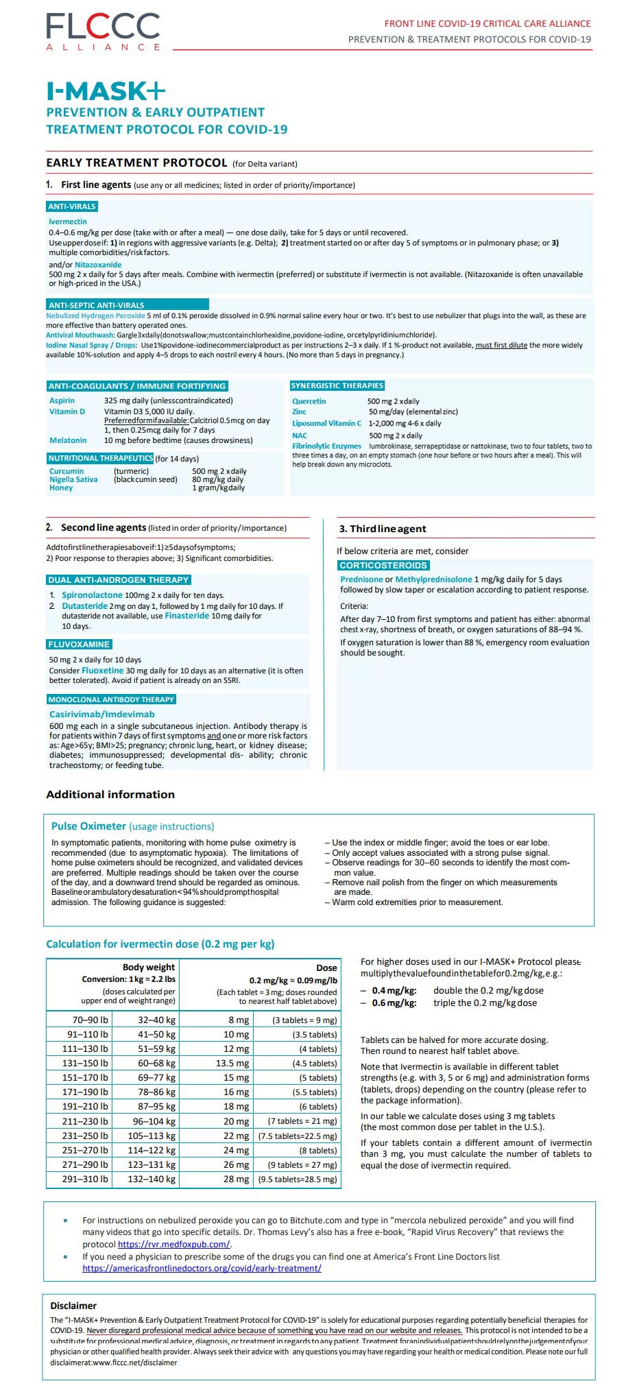
You can find a listing of doctors who can prescribe ivermectin and other necessary medicines on the FLCCC website. There, you can also find downloadable PDFs in several languages for prevention and early at-home treatment, the in-hospital protocol and long-term management guidance for long-haul COVID-19 syndrome. Three other protocols that have great success are:
This is a load of information to review, especially if you are fatigued and sick with COVID or have a family member struggling. So, I reviewed all the protocols and believe the FLCCC one is the easiest and most effective to follow. I’ve posted it below.
However, I’ve altered some of the dosages, and added a few more therapies that they have yet to include, such as:
| Nebulize hydrogen peroxide 5 ml of 0.1% peroxide dissolved in 0.9% normal saline every hour or two. It’s best to use nebulizer that plugs into the wall, as these are more effective than battery operated ones. |
| Intravenous ozone administered by a trained ozone physician. |
| NAC 500 mg twice a day. |
| Make sure the honey is raw honey, not normal honey from the grocery store. Raw honey can be obtained online or at a health food store. |
| Fibrinolytic enzymes like lumbrokinase, serrapeptidase or nattokinase, two to four tablets, two to three times a day, on an empty stomach (one hour before or two hours after a meal). This will help break down any microclots. |
| Decrease zinc dose from 100 mg to 50 mg elemental zinc, but only for three days, then decrease to 15 mg elemental zinc. |
| Increased quercetin from 250 mg to 500 mg. |
| Change vitamin C to liposomal C 1,000 to 2,000 mg four to six times per day. |
Herbs Can Help Lower Blood Pressure
Reproduced from original article:
https://articles.mercola.com/sites/articles/archive/2021/11/26/herbs-and-spices-lower-blood-pressure.aspx
The original Mercola article may not remain on the original site, but I will endeavor to keep it on this site as long as I deem it to be appropriate, and will not be bullied into removing it.
Analysis by Dr. Joseph Mercola Fact Checked November 26, 2021
STORY AT-A-GLANCE
- A controlled feeding study of people with known risk factors for heart disease showed eating 6.5 grams of 24 herbs and spices each day lowered systolic blood pressure
- Heart disease has ranked No. 1 in the top 10 leading causes of death in the U.S. for years; the number of deaths has jumped 9% in just six short years
- Vitamin D deficiency has been associated with high blood pressure in adults, and low levels in infants and children are predictive of high blood pressure in later childhood and teen years
- Salt has been vilified as a primary cause of high blood pressure. Yet, evidence shows it is the sodium/potassium ratio that has a greater impact on blood pressure
- Additional strategies that help lower blood pressure include exercise, stress reduction, inspiratory muscle strength training, meditation and sauna bathing
Heart disease has ranked No. 1 in the top 10 leading causes of death in the U.S. for years. In 2020,1 690,882 people died from heart disease. This is compared to 633,842 who died in 2015, which represents a 9% jump in six short years. One of the key risk factors for heart disease is high blood pressure.2 New data show a diet rich in herbs and spices may help reduce high blood pressure.3
Blood pressure measurements are expressed as two numbers.4 The top number is called the systolic number and the bottom is called the diastolic number. These numbers represent the pressure measured in millimeters of mercury (mmHg) required to move blood through your arteries. The top number is how much pressure is on the artery as the heart beats and the diastolic number is how much pressure remains in the artery between heart beats.
In 2017, the American College of Cardiology,5 in collaboration with the American Heart Association,6 published new guidelines that defined high blood pressure. This moved the measurement to diagnose high blood pressure from 140/90 to 130/80 mmHg.7
Researchers had been noticing a rise in heart disease in individuals whose blood pressure was previously thought to be within normal limits. With the changed guidelines, the American Heart Association8 estimated more than 100 million Americans have high blood pressure.
High blood pressure increases the risk of heart attack, kidney disease, vision loss, stroke and damaged blood vessels.9 New data published in the American Journal of Clinical Nutrition demonstrate how closely associated your diet is to your blood pressure measurement.10
24 Herbs and Spices Over Four Weeks Lowered Blood Pressure
Researchers at Pennsylvania State University11 sought to understand the effect that eating herbs and spices would have on cardiovascular disease. According to the researchers,12 this was the first controlled feeding study to evaluate mixed herbs and spices in a traditional U.S. diet against the risk factors for heart disease.
The researchers recruited 71 people with known risk factors for heart disease. Of those, 63 participants completed the controlled feeding study. The researchers used blood samples and blood pressure to gauge the effect of a low (0.5 grams), moderate (3.2 grams) and high (6.5 grams) intake of herbs and spices.
The participants consumed each diet in random order for a period of four weeks with a two-week washout between each diet.13 The remainder of the diet was based on the average American intake. The herbs and spices included basil, thyme, cinnamon and turmeric.
The researchers discovered that those consuming the diet high in herbs and spices had lower systolic blood pressure then those who consumed the diet with medium or low-dose herbs and spices. The participants wore a blood pressure monitor for 24 hours at the start of the study and at the end of each treatment period.
The researchers were excited by the results because the study did not specifically change the diet to be heart healthy. The only difference was in the number of herbs and spices consumed by the participants. Penny M. Kris-Etherton, professor of nutritional sciences at Penn State Evan Pugh University,14 said in a press release:15
“I think it’s really significant that participants consumed an average American diet throughout the study and we still found these results. We didn’t decrease sodium, we didn’t increase fruits and vegetables, we just added herbs and spices. It begs the next question that if we did alter the diet in these ways, how much better would the results be?”
Vitamin D Deficiency May Be Linked to High Blood Pressure
Several important factors influence your blood pressure, and your vitamin D level is one of them.16 Researchers have found vitamin D insufficiency and deficiency are associated with high blood pressure in adults and now find that low levels in infants and children can increase the risk of high blood pressure later in childhood and during the teen years.17
Vitamin D plays a significant role in several health conditions, and it may be one of the simplest solutions to a wide range of problems. Optimally, you’ll want to obtain vitamin D through sun exposure. However, since many dermatologists and other agencies18,19,20 began telling people to avoid the sun and use liberal amounts of sunscreen, vitamin D deficiency has reached epidemic proportions.21,22,23
In the U.K., the optimal level of vitamin D is 20 ng/mL (50 nmol/L) and higher.24 However, in the U.S., sufficient levels are between 30 ng/mL (75 nmol/L) and 60 ng/mL (150 nmol/L)25 or from 40 ng/mL (100 nmol/L) to 60 ng/mL (150 nmol/L).26 One study27 published in 2018 found 39.92% of the people had a vitamin D level of 20 ng/mL or less and 60.08% had levels of 20 ng/mL or greater.
Since the lowest sufficient level is 30 ng/mL, at least 40% of the population surveyed were deficient in vitamin D, and likely higher. Evidence suggests that low levels are associated with high blood pressure. One literature review28 of 30 randomized clinical trials and 4,744 participants found that vitamin D3 could help reduce systolic and diastolic blood pressure. The effect appeared dependent on the dose, duration and population.
A second review29 of 17 trials with 1,687 participants found supplementation with vitamin D had a statistically significant difference in reducing systolic and diastolic pressure in people who were vitamin D deficient and had high blood pressure.
Low levels of vitamin D also appear to have a predictive value in children. Researchers30 followed 775 children in ages ranging from birth to age 18 from 2005 to 2012 to investigate the effect vitamin D had on the development of high systolic blood pressure. Low vitamin D status was defined as less than 11 ng/mL at birth and less than 25 ng/mL during early childhood.
The researchers31 compared those with low levels of vitamin D to children who were born with adequate levels. They found that children with low levels had about a 60% higher risk of elevated systolic blood pressure from ages 6 to 18. Children who experienced persistently low levels throughout childhood were at double the risk of elevated systolic blood pressure between ages 3 and 18.32
Take Care With Your Sodium/Potassium Ratio
In the U.S. and many other developed countries, salt has been vilified as a primary cause of high blood pressure.33 The idea is with more salt, your body retains more fluid and therefore increases the work of the heart. According to research presented at the American Heart Association meeting in 2013,34 excessive salt contributed to 2.3 million heart-related deaths worldwide in 2010.
However, it is important to understand that sodium and potassium work together to affect your blood pressure. The average reported intake of potassium from food is about half35 of the 4,700 mg recommended.36 Research has demonstrated these low levels of potassium may have a significant impact on blood pressure,37,38,39 especially as it relates to the amount of salt normally found in the Western diet.
Potassium works to relax the walls of your arteries, which keeps your muscles from cramping and lowers your blood pressure.40 Reduction in blood pressure with added potassium has also been associated with a reduced risk of stroke and all-cause mortality.41
It’s recommended that you consume in the range of two to three times more potassium than sodium, depending on whether you currently have a heart condition or diabetes.42,43 But most Americans consume more sodium than potassium.
If researchers are looking only at sodium levels and not the ratio, which is more important than the overall salt intake,44 then it may appear as if salt is driving high blood pressure. Therefore, by lowering your salt intake you automatically improve the ratio.
More Strategies to Help Control Blood Pressure
There are many factors that can positively or negatively affect your blood pressure. As discussed, your diet plays an important role in the nutrients supplied to your arterial system. Several other strategies you may consider include:
| Exercise more — The American Heart Association45 recommends activity and exercise to help manage blood pressure. Studies have also demonstrated the effectiveness of exercise, including as a preventive strategy.46 Regular activity47 can lower blood pressure, reduce heart rest and prevent remodeling from high blood pressure that is pathological and increases the risk of heart failure and mortality. |
| Reduce stress — The American Heart Association48 also recommends managing your stress to help control high blood pressure. Stress stimulates the nervous system to produce hormones that cause vasoconstriction.49 This may cause short-term increases in blood pressure.50
Chronic stress can also affect sleep patterns,51 dietary habits52 and motivation to exercise,53 all of which are key factors in raising blood pressure.54 Acute stress is also associated with broken heart syndrome, which is a potentially life-threatening condition that mimics a heart attack.55 |
| Try inspiratory muscle strength training — One study56 showed using high resistance inspiratory muscle strength training (IMST) could reduce blood pressure measurements as well as aerobic exercise or meditation. IMST was originally developed for critically ill patients with respiratory diseases. The strategy uses a handheld device that provides resistance to the user as they inhale vigorously, thus strengthening muscles. |
| Incorporate meditation — Mind-body practices that trigger your body’s relaxation response,57 such as meditation, play an important role in lowering blood pressure by favorably influencing a recently identified set of genes and biological pathways.58 As the relaxation response is elicited, biochemical changes occur, including decreased oxygen consumption, blood pressure, heart and respiratory rate.59 |
| Use intermittent fasting — This is a form of time restricted eating during which you typically fast for 16 to 18 hours with a window of six to eight hours to eat. Evidence shows that there are several benefits to the cardiovascular system, including lowering blood pressure.60 A recent study61 from Baylor College of Medicine suggested that fasting may help normalize blood pressure by impacting the gut microbiota. |
| Sauna bathing — Sometimes some of the simplest strategies can have a tremendous impact. Sweating in a sauna can help expel toxins, improve blood circulation and improve mitochondrial function. In a video lecture by Rhonda Patrick, Ph.D.,62 she reviews how sauna bathing can be used as an exercise mimetic to increase your longevity.
In this lecture she notes that just a single sauna session has been shown to lower blood pressure, improve heart rate variability, and improve arterial compliance. Some of the positive benefits of the sauna on heart health may have to do with similar physiological changes that also occur during physical exercise. |
| Check your magnesium — Magnesium deficiency can contribute to a significant number of health problems since it is involved in hundreds of biochemical reactions in the body.63 One scientific review64 suggested that low magnesium may be the greatest predictor of heart disease and another65 suggested subclinical deficiency can compromise cardiovascular health.
Low magnesium levels have been linked to a high risk of high blood pressure,66 stroke67 and sudden cardiac death.68 The best way to determine your status is to do an RBC magnesium test. This measures the amount of magnesium in your red blood cells. There are several reasons why you may have insufficient or deficient levels of magnesium, including not getting enough from your diet, sweating, stress and lack of sleep. Seek to eat more magnesium-rich foods and consider high-quality magnesium supplements if necessary. Another way of effectively boosting your level is to use an Epsom salt bath as the magnesium is effectively absorbed through your skin. |
- 1 JAMA Network, 2021; 325(18)
- 2 CDC Heart Disease Facts, Americans at Risk for Heart Disease
- 3, 10, 12 The American Journal of Clinical Nutrition, 2021; doi.org/10.1093/ajcn/nqab291
- 4 American Heart Association, Understanding Blood Pressure Readings, Your blood pressure numbers and what they mean
- 5 American College of Cardiology, May 7, 2018
- 6 Hypertension, 2018;71
- 7 Centers for Disease Control and Prevention, Facts About Hypertension
- 8 American Heart Association, January 31, 2018
- 9 My Cleveland Clinic, What Can Happen if High Blood pressure is not treated?
- 11, 13, 15 EurekAlert! November 8, 2021
- 14 Penn State, Evan Pugh University Professors
- 16, 24 Express, November 15, 2021
- 17, 30 Hypertension, 2019;doi.org/10.1161/HYPERTENSIONAHA.119.13120
- 18 American Academy of Dermatology, Vitamin D Stats and Facts
- 19 UCSF Magazine, Summer 2018
- 20 Centers for Disease Control and Prevention, Sun Safety
- 21 Journal of Bone & Joint Surgery, 2010;92(13):2300 results
- 22 American Journal of Clinical Nutrition, 2008;87(4):1080S
- 23 International Journal of Health Sciences; 2010;4(1)
- 25 ZRT Lab, January 10, 2018
- 26 Grassroots Health, Are You Vitamin D Deficient?
- 27 Cureus, 2018;10(6)
- 28 Nutrition Metabolism and Cardiovascular Disease, 2016;26(8)
- 29 Medicine, 2019;98(19)
- 31, 32 American Heart Association, July 1, 2019
- 33 Nutrients, 2019;11(9)
- 34 Science Daily, March 21, 2013
- 35 USDAFood Surveys Research Group, September 9, 2012
- 36 National Institutes of Health, Potassium, 2nd line under table 2
- 37 Journal of the American Medical Association 1997;277(20):1624
- 38 Journal of Hypertension, 2015;33(8)
- 39 Cell Metabolism, 2015;21(1)
- 40 Harvard Health Publications, January 23, 2017
- 41 Stroke 2014; 45(10):2874
- 42 Autoimmune Association, August 5, 2013
- 43 The American Journal of Clinical Nutrition, 2012;96(2)
- 44 Advances in Nutrition, 2014; 5:712
- 45 American Heart Association, Getting Active to Control High Blood Pressure
- 46 Arq Bras Cardiol. 2016; 106(5)
- 47 Current Hypertension reports, 2015;17(10)
- 48 American Heart Association, Managing Stress to Control High Blood Pressure
- 49 WMJ, 1998;97(11)
- 50 Blood Pressure UK, Stress and Your Blood Pressure
- 51 Sleep Foundation, Stress and Insomnia
- 52 Harvard Health Publishing, February 15, 2021
- 53 Sports Medicine, 2014;44(1) Ab/Concl
- 54 CDC, Prevent High Blood Pressure subheads
- 55 Medical Xpress, October 13, 2021
- 56 Journal of the American Heart Association, 2021;10:e020980
- 57 The Journal of Alternative and Complementary Medicine, 2018;24(5)
- 58 Science Daily, April 4, 2018
- 59 PLOS One May 1, 2013, Intro para 2
- 60 Nutrients, 2019;11(3)
- 61 Science Daily, April 29, 2021
- 62 YouTube, September 10, 2019 Minute 6:54, 11:09, 12:53, 13:56
- 63 NIH.gov Magnesium Fact Sheet for Professionals
- 64 New Hope Network January 31, 2013
- 65 Open Heart 2018:e000668 (PDF)
- 66 Hypertension, 2016;68
- 67 BMC Medicine, 2016;14(210)
- 68 American Heart Journal, 2010;160(3)
Summary of Recent Research with Three Nutrients in the War Against Cancer
Reproduced from original OMNS article (OrthoMolecular News Service):
Subscribe to the free Orthomolecular Newsletter: http://orthomolecular.org/subscribe.html
Go to the OMNS Archive: http://orthomolecular.org/resources/omns/index.shtml
This site is non-profit, existing only to help people improve health and immunity
Updated 19th November 2021
Orthomolecular Medicine News Service, November 11, 2021
by Michael Passwater
OMNS (Nov. 11, 2021) We have much to learn about preventing and treating cancer. Each disease in the cohort of cancers is potentially a different disease related to the specific genetic mutations that drive it. In the interest of promoting further efforts to improve our understanding of these complex diseases, here is an update on research involving three essential nutrients: vitamin C, selenium (selenite and methyl selenocysteine), and vitamin E (delta tocotrienol). Although much progress has been made in the fight against some types of cancer, many types still remain literally a death sentence. Each year, more than 1.3 million people are diagnosed with one of the ten most deadly types of cancer in the United States and over 400,000 people die from these dreaded diseases.
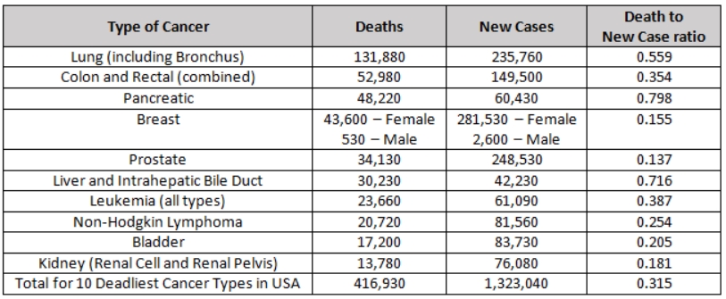
Ascorbic Acid (Vitamin C)
By definition, a vitamin is essential for life, and vitamin C (ascorbic acid) is no exception. The health benefits of vitamin C are so extensive that it would be an important tool in the battle against cancer even without its powerful anti-tumor effect. Vitamin C is important for many aspects of immune and endothelial health; synthesis of catecholamines, carnitine, neuropeptides, neurotransmitters, collagen, and elastin; breakdown of L-tyrosine and hypoxic inducible factor 1-alpha (HIF-1a); epigenomic regulation; somatic stem cell reprogramming; and redox regulation including scavenging damaging free radicals, breaking chains of lipid peroxidation, and recycling other antioxidants. [2-8]
In 1976, Cameron and Pauling reported longer survival in terminal cancer patients given vitamin C. [9] Was this from improvement to the patients’ constitution, was there an anti-tumor effect, or both? We now know that high doses of vitamin C can have a prooxidant effect, especially in combination with iron, which can selectively kill tumor cells containing less catalase than healthy human cells. [10, 11] The catalase enzyme is necessary for nearly all cells to remove hydrogen peroxide that can cause free radicals and damage the cell’s biomolecules, including proteins and DNA. And vitamin C can also disrupt the “Warburg Effect” in tumor cells. [12,13,14] The Warburg Effect is the tendency of tumor cells to switch from using mitochondrial oxidative phosphorylation to aerobic glycolysis for the production of cellular energy (ATP). While greatly reducing the efficiency of energy production (2 ATP molecules generated per glucose molecule vs. 36 ATP molecules generated per glucose molecule), aerobic glycolysis enhances the ability of the cell to proliferate. Efficiency is only important when resources are scarce. In a glucose rich environment, 2 ATP molecules per glucose will work just fine, and allows more of the glucose, along with the amino acid glutamine, to provide the structures necessary to make additional tumor cells. This primitive, yet streamlined, tumor metabolism allows abundantly available glucose and glutamine to supply the biochemical needs of cell growth and division. Many tumor cells over-express glucose transporters, particularly GLUT1 to increase intake of glucose. Ascorbic acid, and especially oxidized ascorbic acid (dehydroascorbic acid) is similar in molecular shape to glucose, and can enter cells through these membrane transport channels. Not only does this disrupt the supply of glucose to the tumor cell, it also allows increased vitamin C concentrations within the cell where it can cause epigenetic effects including increasing TET enzyme activity, re-expression of tumor suppressor genes, or cause cell death through metabolic disruption. Depletion of intracellular glutathione leads to inactivation of glyceraldehyde 3-phosphate dehydrogenase and inhibition of glycolysis. In the setting of high vitamin C concentrations, the reliance of some tumor cells on aerobic glycolysis becomes their “Achilles’ heel”. [15]
In recent years, cell culture studies and human clinical trials have confirmed that tumor cells with KRAS and BRAF mutations can be selectively killed by appropriate doses of vitamin C. [16] KRAS and BRAF mutations are common in solid tumors, especially pancreatic, colon, and lung cancers. Synergistic effects have been seen by combining vitamin C with some chemotherapy drugs, radiation treatment, or in combination with a fasting-mimicking diet. [17-23] Mark Levine, Channing Paller, Tami Tamashiro, Thomas Luechtefeld and Amy Gravell recently reviewed 53 cancer clinical trials involving IV and/or oral vitamin C. [15] These trials give a clear signal of safety when patients with glucose 6 phosphate dehydrogenase (G6PD) deficiency are excluded from high dose IVC administration. Most studies were small, and involved a wide variety of late stage cancers. Nonetheless, encouraging signals were seen, including a few patients surviving years later eventually dying of causes other than cancer. Studies involving pancreatic ductal adenocarcinoma (PDAC) show a median survival following diagnosis of 3.5 months untreated, 6.7 months when treated with gemcitabine, 8.5 – 13 months when treated with gemcitabine and nab-paclitaxel, and 15.1 months when IVC with or without gemcitabine was used. [17,24] It is likely that with refinement of dosing and route of administration, earlier intervention, and improved knowledge of which tumors are most susceptible to high dose vitamin C therapy, more consistent positive results will be seen.
Three promising active clinical trials:
A Phase II Trial of Pharmacological Ascorbate, Gemcitabine, and Nab-Paclitaxel for Metastatic Pancreatic Cancer (PACMAN 2.1; University of Iowa Holden Comprehensive Cancer Center) [25]
A Phase II Study of High Dose Vitamin C Intravenous Infusion in Patients with Resectable or Metastatic Solid Tumor Malignancies (Weill Cornell Medicine, NYC) [26]
To download an informative description of “IV Vitamin C for Cancer Care”, please click https://riordanclinic.org/wp-content/uploads/2017/09/IVChandout.pdf.
(Riordan Clinic, Kansas, >250,000 IVC treatments provided) [27]
Selenium
Selenium is a micronutrient essential for human health. There are 25 human selenoproteins known to be involved in numerous functions throughout the body, including brain, blood vessel, heart, and immune system health. These proteins perform diverse functions including antioxidant and redox recycling, gene “proof-reading”, vitamin D metabolism, and hemostasis. Like vitamin C, even if there were no direct tumor effect of selenium, it would be an important tool in the battle for wellness and against cancer. [28-31]
HLA-E is a “camouflage protein” expressed by some tumors to hide from the immune system. The protein fools the immune system into thinking the tumor cell is a normal cell. A decade ago, selenite was shown to reduce – nearly eliminate – HLA-E expression on tumor cells. The tumor cells then became susceptible to destruction by Natural Killer cells (immune cells which clear unhealthy cells from the body). [32] A recent study of 243 PDAC patients showed that higher HLA-E and HLA-G (a similar protein) expression was associated with shorter survival. [33] In addition to optimizing tissue health throughout the body, the reduction of camouflage membrane proteins HLA-E and HLA-G by selenite may be a useful tool in the battle against cancer. Of note, selenomethionine (SeMet) had no effect on tumor cell HLA-E expression. Selenomethionine was the form of selenium used in the SELECT trial which did not show a benefit of selenium against prostate cancer prevention. In addition to the unfortunate choice of selenium supplement, the trial had other flaws, including a lack of difference in selenium levels between the control and test groups. Methyl selenocysteine (MSC) was the form of selenium used in the Nutritional Prevention of Cancer (NPC) trial which showed benefit. MSC is also the dominant form of selenium in broccoli, cabbage, onions and garlic. In 2014, MSC was shown to protect against the toxicity of four chemotherapy agents (cyclophosphamide, cisplatin, oxalplatin, and irinotecan), and enhance antitumor activity. [34]
More recently, a phase I study at Stanford using selenite and radiation therapy in humans validated the further study of selenite against cancer. [35] Additionally, in vitro and in vivo (mice) studies at the University of Grenada showed a strong antitumor effect of selenite against pancreatic cancer, alone and in combination with gemcitabine. [36]
Delta Tocotrienol
Vitamin E is an essential nutrient for humans. Vitamin E is a lipid soluble antioxidant. There are 8 different molecules in the vitamin E family pertinent to humans. It comprises 4 tocopherols (alpha, beta, gamma, delta), and 4 tocotreinols (alpha, beta, gamma, delta). Tocotreinols are smaller than tocopherols and are unsaturated. Tocotrienols have a half-life of approximately 4 hours while tocopherols have a half-life of 20 hours. Delta tocotrienol (VEDT = Vitamin E Delta Tocotrienol) is the smallest member of the vitamin E family, as it has the shortest “tail”. Its small size allows delta tocotrienol greater mobility within lipid layers of cell membranes.
Studies have shown that tocotreinols inhibit Nuclear factor kappa B (NF-kB) activity and human pancreatic cancer cell survival, with VEDT having the strongest effect. [37] NF-kB is involved in immune and inflammatory responses, and in the regulation of cellular gene expression, proliferation, differentiation, and survival. VEDT has also been shown to enhance gemcitabine activity in pancreatic cancer cells. [38-40] Furthermore, VEDT has been shown to inhibit PDAC stem-like cells. VEDT significantly inhibited growth and metastases of these cells, including inhibiting the growth and metastasis of gemcitabine resistant PDAC stem-cell-like cells. [41] A phase I human clinical trial at Moffitt Cancer Center in Tampa, Florida showed 200 to 1600 mg VEDT taken orally daily for 2 weeks was well tolerated and significantly induced apoptosis (cell death) in pancreatic ductal cancer cells. [42] An anti-tumor effect, as well as a reduction in side effects from chemotherapy, has also been shown in the setting of other solid tumors. [43-56]
Note that to prevent the larger tocopherols from blocking absorption of the smaller tocotreinols, it is best to avoid taking tocopherols at the same time as tocotrienols. [57-63] Twice daily dosing of tocotrienols have been shown to produce a steady state after three days.
Summary
The redox synergy of vitamin C, vitamin E, and selenium-containing glutathione peroxidases has been explored since the 1960s and 1970s. Studies focusing on single nutrient interventions, with and without chemotherapy and radiation, have suggested benefit in humans, with increasingly specific mechanistic and treatment details discovered each decade. Along the way, a reassuring safety profile of nutrient interventions with and without traditional chemotherapy and radiation interventions has been established. Further studies to better define the most effective match of treatment for each tumor type, along with the best route, dose, and combination of treatments are important next steps to improve the reliability and effectiveness of preventing and treating the many types of cancer. Combining nutrients known to be synergistic together, while limiting glucose and glutamine central to the metabolism of many tumor cells, may optimize the effectiveness of treatment strategies.
With nutrient therapy – orthomolecular medicine – one does not need to choose between attacking the tumor and strengthening the host. The treatments often impact both simultaneously.
Foundational Support
NAD deficiency is associated with greater risk for mutagenesis with cancer and this is likely best avoided using daily niacin, e.g. start with 3×25-100mg/d to adapt to the flush, and then slowly work up to 3×500-1,000mg/d. [64] You can start by purchasing 100 mg tablets and break them into halves or quarters at first.
For cancer patients, chemotherapy commonly causes NAD deficiency, which is best rescued with niacinamide; e.g. 3x500mg/d. [64]
Dietary relevance, glutamine restriction with niacinamide; glucose restriction and ketogenic diet is recommended. [64]
Vitamin D3 to maintain plasma vitamin D > 40 ng/ml. Test a dose of 125 mcg/d (5000 IU) D3 with 100 mcg/d K2 for 4 months before getting a blood test for vitamin D.
100 – 200 mcg/d selenium as methyl-selenocysteine or selenium yeast
300 mg/d alpha lipoic acid
1-2 g vitamin C with each meal
Exercise
Cultivate a cheerful heart [65]
Advanced Support
Vitamin C in divided doses throughout the day to bowl tolerance; consider mixtures of lipid- and water-soluble forms
IVC if G6PD levels are adequate and IVC is available from a licensed medical provider [66]
Sodium selenite if available from a licensed medical provider
Delta tocotrienol 300 – 1600 mg/d [42]
(Michael Passwater is certified by the American Society for Clinical Pathology as a Medical Technologist, a specialist in Immunohematology, and a diplomate in Laboratory Management. He has worked in clinical laboratories for 28 years, and has his degree in Medical Technology from the University of Delaware.)
References & Further Reading
1. National Cancer Institute. Common Cancer Types. https://www.cancer.gov/types/common-cancers
2. Oudemans-van Straaten HM, Spoelstra-de Man AME, de Waard MC. (2014) Vitamin C revisited. Critical Care 18:460-473. https://pubmed.ncbi.nlm.nih.gov/25185110
3. Manning J, Mitchell B, Appaduras DA, May JM, et al. (2013) Vitamin C Promotes Maturation of T-Cells. Antioxid Redox Signal. 19:2054-2067. https://pubmed.ncbi.nlm.nih.gov/23249337
4. Ladumer A, Schmitt CA, Schachner D, et al. (2012) Ascorbate stimulates endothelial nitric oxide synthase enzyme activity by rapid modulation of its phosphorylation status. Free Radic Biol Med. 2012 May 15; 52:2082-2090. https://pubmed.ncbi.nlm.nih.gov/22542797
5. May JM, Qu ZC. (2010) Ascorbic Acid Prevents Increased Endothelial Permeability Caused by Oxidized Low Density Lipoprotein. Free Radical Res. 44:1359-1368. https://pubmed.ncbi.nlm.nih.gov/20815791
6. Deicher R, Ziai F, Begknayer C, et al. (2005) Low Total Vitamin C Plasma Level Is a Risk Factor for Cardiovascular Morbidity and Mortality in Hemodialysis Patients. J Am Soc Nephrol. 16:1811-1818. https://pubmed.ncbi.nlm.nih.gov/15814831
7. Heller R, Munscher-Paulig F, Grabner R, Till V. (1999) L-Ascorbic Acid Potentiates Nitric Oxide Synthesis in Endothelial Cells. J Biol Chem, 274:8254-8260. https://pubmed.ncbi.nlm.nih.gov/10075731
8. Leibovitz B, Siegel BV. (1978) Ascorbic acid, neutrophil function, and the immune response. Int J Vitam Nutr Res. 48:159-164. https://pubmed.ncbi.nlm.nih.gov/357320
9. Cameron E, Pauling L. (1976) Supplemental ascorbate in the supportive treatment of cancer: Prolongation of survival times in terminal human cancer. Proc Natl Acad Sci USA. 73: 3685-3689. https://pubmed.ncbi.nlm.nih.gov/1068480
10. Schoenfeld JD, Sibenaller ZA Mapuskar KA, et al (2017) O2- and H2O2-Mediated Disruption of Fe Metabolism Causes the Differential Susceptibility of NSCLC and GBM Cancer Cells to Pharmacological Ascorbate. Cancer Cell 31: 487-500. https://pubmed.ncbi.nlm.nih.gov/28366679
11. O’Leary BR, Alexander MS, Du J, et al (2020) Pharmacological ascorbate inhibits pancreatic cancer metastases via a peroxide-mediated mechanism. Sci Rep 10:17649 https://pubmed.ncbi.nlm.nih.gov/33077776.
12. Aguilera O, Muñoz-Sagastibelza M, Torrejón E, et al. (2016) Vitamin C uncouples the Warburg metabolic switch in KRAS mutant colon cancer. Oncotarget, 7:47954-47965. https://pubmed.ncbi.nlm.nih.gov/27323830
13. Suzuki T, Kishikawa T, Sato T, et al. (2021) Mutant KRAS drives metabolic reprogramming and autophagic flux in premalignant pancreatic cells. Cancer Gene Ther. Online ahead of print. https://pubmed.ncbi.nlm.nih.gov/33833413
14. Gonzalez MJ, Seyfried T; Nicolson GL et al. (2018) Mitochondrial Correction: A New Therapeutic Paradigm for Cancer and Degenerative Diseases. J Orthomolecular Med 33: 4. https://riordanclinic.org/journal-article-archive/mitochondrial-correction-a-new-therapeutic-paradigm-for-cancer-and-degenerative-diseases
15. Chen Q, Vissers MCM (2020) Cancer and Vitamin C. CRC Press. ISBN-13: 978-0367858049
16. Yun J, Mullarky E, Lu C, et al. (2015) Vitamin C selectively kills KRAS and BRAF mutant colorectal cancer cells by targeting GAPDH. Science 350:1391-1396. https://pubmed.ncbi.nlm.nih.gov/26541605
17. Polireddy K, Dong R, Reed G, et al. (2017) High Dose Parenteral Ascorbate Inhibited Pancreatic Cancer Growth and Metastasis: Mechanisms and a Phase I/IIa study. Sci Rep 7: 17188 https://pubmed.ncbi.nlm.nih.gov/29215048
18. Alexander MS, Wilkes JG, Schroeder SR, Buettner GR. (2018) Pharmacologic ascorbate reduces radiation-induced normal tissue toxicity and enhances tumor radiosensitization in pancreatic cancer. Cancer Res. 78:6838-6851. https://pubmed.ncbi.nlm.nih.gov/30254147.
19. Drisko JA, Serrano OK, Spruce LR, Chen Q, Levine M. (2018) Treatment of pancreatic cancer with intravenous vitamin C: a case report. Anti-Cancer Drugs 29:373-379. https://pubmed.ncbi.nlm.nih.gov/29438178
20. Welsh JL, Wagner BA, van’t Erve TJ, et al. (2013) Pharmacological ascorbate with gemcitabine for the control of metastatic and node-positive pancreatic cancer (PACMAN): results from a phase I clinical trial. Cancer Chemother. Pharmacol. 71:765-775. https://pubmed.ncbi.nlm.nih.gov/23381814
21. Monti DA, Mitchell E, Bazzan AJ, et al. (2012) Phase I evaluation of intravenous ascorbic acid in combination with gemcitabine and erlotinib in patients with metastatic pancreatic cancer. PLoS ONE 7(1):e29794. https://pubmed.ncbi.nlm.nih.gov/22272248
22. Mikirova N, Casciari J, Hunninghake R. (2019) Continuous Intravenous Vitamin C in the Cancer Treatment: Reevaluation of a Phase I Clinical Study. Functional Foods in Health and Disease. https://riordanclinic.org/journal-article-archive/continuous-intravenous-vitamin-c-in-the-cancer-treatment-reevaluation-of-a-phase-i-clinical-study
23. Di Tano M, Raucci F, Vernieri C, et al. (2020) Synergistic effect of fasting-mimicking diet and vitamin C against KRAS mutated cancers. Nat Comm 11:2332 https://pubmed.ncbi.nlm.nih.gov/32393788
24. Carr AC, Cook J. (2018) Intravenous Vitamin C for Cancer Therapy — Identifying the Current Gaps in Our Knowledge. Front. Physiol. 9:1182. https://pubmed.ncbi.nlm.nih.gov/30190680
25. Cullen JJ (2021) A Phase II Trial of Pharmacological Ascorbate, Gemcitabine, and Nab-Paclitaxel for Metastatic Pancreatic Cancer (PACMAN 2.1) https://clinicaltrials.gov/ct2/show/NCT02905578
26. Shah MA, Khan U. (2021) A Phase II Study of High Dose Vitamin C Intravenous Infusion in Patients with Resectable or Metastatic Solid Tumor Malignancies. https://jcto.weill.cornell.edu/open_clinical_trials/a-phase-ii-study-of-high-dose-vitamin-c-intravenous-infusion-in-patients-with-resectable-or-metastatic-solid-tumor-malignancies
27. Riordan Clinic (2021) High Dose IV Vitamin C (IVC) https://riordanclinic.org/what-we-do/high-dose-iv-vitamin-c
28. Grimble RF. (2001) Nutritional modulation of immune function. Proc. Nutr. Soc. 60:389-397. https://pubmed.ncbi.nlm.nih.gov/11681814
29. Arthur JR, McKenzie RC, Beckett GJ. (2003) Selenium in the immune system. J. Nutr. 133:1457S-1459S. https://pubmed.ncbi.nlm.nih.gov/12730442
30. Guillin OM, Vindry C, Ohlmann T, Chavatte L. (2019) Selenium, Selenoproteins, and Viral Infection. Nutrients, 11:2101. https://pubmed.ncbi.nlm.nih.gov/31487871
31. Huang Z, Rose AH, Hoffman PR. (2012) The Role of Selenium in Inflammation and Immunity: From Molecular Mechanisms to Therapeutic Opportunities. Antioxid Redox Signal. 16:705-743. https://pubmed.ncbi.nlm.nih.gov/21955027
32. Enqvist M, Nilsonne G, Hammarfjord O, et al, (2011) Selenite Induces Posttranscriptional Blockade of HLA-E Expression and Sensitizes Tumor Cells to CD94/NKG2A-Positive NK Cells. J Immunol. 187:3546-3554. https://pubmed.ncbi.nlm.nih.gov/21890659
33. Hiraoka N, Ino Y, Hori S, et al. (2020) Expression of classical human leukocyte antigen class I antigens, HLA-E and HLA-G, is adversely prognostic in pancreatic cancer patients Cancer Sci. 111:3057-3070. https://pubmed.ncbi.nlm.nih.gov/32495519
34. Cao S, Durrani FA, Tóth K, Rustum YM. (2014) Se-methylselenocysteine offers selective protection against toxicity and potentiates the antitumour activity of anticancer drugs in preclinical animal models Br J Cancer 110:1733-1743. https://pubmed.ncbi.nlm.nih.gov/24619073
35. Knox SJ, Jayachandran P, Keeling CA, et al. (2019) Results from a Phase 1 Study of Sodium Selenite in Combination with Palliative Radiation Therapy in Patients with Metastatic Cancer. Transl Oncol. 12:1525-1531. https://pubmed.ncbi.nlm.nih.gov/31454725
36. Doello K, Mesas C, Quiñonero F, et al. (2021) The Antitumor Activity of Sodium Selenite Alone and in Combination with Gemcitabine in Pancreatic Cancer: An In Vitro and In Vivo Study. Cancers. 13:3169. https://pubmed.ncbi.nlm.nih.gov/34201986
37. Husain K, Francois RA, Yamauchi T, et al. (2011) Vitamin E delta-tocotrienol augments the antitumor activity of gemcitabine and suppresses constitutive NF-kappaB activation in pancreatic cancer. Mol Cancer Ther. 10:2363-2372. https://pubmed.ncbi.nlm.nih.gov/21971120
38. Malafa MP, Sebti S, (2014) Delta-Tocotrienol Treatment and Prevention of Pancreatic Cancer. Lee Moffitt Cancer Center & Research Institute, University of South Florida (Tampa): US Patent US 8,846,653. https://patentimages.storage.googleapis.com/c7/ff/ef/b836e04b18be57/US8846653.pdf
39. Hussein D, Mo H. (2009) d-Delta-tocotrienol-mediated suppression of the proliferation of human PANC-1, MIA PaCa-2, and BxPC-3 pancreatic carcinoma cells. Pancreas. 38:e124-e136. https://pubmed.ncbi.nlm.nih.gov/19346993
40. Husain K, Centeno BA, Chen D-T, et al. (2013) Vitamin E delta-tocotrienol prolongs survival in the LSLKrasG12D/+;LSL-Trp53R172H/+;Pdx-1-Cre (KPC) transgenic mouse model of pancreatic cancer. Cancer Prev Res (Phila). 6:1074-83. https://pubmed.ncbi.nlm.nih.gov/23963802
41. Husain K, Centeno BA, Coppola D, et al. (2017) d-Tocotrienol, a natural form of vitamin E, inhibits pancreatic cancer stem-like cells and prevents pancreatic cancer metastasis. Oncotarget. 8:31554-31567. https://pubmed.ncbi.nlm.nih.gov/28404939
42. Springett GM, Husain K, Neuger A, et al. (2015) A Phase I Safety, Pharmacokinetic, and Pharmacodynamic Presurgical Trial of Vitamin E d-tocotrienol in Patients with Pancreatic Ductal Neoplasia EBioMedicine 2:1987-1995. https://pubmed.ncbi.nlm.nih.gov/26844278
43. Guthrie N, Gapor A, Chambers AF, Carroll KK. (1997) Inhibition of proliferation of estrogen receptor-negative MDA-MB-435 and -positive MCF-7 human breast cancer cells by palm oil tocotrienols and tamoxifen, alone and in combination. J Nutr. 127:544S-548S. https://pubmed.ncbi.nlm.nih.gov/9082043
44. Nesaretnam K, Stephen R, Dils R, Darbre P. (1998) Tocotrienols inhibit the growth of human breast cancer cells irrespective of estrogen receptor status. Lipids. 33:461-469. https://pubmed.ncbi.nlm.nih.gov/9625593
45. Shun M-C, Yu W, Gapor A, et al. (2004) Pro-apoptotic mechanisms of action of a novel vitamin E analog (alpha-TEA) and a naturally occurring form of vitamin E (delta-tocotrienol) in MDA-MB-435 human breast cancer cells. Nutr Cancer. 48:95-105. https://pubmed.ncbi.nlm.nih.gov/15203383
46. Kaneko S, Sato C, Shiozawa N, et al. (2018) Suppressive Effect of Delta-Tocotrienol on Hypoxia Adaptation of Prostate Cancer Stem-like Cells. Anticancer Res. 38:1391-1399. https://pubmed.ncbi.nlm.nih.gov/15203383
47. Ji X, Wang Z, Geamanu A, et al. (2012) Delta-tocotrienol suppresses Notch-1 pathway by upregulating miR-34a in nonsmall cell lung cancer cells. Int J Cancer. 131: 2668-2677. https://pubmed.ncbi.nlm.nih.gov/22438124
48. Ji X, Wang Z, Sarkar FH, Gupta SV. (2012) Delta-tocotrienol augments cisplatin-induced suppression of non-small cell lung cancer cells via inhibition of the Notch-1 pathway. Anticancer Res. 32:2647-2655. https://pubmed.ncbi.nlm.nih.gov/22753722
49. Nasr M, Nafee N, Saad H, Kazem A. (2014) Improved antitumor activity and reduced cardiotoxicity of epirubicin using hepatocyte-targeted nanoparticles combined with tocotrienols against hepatocellular carcinoma in mice. Eur J Pharm Biopharm. 88:216-225. https://pubmed.ncbi.nlm.nih.gov/24813390
50. Wada S, Naito Y, Matsushita Y, et al. (2017) Delta-tocotrienol suppresses tumorigenesis by inducing apoptosis and blocking the COX-2/PGE2 pathway that stimulates tumor-stromal interactions in colon cancer. J Funct Foods. 35:428-435. https://www.sciencedirect.com/science/article/pii/S1756464617303183
51. Shibata A, Nakagawa K, Tsuduki T, Miyazawa T. (2015) Delta-Tocotrienol treatment is more effective against hypoxic tumor cells than normoxic cells: potential implications for cancer therapy. J Nutr Biochem. 26:832-840. https://pubmed.ncbi.nlm.nih.gov/25979648
52. Zhang J-S, Li D-M, He N, et al. (2011) A paraptosis-like cell death induced by delta-tocotrienol in human colon carcinoma SW620 cells is associated with the suppression of the Wnt signaling pathway. Toxicology. 285:8-17. https://pubmed.ncbi.nlm.nih.gov/21453743
53. Sun W, Wang Q, Chen B, et al. (2008) Gamma-tocotrienol-induced apoptosis in human gastric cancer SGC-7901 cells is associated with a suppression in mitogen-activated protein kinase signalling. Br J Nutr. 99:1247-1254. https://pubmed.ncbi.nlm.nih.gov/18081943
54. Sun W, Xu W, Liu H, et al. (2009) gamma-Tocotrienol induces mitochondria-mediated apoptosis in human gastric adenocarcinoma SGC-7901 cells. J Nutr Biochem. 20:276-284. https://pubmed.ncbi.nlm.nih.gov/18602811
55. Satyamitra MM, Kulkarni S, Ghosh SP, et al. (2011) Hematopoietic Recovery and Amelioration of Radiation-Induced Lethality by the Vitamin E Isoform delta-Tocotrienol. Radiat Res. 175:736-745. https://pubmed.ncbi.nlm.nih.gov/21434782
56. Constantinou C, Charalambous C, Kanakis D. (2020) Vitamin E and cancer: an update on the emerging role of gamma and delta tocotrienols. Eur J Nutr. 59:845-857. https://pubmed.ncbi.nlm.nih.gov/31016386
57. Drotleff AM, Bohnsack C, Schneider I, et al. (2014) Human oral bioavailability and pharmacokinetics of tocotrienols from tocotrienol-rich (tocopherol-low) barley oil and palm oil formulations. J Funct Foods. 7:150-160. https://www.sciencedirect.com/science/article/pii/S1756464614000024
58. Szewczyk K, Chojnacka A, Górnicka M. (2021) Tocopherols and Tocotrienols — Bioactive Dietary Compounds; What Is Certain, What Is Doubt? Int J Mol Sci. 22:6222. https://pubmed.ncbi.nlm.nih.gov/34207571
59. Trias AM, Tan B. (2013) Alpha-tocopherol: a detriment to tocotrienol benefits. In: Tan B, Watson RR, Preedy VR, eds. Tocotrienols: Vitamin E Beyond Tocopherols. 2nd ed. Boca Raton, FL: CRC Press; 2013. pp 61-78. ISBN-13: 978-1138199729
60. Qureshi AA, Pearce BC, Nor RM, et al. (1996) Dietary alpha-tocopherol attenuates the impact of gamma-tocotrienol on hepatic 3-hydroxy-3-methylglutaryl coenzyme A reductase activity in chickens. J Nutr. 126:389-394. https://pubmed.ncbi.nlm.nih.gov/8632210
61. Shibata A, Kawakami Y, Kimura T, et al. (2016) Alpha-tocopherol attenuates the triglyceride- and cholesterol-lowering effects of rice bran tocotrienol in rats fed a Western diet. J Agric Food Chem. 64:5361-5366. https://pubmed.ncbi.nlm.nih.gov/27295311
62. Shibata A, Nakagawa K, Tsuduki T, Miyazawa T. (2015) Alpha-tocopherol suppresses antiangiogenic effect of delta-tocotrienol in human umbilical vein endothelial cells. J Nutr Biochem, 26:345-50. https://pubmed.ncbi.nlm.nih.gov/25662730
63. Passwater R (2019) More Than Vitamin E: The Story & Science Behind Tocotrienols. Whole Foods Magazine. https://wholefoodsmagazine.com/columns/vitamin-connection/more-than-vitamin-e-the-story-science-behind-tocotrienols-part-1-tocotrienols-no-longer-the-obscure-member-of-the-vitamin-e-family
64. Penberthy WT, Saul AW, Smith RG, (2021) Niacin and Cancer How vitamin B-3 protects and even helps repair your DNA. Orthomolecular Medicine News Service. http://orthomolecular.org/resources/omns/v17n05.shtml
65. Diener E,Chan MY. (2011) Happy People Live Longer: Subjective Well-Being Contributes to Health and Longevity. Applied Psychology: Health and Well-Being. 3:1-43. https://doi.org/10.1111/j.1758-0854.2010.01045.x
66. Yuen RCF (2021) High Dose Vitamin C for Cancer: The Struggle with “Non-Evidence-Based” Medical Practice. Orthomolecular Medicine News Service, http://orthomolecular.org/resources/omns/v17n20.shtml
Nutritional Medicine is Orthomolecular Medicine
Orthomolecular medicine uses safe, effective nutritional therapy to fight illness. For more information: http://www.orthomolecular.org
Find a Doctor
To locate an orthomolecular physician near you: http://orthomolecular.org/resources/omns/v06n09.shtml
The peer-reviewed Orthomolecular Medicine News Service is a non-profit and non-commercial informational resource.
Editorial Review Board:
Albert G. B. Amoa, MB.Ch.B, Ph.D. (Ghana)
Seth Ayettey, M.B., Ch.B., Ph.D. (Ghana)
Ilyès Baghli, M.D. (Algeria)
Ian Brighthope, MBBS, FACNEM (Australia)
Gilbert Henri Crussol, D.M.D. (Spain)
Carolyn Dean, M.D., N.D. (USA)
Ian Dettman, Ph.D. (Australia)
Damien Downing, M.B.B.S., M.R.S.B. (United Kingdom)
Susan R. Downs, M.D., M.P.H. (USA)
Ron Ehrlich, B.D.S. (Australia)
Hugo Galindo, M.D. (Colombia)
Martin P. Gallagher, M.D., D.C. (USA)
Michael J. Gonzalez, N.M.D., D.Sc., Ph.D. (Puerto Rico)
William B. Grant, Ph.D. (USA)
Claus Hancke, MD, FACAM (Denmark)
Tonya S. Heyman, M.D. (USA)
Suzanne Humphries, M.D. (USA)
Ron Hunninghake, M.D. (USA)
Bo H. Jonsson, M.D., Ph.D. (Sweden)
Dwight Kalita, Ph.D. (USA)
Felix I. D. Konotey-Ahulu, MD, FRCP, DTMH (Ghana)
Jeffrey J. Kotulski, D.O. (USA)
Peter H. Lauda, M.D. (Austria)
Alan Lien, Ph.D. (Taiwan)
Homer Lim, M.D. (Philippines)
Stuart Lindsey, Pharm.D. (USA)
Victor A. Marcial-Vega, M.D. (Puerto Rico)
Charles C. Mary, Jr., M.D. (USA)
Mignonne Mary, M.D. (USA)
Jun Matsuyama, M.D., Ph.D. (Japan)
Joseph Mercola, D.O. (USA)
Jorge R. Miranda-Massari, Pharm.D. (Puerto Rico)
Karin Munsterhjelm-Ahumada, M.D. (Finland)
Tahar Naili, M.D. (Algeria)
W. Todd Penberthy, Ph.D. (USA)
Zhiyong Peng, M.D. (China)
Isabella Akyinbah Quakyi, Ph.D. (Ghana)
Selvam Rengasamy, MBBS, FRCOG (Malaysia)
Jeffrey A. Ruterbusch, D.O. (USA)
Gert E. Schuitemaker, Ph.D. (Netherlands)
T.E. Gabriel Stewart, M.B.B.CH. (Ireland)
Thomas L. Taxman, M.D. (USA)
Jagan Nathan Vamanan, M.D. (India)
Garry Vickar, M.D. (USA)
Ken Walker, M.D. (Canada)
Anne Zauderer, D.C. (USA)
Andrew W. Saul, Ph.D. (USA), Editor-In-Chief
Associate Editor: Robert G. Smith, Ph.D. (USA)
Editor, Japanese Edition: Atsuo Yanagisawa, M.D., Ph.D. (Japan)
Editor, Chinese Edition: Richard Cheng, M.D., Ph.D. (USA)
Editor, French Edition: Vladimir Arianoff, M.D. (Belgium)
Editor, Norwegian Edition: Dag Viljen Poleszynski, Ph.D. (Norway)
Editor, Arabic Edition: Moustafa Kamel, R.Ph, P.G.C.M (Egypt)
Editor, Korean Edition: Hyoungjoo Shin, M.D. (South Korea)
Editor, Spanish Edition: Sonia Rita Rial, PhD (Argentina)
Contributing Editor: Thomas E. Levy, M.D., J.D. (USA)
Assistant Editor: Helen Saul Case, M.S. (USA)
Technology Editor: Michael S. Stewart, B.Sc.C.S. (USA)
Associate Technology Editor: Robert C. Kennedy, M.S. (USA)
Legal Consultant: Jason M. Saul, JD (USA)
Comments and media contact: drsaul@doctoryourself.com OMNS welcomes but is unable to respond to individual reader emails. Reader comments become the property of OMNS and may or may not be used for publication.
Canceling the Spike Protein, Striking Visual Evidence
Reproduced from original OMNS article (OrthoMolecular News Service):
http://orthomolecular.org/resources/omns/v17n24.shtml
Subscribe to the free Orthomolecular Newsletter: http://orthomolecular.org/subscribe.html
Go to the OMNS Archive: http://orthomolecular.org/resources/omns/index.shtml
Editorial by Thomas E. Levy, MD, JD
OMNS (Oct. 18, 2021) No issue in the history of medicine has been as strident and polarized as that of the risk/benefit profiles of the various COVID vaccines being administered around the world. This article does not seek to clarify this issue to the satisfaction of either the pro-vaccine or the anti-vaccine advocates. However, all parties should realize that some toxicity does result in some vaccinated individuals some of the time, and that such toxicity can sometimes be unequivocally attributed to the preceding administration of the vaccine. Whether this toxicity occurs often enough and with great enough severity in vaccinated persons to be of greater concern than dealing with the contraction and evolution of COVID infections remains the question for many people.
Practically speaking, it does not matter whether an adverse event that occurs after a vaccination gets “blamed” on the vaccination. Such a matter may never get resolved. The issue of greatest concern is whether that adverse event can be clinically resolved if not effectively prevented, and whether any long-term damage to the body can be prevented once an adverse event is recognized. The remainder of this article will address the etiologies of such damage along with measures that can mitigate or even resolve such damage.
Toxins and Oxidative Stress
All toxins ultimately inflict their damage by directly oxidizing biomolecules, or by indirectly resulting in the oxidation of those biomolecules (proteins, sugars, fats, enzymes, etc.). When biomolecules becomes oxidized (lose electrons) they can no longer perform their normal chemical or metabolic functions. No toxin can cause any clinical toxicity unless biomolecules end up becoming oxidized. The unique array of biomolecules that become oxidized determines the nature of the clinical condition resulting from a given toxin exposure. There is no “disease” present in a cell involved in a given medical condition beyond the distribution and degree of biomolecules that are oxidized. Rather than “causing” disease, the state of oxidation in a grouping of biomolecules IS the disease.
When antioxidants can donate electrons back to oxidized biomolecules (reduction), the normal function of these biomolecules is restored (Levy, 2019). This is the reason why sufficient antioxidant therapy, such as can be achieved by highly-dosed intravenous vitamin C, has proven to be so profoundly effective in blocking and even reversing the negative clinical impact of any toxin or poison. There exists no toxin against which vitamin C has been tested that has not been effectively neutralized (Levy, 2002). There is no better way to save a patient clinically poisoned by any agent than by immediately administering a sizeable intravenous infusion of sodium ascorbate. The addition of magnesium chloride to the infusion is also important to protect against sudden life-threatening arrhythmias that can occur before a sufficient number of the newly-oxidized biomolecules can be reduced and any remaining toxin is neutralized and excreted.
Abnormal Blood Clotting
Both the COVID vaccine and the COVID infection have been documented to provoke increased blood clotting [thrombosis] (Biswas et al., 2021; Lundstrom et al., 2021). Viral infections in general have been found to cause coagulopathies resulting in abnormal blood clotting (Subramaniam and Scharrer, 2018). Critically ill COVID ICU patients demonstrated elevated D-dimer levels roughly 60% of the time (Iba et al., 2020). An elevated D-dimer test result is almost an absolute confirmation of abnormal blood clotting taking place somewhere in the body. Such clots can be microscopic, at the capillary level, or much larger, even involving the thrombosis of large blood vessels. Higher D-dimer levels that persist in COVID patients appear to directly correlate with significantly increased morbidity and mortality (Naymagon et al., 2020; Paliogiannis et al., 2020; Rostami and Mansouritorghabeh, 2020).
Platelets, the elements of the blood that can get sticky and both initiate and help grow the size of blood clots, will generally demonstrate declining levels in the blood at the same time D-dimer levels are increasing, since their stores are being actively depleted. A post-vaccination syndrome known as vaccine-induced prothrombotic immune thrombocytopenia (VIPIT) with these very findings has been described (Favaloro, 2021; Iba et al., 2021; Scully et al., 2021; Thaler et al., 2021). Vaccinations have also been documented to cause bleeding syndromes due to autoimmune reactions resulting in low platelet levels (Perricone et al., 2014).
This can create some confusion clinically, as chronically low platelet levels by themselves can promote clinical syndromes of increased bleeding rather than increased blood clotting. As such, some primary low platelet disorders require pro-coagulation measures to stop bleeding, while other conditions featuring primary increased thrombosis with the secondary rapid consumption of platelet stores end up needing anticoagulation measures to stop that continued consumption of platelets (Perry et al., 2021). Significant thrombosis post-vaccination in the absence of an elevated D-dimer level or low platelet count has also been described (Carli et al., 2021). In platelets taken from COVID patients, platelet stickiness predisposing to thrombosis has been shown to result from spike protein binding to ACE2 receptors on the platelets (Zhang et al., 2021).
Of note, a D-dimer test that is elevated due to increased blood clotting will usually only stay elevated for a few days after the underlying pathology provoking the blood clotting has been resolved. Chronic, or “long-haul” COVID infections, often demonstrate persistent evidence of blood clotting pathology. In one study, 25% of convalescent COVID patients who were four months past their acute COVID infections demonstrated increased D-dimer levels. Interestingly, these D-dimer elevations were often present when the other common laboratory parameters of abnormal blood clotting had returned to normal. These other tests included prothrombin time, partial thromboplastin time, fibrinogen level, and platelet count. Inflammation parameters, including C-reactive protein and interleukin-6, typically also had returned to normal (Townsend et al., 2021).
Persistent evidence of blood clotting (increased D-dimer levels) in chronic COVID patients might be a reliable way to determine the persistent presence/production of the COVID spike protein. Another way, discussed below, might be dark field microscopy to look for rouleaux formation of the red blood cells (RBCs). At the time of the writing of this article, the correlation between an increased D-dimer level and rouleaux formation of the RBCs remains to be determined. Certainly, the presence of both should trigger the greatest of concern for the development of significant chronic COVID and post-COVID vaccination complications.
Is Persistent Spike Protein the Culprit?
Spike proteins are the spear-like appendages attached to and completely surrounding the central core of the COVID virus, giving the virion somewhat of a porcupine-like appearance. Upon binding to the angiotensin converting enzyme 2 (ACE2) receptors on the cell membranes of the target cells, dissolving enzymes are released that then permit entry of the complete COVID virus into the cytoplasm, where replication of the virus can ensue (Belouzard et al., 2012; Shang et al., 2020).
Concern has been raised regarding the dissemination of the spike protein throughout the body after vaccination. Rather than staying localized at the injection site in order to provoke the immune response and nothing more, spike protein presence has been detected throughout the body of some vaccinated individuals. Furthermore, it appears that some of the circulating spike proteins simply bind the ACE2 receptors without entering the cell, inducing an autoimmune response to the entire cell-spike protein entity. Depending on the cell type that binds the spike protein, any of a number of autoimmune medical conditions can result.
While the underlying pathology remains to be completely defined, one explanation for the problems with thrombotic tendencies and other symptomatology seen with chronic COVID and post-vaccination patients relates directly to the persistent presence of the spike protein part of the coronavirus. Some reports assert that the spike protein can continue to be produced after the initial binding to the ACE2 receptors and entry into some of the cells that it initially targets. The clinical pictures of chronic COVID and post-vaccine toxicity appear very similar, and both are likely due to this continued presence, and body-wide dissemination, of the spike protein (Mendelson et al., 2020; Aucott and Rebman, 2021; Levy, 2021; Raveendran, 2021).
Although they are found on many different types of cells throughout the body, the ACE2 receptors on the epithelial cells lining the airways are the first targets of the COVID virus upon initial encounter when inhaled (Hoffman et al., 2020). Furthermore, the concentration of these receptors is especially high on lung alveolar epithelial cells, further causing the lung tissue to be disproportionately targeted by the virus (Alifano et al., 2020). Unchecked, this avid receptor binding and subsequent viral replication inside the lung cells leads directly to low blood oxygen levels and the adult respiratory distress syndrome [ARDS] (Batah and Fabro, 2021). Eventually there is a surge of intracellular oxidation known as the cytokine storm, and death from respiratory failure results (Perrotta et al., 2020; Saponaro et al., 2020; Hu et al., 2021).
COVID, Vaccination, and Oxidative Stress
Although some people have prompt and clear-cut negative side effects after COVID vaccination, many appear to do well and feel completely fine after their vaccinations. Is this an assurance that no harm was done, or will be done, by the vaccine in such individuals? Some striking anecdotal evidence suggests otherwise, while also indicating that there exist good options for optimal protection against side effects in both the short- and long-term.
Under conditions of inflammation and systemically increased oxidative stress, RBCs can aggregate to varying degrees, sometimes sticking together like stacks of coins with branching of the stacks seen when the stickiness is maximal. This is known as rouleaux formation of the RBCs (Samsel and Perelson, 1984). When this rouleaux formation is pronounced, increased blood viscosity (thickness) is seen, and there is increased resistance to the normal, unimpeded flow of blood, especially in the microcirculation (Sevick and Jain, 1989; Kesmarky et al., 2008; Barshtein et al., 2020; Sloop et al., 2020).
With regard to the smallest capillaries through which the blood must pass, it needs to be noted that individual RBCs literally need to fold slightly to pass from the arterial to the venous side, as the capillary diameter at its narrowest point is actually less than the diameter of a normal RBC, or erythrocyte. It is clear that any aggregation of the RBCs, as is seen with rouleaux formation, will increase resistance to normal blood flow, and it will be more pronounced as the caliber of the blood vessel decreases. Not surprisingly, rouleaux formation of the RBCs is also associated with an impaired ability of the blood to optimally transport oxygen, which notably is another feature of COVID spike protein impact (Cicco and Pirrelli, 1999). Increased RBC aggregation has been observed in a number of different microcirculatory disorders, and it appears to be linked to the pathophysiology in these disorders.
Rouleaux formation is easily visualized directly with dark field microscopy. When available, feedback is immediate, and there is no need to wait for a laboratory to process a test specimen. It is a reliable indicator of abnormal RBC stickiness and increased blood viscosity, typically elevating the erythrocyte sedimentation test (ESR), an acute phase reactant test that consistently elevates along with C-reactive protein in a setting of generalized increased oxidative stress throughout the body (Lewi and Clarke, 1954; Ramsay and Lerman, 2015). As such, it can never be dismissed as an incidental and insignificant finding, especially in the setting of a symptom-free individual post-vaccination appearing to be normal and presumably free of body-wide increased inflammation and oxidative stress. States of advanced degrees of increased systemic oxidative stress, as is often seen in cancer patients, can also display rouleaux formation among circulating neoplastic cells and not just the RBCs (Cho, 2011).
Rouleaux Formation Post-COVID Vaccination
The dark field blood examinations seen below come from a 62-year-old female who had received the COVID vaccination roughly 60 days earlier. The first picture reveals mild rouleaux formation of the blood. After a sequence of six autohemotherapy ozone passes, the second picture shows a completely normal appearance of the RBCs.
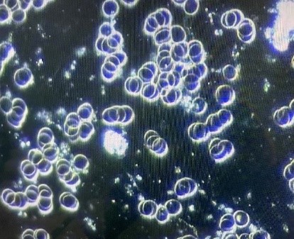

A second patient, a young adult male who received his vaccination 15 days earlier without any side effects noted and feeling completely well at the time, had the dark field examination of his blood performed. This first examination seen below revealed severe rouleaux formations of the RBCs with extensive branching, appearing to literally involve all of the RBCs visualized in an extensive review of multiple different microscopic fields. He then received one 400 ml ozonated saline infusion followed by a 15,000 mg infusion of vitamin C. The second picture reveals a complete and immediate resolution of the rouleaux formation seen on the first examination. Furthermore, the normal appearance of the RBCs was still seen 15 days later, giving some reassurance that the therapeutic infusions had some durability, and possibly permanency, in their positive impact.
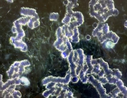
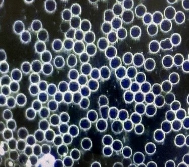
A third adult who received the vaccination 30 days earlier also had severe rouleaux formation on her dark field examination, and this was also completely resolved after the ozonated saline infusion followed by the vitamin C infusion. Of note, similar abnormal dark field microscopy findings were found in other individuals following Pfizer, Moderna, or Johnson & Johnson COVID vaccinations.
Preventing and Treating Chronic COVID and COVID Vaccine Complications
In addition to the mechanisms already discussed by which the spike protein can inflict damage, it appears the spike protein itself is significantly toxic. Such intrinsic toxicity (ability to cause the oxidation of biomolecules) combined with the apparent ability of the spike protein to replicate itself like a complete virus greatly increases the amount of toxic damage that can potentially be inflicted. A potent toxin is bad enough, but one that can replicate and increase its quantity inside the body after the initial encounter represents a unique challenge among toxins. And if the mechanism of replication can be sustained indefinitely, the long-term challenge to staying healthy can eventually become insurmountable. Nevertheless, this toxicity also allows it to be effectively targeted by high enough doses of the ultimate antitoxin, vitamin C, as discussed above. And even the continued production of spike protein can be neutralized by a daily multi-gram dosing of vitamin C, which is an excellent way to support optimal long-term health, anyway.
As was noted in an earlier article (Levy, 2021), there appear to be multiple ways to deal with spike protein effectively. The approaches to preventing and treating chronic COVID and COVID vaccine complications are similar, except that it would appear that a completely normal D-dimer blood test combined with a completely normal dark field examination of the blood could give the reassurance that the therapeutic goal has been achieved.
Until more data is accumulated on these approaches, it is probably advisable, if possible, to periodically reconfirm the normalcy of both the D-dimer blood test and the dark field blood examination to help assure that no new spike protein synthesis has resumed. This is particularly important since some patients who are clinically normal and symptom-free following COVID infection have been found to have the COVID virus persist in the fecal matter for an extended period of time (Chen et al., 2020; Patel et al., 2020; Zuo et al., 2020). Any significant immune challenge or new pathogen exposure facilitating a renewed surge of COVID virus replication could result in a return of COVID symptoms in such persons if the virus cannot be completely eliminated from the body.
Suggested Protocol (to be coordinated with the guidance of your chosen health care provider):
- For individuals who are post-vaccination or symptomatic with chronic COVID, vitamin C should be optimally dosed, and it should be kept at a high but lesser dose daily indefinitely.
- Ideally, an initial intravenous administration of 25 to 75 grams of vitamin C should be given depending on body size. Although one infusion would likely resolve the symptoms and abnormal blood examination, several more infusions can be given if feasible over the next few days.
- An option that would likely prove to be sufficient and would be much more readily available to larger numbers of patients would be one or more rounds of vitamin C given as a 7.5 gram IV push over roughly 10 minutes, avoiding the need for a complete intravenous infusion setup, a prolonged time in a clinic, and substantially greater expense (Riordan-Clinic-IVC-Push-Protocol, 10.16.14.pdf).
- Additionally, or alternatively if IV is not available, 5 grams of liposome-encapsulated vitamin C (LivOn Labs) can be given daily for at least a week.
- When none of the above three options are readily available, a comparable positive clinical impact will be seen with the proper supplementation of regular forms of oral vitamin C as sodium ascorbate or ascorbic acid. Either of these can be taken daily in three divided doses approaching bowel tolerance after the individual determines their own unique needs (additional information, see Levy, vitamin C Guide in References; Cathcart, 1981).
- An excellent way to support any or all of the above measures for improving vitamin C levels in the body is now available and very beneficial clinically. A supplemental polyphenol that appears to help many to overcome the epigenetic defect preventing the internal synthesis of vitamin C in the liver can be taken once daily. This supplement also appears to provide the individual with the ability to produce and release even greater amounts of vitamin C directly into the blood in the face of infection and other sources of oxidative stress (www.formula216.com).
- Hydrogen peroxide (HP) nebulization (Levy, 2021, free eBook) is an antiviral and synergistic partner with vitamin C, and it is especially important in dealing with acute or chronic COVID, or with post-COVID vaccination issues. As noted above, the COVID virus can persist in the stool. In such cases, a chronic pathogen colonization (CPC) of COVID in the throat continually supplying virus that is swallowed into the gut is likely present as well, even when the patient seems to be clinically normal. This will commonly be the case when specific viral eradication measures were not taken during the clinical course of the COVID infection. HP nebulization will clear out this CPC, which will stop the continued seeding of the COVID virus in the gut and stool as well. Different nebulization approaches are discussed in the eBook.
- When available, ozonated saline and/or ozone autohemotherapy infusions are excellent. Conceivably, this approach alone might suffice to knock out the spike protein presence, but the vitamin C and HP nebulization approaches will also improve and maintain health in general. Ultraviolet blood irradiation and hyperbaric oxygen therapy will likely achieve the same therapeutic effect if available.
- Ivermectin, hydroxychloroquine, and chloroquine are especially important in preventing new binding of the spike protein to the ACE2 receptors that need to be bound in order for either the spike protein alone or for the entire virus to gain entry into the target cells (Lehrer and Rheinstein, 2020; Wang et al., 2020; Eweas et al., 2021). These agents also appear to have the ability to directly bind up any circulating spike protein before it binds any ACE2 receptors (Fantini et al., 2020; Sehailia and Chemat, 2020; Saha and Raihan, 2021). When the ACE2 receptors are already bound, the COVID virus cannot enter the cell (Pillay, 2020). These three agents also serve as ionophores that promote intracellular accumulation of zinc that is needed to kill/inactivate any intact virus particles that might still be present.
- Many other positive nutrients, vitamins, and minerals are supportive of defeating the spike protein, but they should not be used to the exclusion of the above, especially the combination of highly-dosed vitamin C and HP nebulization.
Recap
As the pandemic continues, there is an increasing number of chronic COVID patients and patients post-COVID vaccination with a number of different symptoms. Furthermore, there is increasing number of vaccinated individuals who still end up contracting a COVID infection. This is resulting in a substantial amount of morbidity and mortality around the world. The presence and persistence of the COVID spike protein, along with the chronic colonization of the COVID virus itself in the aerodigestive tract as well as in the lower gut, appear to be major reasons for illness in this group of patients.
Persistent elevation of D-dimer protein in the blood and the presence of rouleaux formation of the RBCs, especially when advanced in degree, appear to be reliable markers of persistent spike protein-related illness. The measures noted above, particular the vitamin C and HP nebulization, should result in the disappearance of the D-dimer in the blood while normalizing the appearance of the RBCs examined with dark field microscopy. Even though new research is taking place daily that may modify therapeutic recommendations, it appears that taking the measures to eliminate D-dimer from the blood and to maintain a consistently normal morphological appearance of the blood is a very practical and efficient way to curtail the ongoing morbidity and mortality secondary to the persistent spike protein presence seen in chronic COVID and in post-COVID vaccination patients.
There are many vaccinated individuals who feel well yet remain cautious about potential future side effects, and who really have no easy access to D-dimer testing or dark field examination of their blood. Such persons can follow a broad-spectrum supplementation regimen featuring vitamin C, magnesium chloride, vitamin D, zinc, and a good multivitamin/multimineral supplement free of iron, copper, and calcium. Periodic but regular HP nebulization should be included as well. This regimen will offer good spike protein protection while optimizing long-term health. Furthermore, such a long-term supplementation regimen is advisable regardless of how much of the protocol discussed above is followed.
(OMNS Contributing Editor Dr. Thomas E. Levy is board certified in internal medicine and cardiology. He is also an attorney, admitted to the bar in Colorado and in the District of Columbia. The views presented in this article are the author’s and not necessarily those of all members of the Orthomolecular Medicine News Service Editorial Review Board.)
References
Alifano M, Alifano P, Forgez P, Iannelli A (2020) Renin-angiotensin system at the heart of the COVID-19 pandemic. Biochemie 174:30-33. PMID: 32305506
Aucott J, Rebman A (2021) Long-haul COVID: heed the lessons from other infection-triggered illnesses. Lancet 397:967-968. PMID: 33684352
Barshtein G, Waynblum D, Yedgar S (2020) Kinetics of linear rouleaux formation studied by visual monitoring of red cell dynamic organization. Biophysical Journal 78:2470-2474. PMID: 10777743
Batah S, Fabro A (2021) Pulmonary pathology of ARDS in COVID-19: a pathological review for clinicians. Respiratory Medicine 176:106239. PMID: 33246294
Belouzard S, Millet J, Licitra B, Whittaker G (2012) Mechanisms of coronavirus cell entry mediated by the viral spike protein. Viruses 4:1011-1033. PMID: 22816037
Biswas S, Thakur V, Kaur P et al. (2021) Blood clots in COVID-19 patients: simplifying the curious mystery. Medical Hypotheses 146:110371. PMID: 33223324
Carli G, Nichele I, Ruggeri M, Barra S, Tosetto A (2021) Deep vein thrombosis (DVT) occurring shortly after the second dose of mRNA SARS-CoV-2 vaccine. Internal and Emergency Medicine 16:803-804. PMID: 336876791
Cathcart R (1981) Vitamin C, titrating to bowel tolerance, anascorbemia, and acute induced scurvy. Medical Hypotheses 7:1359-1376. PMID: 7321921
Chen Y, Chen L, Deng Q et al. (2020) The presence of SARS-CoV-2 RNA I the feces of COVID-19 patients. Journal of Medical Virology 92:833-840. PMID: 32243607
Cho S (2011) Plasma cell leukemia with rouleaux formation involving neoplastic cells and RBC. The Korean Journal of Hematology 46:152. PMID: 22065968
Cicci G, Pirrelli A (1999) Red blood cell (RBC) deformability, RBC aggregability and tissue oxygenation in hypertension. Clinical Hemorheology and Microcirculation 21:169-177. PMID: 10711739
Eweas A, Alhossary A, Abdel-Moneim A (2021) Molecular docking reveals ivermectin and remdesivir as potential repurposed drugs against SARS-CoV-2. Frontiers in Microbiology 11:592908. PMID: 33746908
Fantini J, Di Scala C, Chahinian H, Yahi N (2020) Structural and molecular modelling studies reveal a new mechanism of action of chloroquine and hydroxychloroquine against SARS-CoV-2 infection. International Journal of Antimicrobial Agents 55:105960. PMID: 32251731
Favaloro E (2021) Laboratory testing for suspected COVID-19 vaccine-induced (immune) thrombotic thrombocytopenia. International Journal of Laboratory Hematology 43:559-570. PMID: 34138513
Hoffman M, Kleine-Weber H, Schroeder S et al. (2020) SARS-CoV-2 entry depends on ACE 2 and TMPRSS2 and is blocked by a clinically proven protease inhibitor. Cell 181:271-280. PMID: 32142651
Hu B, Huang S, Yin L (2021) The cytokine storm and COVID-19. Journal of Medical Virology 93:250-256. PMID: 32592501
Iba T, Levy J, Levi M et al. (2020) Coagulopathy of coronavirus disease 2019. Critical Care Medicine 48:1358-1364. PMID: 32467443
Iba T, Levy J, Warkentin T (2021) Recognizing vaccine-induced immune thrombotic thrombocytopenia. Critical Care Medicine [Online ahead of print]. PMID: 34259661
Kesmarky G, Kenyeres P, Rabai M, Toth K (2008) Plasma viscosity: a forgotten variable. Clinical Hemorheology and Microcirculation 39:243-246. PMID: 18503132
Lehrer S, Rheinstein P (2020) Ivermectin docks to the SARS-CoV-2 spike receptor-binding domain attached to ACE 2. In Vivo 34:3023-3026. PMID: 32871846
Levy T Guide-to-Optimal-Admin-of-IVC-10-18-2021.pdf
Levy T (2002) Curing the Incurable. Vitamin C, Infectious Diseases, and Toxins Henderson, NV: MedFox Publishing
Levy T (2019) Magnesium, Reversing Disease Chapter 12, Henderson, NV: MedFox Publishing
Levy T (2021) Resolving “Long-Haul COVID” and vaccine toxicity: neutralizing the spike protein. Orthomolecular Medicine News Service, June 21, 2021. http://orthomolecular.org/resources/omns/v17n15.shtml
Levy T (2021) Rapid Virus Recovery: No need to live in fear! Henderson, NV: MedFox Publishing. Free eBook download (English or Spanish) available at https://rvr.medfoxpub.com/
Lewi S, Clarke K (1954) Rouleaux formation intensity and E.S.R. British Medical Journal 2:336-338. PMID: 13182211
Lundstrom K, Barh D, Uhal B et al. (2021) COVID-19 vaccines and thrombosis-roadblock or dead-end street? Biomolecules 11:1020. PMID: 34356644
Mendelson M, Nel J, Blumberg L et al. (2020) Long-COVID: an evolving problem with an extensive impact. South African Medical Journal 111:10-12. PMID: 33403997
Naymagon L, Zubizarreta N, Feld J et al. (2020) Admission D-dimer levels, D-dimer trends, and outcomes in COVID-19. Thrombosis Research 196:99-105. PMID: 32853982
Paliogiannis P, Mangoni A, Dettori P et al. (2020) D-dimer concentrations and COVID-19 severity: a systematic review and meta-analysis. Frontiers in Public Health 8:432. PMID: 32903841
Patel K, Patel P, Vunnam R et al. (2020) Gastrointestinal, hepatobiliary, and pancreatic manifestations of COVID-19. Journal of Clinical Virology 128:104386. PMID: 32388469
Perricone C, Ceccarelli F, Nesher G et al. (2014) Immune thrombocytopenic purpura (ITP) associated with vaccinations: a review of reported cases. Immunologic Research 60:226-235. PMID: 25427992
Perrotta F, Matera M, Cazzola M, Bianco A (2020) Severe respiratory SARS-CoV2 infection: does ACE2 receptor matter? Respiratory Medicine 168:105996. PMID: 32364961
Perry R, Tamborska A, Singh B et al. (2021) Cerebral venous thrombosis after vaccination against COVID-19 in the UK: a multicentre cohort study. Lancet Aug 6. Online ahead of print. PMID: 34370972
Pillay T (2020) Gene of the month: the 2019-nCoV/SARS-CoV-2 novel coronavirus spike protein. Journal of Clinical Pathology 73:366-369. PMID: 32376714
Ramsay E, Lerman M (2015) How to use the erythrocyte sedimentation rate in paediatrics. Archives of Disease in Childhood. Education and Practice Edition 100:30-36. PMID: 25205237
Raveendran A (2021) Long COVID-19: Challenges in the diagnosis and proposed diagnostic criteria. Diabetes & Metabolic Syndrome: Clinical Research & Reviews 15:145-146. PMID: 33341598
Rostami M, Mansouritorghabeh H (2020) D-dimer level in COVID-19 infection: a systematic review. Expert Review of Hematology 13:1265-1275. PMID: 32997543
Saha J, Raihan M (2021) The binding mechanism of ivermectin and levosalbutamol with spike protein of SARS-CoV-2. Structural Chemistry Apr 12. Online ahead of print. PMID: 33867777
Samsel R, Perelson A (1984) Kinetics of rouleau formation. II. Reversible reactions. Biophysical Journal 45:805-824. PMID: 6426540
Saponaro F, Rutigliano G, Sestito S et al. (2020) ACE 2 in the era of SARS-CoV-2: controversies and novel perspectives. Frontiers in Molecular Biosciences 7:588618. PMID: 33195436
Scully M, Singh D, Lown R et al. (2021) Pathologic antibodies to platelet factor 4 after ChAdOx1 nCoV-19 vaccination. The New England Journal of Medicine 384:2202-2211. PMID: 33861525
Sehailia M, Chemat S (2021) Antimalarial-agent artemisinin and derivatives portray more potent binding of Lys353 and Lys31-binding hotspots of SARS-CoV-2 spike protein than hydroxychloroquine: potential repurposing of artenimol for COVID-19. Journal of Biomolecular Structure & Dynamics 39:6184-6194. PMID: 32696720
Sevick E, Jain R (1989) Viscous resistance to blood flow in solid tumors: effect of hemocrit on intratumor blood viscosity. Cancer Research 49:3513-3519. PMID: 2731173
Shang J, Wan Y, Luo C et al. (2020) Cell entry mechanisms of SARS-CoV-2. Proceedings of the National Academy of Sciences of the United States of America 117:11727-11734. PMID: 32376634
Sloop G, De Mast Q, Pop G et al. (2020) The role of blood viscosity in infectious diseases. Cureus 12:e7090. PMID: 32226691
Subramaniam S, Scharrer I (2018) Procoagulant activity during viral infections. Frontiers in Bioscience 23:1060-1081. PMID: 28930589
Thaler J, Ay C, Gleixner K et al. (2021) Successful treatment of vaccine-induced prothrombotic immune thrombocytopenia (VIPIT). Journal of Thrombosis and Haemostasis 19:1819-1822. PMID: 33877735
Townsend L, Fogarty H, Dyer A et al. (2021) Prolonged elevation of D-dimer levels in convalescent COVID-19 patients is independent of the acute phase response. Journal of Thrombosis and Haemostasis 19:1064-1070. PMID: 33587810
Wang N, Han S, Liu R et al. (2020) Chloroquine and hydroxychloroquine as ACE2 blockers to inhibit viropexis of 2019-nCoV spike pseudotyped virus. Phytomedicine: International Journal of Phytotherapy and Phytopharmacology 79:153333. PMID: 32920291
Zhang S, Liu Y, Want X et al. (2021) SARS-Cov-2 binds platelet ACE2 to enhance thrombosis in COVID-19. Journal of Hematology & Oncology 13:120. PMID: 32887634
Zuo T, Zhang F, Lui G et al. (2020) Alterations in gut microbiota of patients with COVID-19 during time of hospitalization. Gastroenterology 159:944-955. PMID: 32442562
Nutritional Medicine is Orthomolecular Medicine
Orthomolecular medicine uses safe, effective nutritional therapy to fight illness. For more information: http://www.orthomolecular.org
Find a Doctor
To locate an orthomolecular physician near you: http://orthomolecular.org/resources/omns/v06n09.shtml
The peer-reviewed Orthomolecular Medicine News Service is a non-profit and non-commercial informational resource.
Editorial Review Board:
Albert G. B. Amoa, MB.Ch.B, Ph.D. (Ghana)
Seth Ayettey, M.B., Ch.B., Ph.D. (Ghana)
Ilyès Baghli, M.D. (Algeria)
Ian Brighthope, MBBS, FACNEM (Australia)
Gilbert Henri Crussol, D.M.D. (Spain)
Carolyn Dean, M.D., N.D. (USA)
Ian Dettman, Ph.D. (Australia)
Damien Downing, M.B.B.S., M.R.S.B. (United Kingdom)
Susan R. Downs, M.D., M.P.H. (USA)
Ron Ehrlich, B.D.S. (Australia)
Hugo Galindo, M.D. (Colombia)
Martin P. Gallagher, M.D., D.C. (USA)
Michael J. Gonzalez, N.M.D., D.Sc., Ph.D. (Puerto Rico)
William B. Grant, Ph.D. (USA)
Claus Hancke, MD, FACAM (Denmark)
Tonya S. Heyman, M.D. (USA)
Suzanne Humphries, M.D. (USA)
Ron Hunninghake, M.D. (USA)
Bo H. Jonsson, M.D., Ph.D. (Sweden)
Dwight Kalita, Ph.D. (USA)
Felix I. D. Konotey-Ahulu, MD, FRCP, DTMH (Ghana)
Jeffrey J. Kotulski, D.O. (USA)
Peter H. Lauda, M.D. (Austria)
Alan Lien, Ph.D. (Taiwan)
Homer Lim, M.D. (Philippines)
Stuart Lindsey, Pharm.D. (USA)
Victor A. Marcial-Vega, M.D. (Puerto Rico)
Charles C. Mary, Jr., M.D. (USA)
Mignonne Mary, M.D. (USA)
Jun Matsuyama, M.D., Ph.D. (Japan)
Joseph Mercola, D.O. (USA)
Jorge R. Miranda-Massari, Pharm.D. (Puerto Rico)
Karin Munsterhjelm-Ahumada, M.D. (Finland)
Tahar Naili, M.D. (Algeria)
W. Todd Penberthy, Ph.D. (USA)
Zhiyong Peng, M.D. (China)
Isabella Akyinbah Quakyi, Ph.D. (Ghana)
Selvam Rengasamy, MBBS, FRCOG (Malaysia)
Jeffrey A. Ruterbusch, D.O. (USA)
Gert E. Schuitemaker, Ph.D. (Netherlands)
T.E. Gabriel Stewart, M.B.B.CH. (Ireland)
Thomas L. Taxman, M.D. (USA)
Jagan Nathan Vamanan, M.D. (India)
Garry Vickar, M.D. (USA)
Ken Walker, M.D. (Canada)
Raymond Yuen, MBBS, MMed (Singapore)
Anne Zauderer, D.C. (USA)
Andrew W. Saul, Ph.D. (USA), Editor-In-Chief
Associate Editor: Robert G. Smith, Ph.D. (USA)
Editor, Japanese Edition: Atsuo Yanagisawa, M.D., Ph.D. (Japan)
Editor, Chinese Edition: Richard Cheng, M.D., Ph.D. (USA)
Editor, French Edition: Vladimir Arianoff, M.D. (Belgium)
Editor, Norwegian Edition: Dag Viljen Poleszynski, Ph.D. (Norway)
Editor, Arabic Edition: Moustafa Kamel, R.Ph, P.G.C.M (Egypt)
Editor, Korean Edition: Hyoungjoo Shin, M.D. (South Korea)
Editor, Spanish Edition: Sonia Rita Rial, PhD (Argentina)
Contributing Editor: Thomas E. Levy, M.D., J.D. (USA)
Assistant Editor: Helen Saul Case, M.S. (USA)
Technology Editor: Michael S. Stewart, B.Sc.C.S. (USA)
Associate Technology Editor: Robert C. Kennedy, M.S. (USA)
Legal Consultant: Jason M. Saul, JD (USA)
Comments and media contact: drsaul@doctoryourself.com OMNS welcomes but is unable to respond to individual reader emails. Reader comments become the property of OMNS and may or may not be used for publication.
Dr. Pierre Kory: ‘COVID-19 Is Highly Treatable’
Reproduced from original article:
https://articles.mercola.com/sites/articles/archive/2021/11/13/covid-19-highly-treatable.aspx
The original Mercola article may not remain on the original site, but I will endeavor to keep it on this site as long as I deem it to be appropriate, and will not be bullied into removing it.
Analysis by Dr. Joseph Mercola Fact Checked November 13, 2021
STORY AT-A-GLANCE
- New York pulmonologist Dr. Pierre Kory, an unapologetic champion of evidence-based medicine, has had remarkable success treating patients with ivermectin and other therapies during the pandemic
- His efforts to get the word out on this treatment protocol as part of the Front Line COVID-19 Critical Care Working Group (FLCCC) have largely been stifled by censorship, ridicule and colleagues — brainwashed by the official narrative — unwilling to accept the science
- Kory is driven to share what he’s learned with as many people as possible because he believes that everyone should feel empowered to stay healthy; there are dozens of treatment options available for viral infections, about 90% of which are repurposed, cost pennies and are readily available
- Kory wants everyone to know that COVID-19 is a highly treatable disease, and if you’re infected, the earlier you begin treatment, the better
- FLCCC’s I-MASK+ protocol can be downloaded in full, giving you step-by-step instructions on how to prevent and treat the early symptoms of COVID-19
New York pulmonologist Dr. Pierre Kory, an unapologetic champion of evidence-based medicine, has had remarkable success treating patients with ivermectin and other therapies during the pandemic. His efforts to get the word out on this treatment protocol as part of the Front Line COVID-19 Critical Care Working Group (FLCCC) have largely been stifled by censorship, ridicule and colleagues — brainwashed by the official narrative — unwilling to accept the science.
Kory spoke with Dr. Chris Martenson, host of the Peak Prosperity podcast, about his incredible experiences over the last nearly two years.1 On December 8, 2020, Kory testified to the Senate Committee on Homeland Security and Governmental Affairs, which held a hearing on “Early Outpatient Treatment: An Essential Part of a COVID-19 Solution.”
He called on the NIH, CDC and FDA to review the expansive data on ivermectin to prevent COVID-19, keep those with early symptoms from progressing and help critically ill patients recover.2,3 As he told Martenson, due to their promising results, he believed early on that “the pandemic has been solved,” until he realized that those in power weren’t open to hearing what he had to say.
Despite his impassioned pleas and astonishing science to back them up, the treatment not only was ignored by the Senate committee but promptly eviscerated.4 Now, he feels his colleagues in the health care field are living in one of two worlds — by either not following the data or putting patients first because they’re afraid of losing their job or status, or by risking everything to put patients first. He’s become estranged from many colleagues who he says “don’t get it.”
There Is Treatment Available for Viruses
Kory’s eyes have been opened to the reality that many people only hear or believe what public health officials tell them, whether it’s because they’re overworked and don’t have time to delve into the real data or because they’re following with blind trust. Many of Kory’s colleagues have gone along with those they believe to be authoritative experts, even when their guidance defies logic and commonsense. Kory’s trust in the “experts,” however, started to erode the more that he learned.
One of Kory’s role models is Dr. Paul Marik, a critical care doctor at Sentara Norfolk General Hospital in East Virginia, who is renowned for his work in creating the “Marik Cocktail,” which significantly reduces death rates from sepsis using inexpensive, safe, generic medications.5
Marik was one of a small group of critical care physicians who formed FLCCC, which developed a highly effective COVID-19 treatment protocol known as MATH+.6 Marik is so in tune with science that if he reads a new study and has questions, he’ll contact the first author on the paper to get direct answers.
Right off the bat, the MATH+ protocol led to high survival rates. Out of more than 100 hospitalized COVID-19 patients treated with the MATH+ protocol by mid-April 2020, only two died. Both were in their 80s and had advanced chronic medical conditions.7
After several tweaks and updates, the prophylaxis and early outpatient treatment protocol is now known as I-MASK+8 while the hospital treatment has been renamed I-MATH+,9 due to the addition of ivermectin.
Kory is now a public face of FLCCC, and he’s forged a global network of colleagues who are willing to adapt to new information in any way they can to help patients. One of Kory’s biggest revelations involved the treatment of viruses — specifically, the fact that there are dozens of treatment options available, about 90% of which are repurposed, cost pennies and are readily available:10
“I went into this pandemic believing what I’ve been taught my whole career, which is that there is no specific antiviral therapy … I mean, you get a cold, you just rest … and now here I am 18 months later — oh my gosh — there are literally two dozen compounds and now we have trial evidence showing pretty profound large magnitude benefits, either in the duration of symptoms, the duration of viral transmission, hospitalization and death.
We have a number of molecules that actually reduce mortality in what’s turned out to be a deadly viral disease. This isn’t the common cold, we’re clear on that.
I went from, there’s nothing to do for a virus to now, anytime I have a cold going forward, or any of my children, or any other virus that comes at us, we already have a whole armory of stuff that we can employ. And that data for those — which are best, which should be employed — is only going to increase.”
Giving Patients Agency Over Their Own Health
Marteson said that, since learning about accessible treatment options, “I feel like I have agency in my own health that I didn’t have before.”11 Kory mentions natural options like curcumin and nigella sativa, or black cumin, which he would have laughed off years ago, but now realizes they have multiple mechanisms by which they fight viruses:12
“Reading about something like curcumin or nigella sativa, which if someone told me a year ago to take something like nigella sativa — black cumin seed — it would save your life in a viral disease, I would have literally burst out laughing … but when you look, there’s literally 10 years of lots of little trials and studies that have evaluated and defined multiple mechanisms of black cumin seed — immunomodulatory, anti-inflammatory, antiviral.
So you have all of these building blocks, and then you have this trial from Pakistan — large randomized controlled trial with really large magnitude benefits — of literally nigella sativa and honey. And then you find out about honey. Honey also has pleotropic properties.”
Kory is driven to share what he’s learned with as many people as possible, because he believes that everyone should feel empowered to stay healthy, similar to what I have long advised — to take control of your health. He told Marteson:13
“It’s so satisfying because now we have agency, and so many people have agency by learning this knowledge of things that are readily available, cheap, don’t need a prescription, that you can actually treat yourself with very safe compounds. Not only is that agency so satisfying, but boy does it seem critical for the future. Is this going to be the last viral pandemic?”
His index case with ivermectin — the first person with COVID-19 whom he treated with the drug — is also etched in his memory. The patient — a “slightly older, slightly overweight” woman — was two weeks into COVID-19 and still having fevers and night sweats, so still quite sick. He treated her with ivermectin and she woke up in the morning feeling great:14
“Literally I saw what could only be described as a phenomenal response to a medication. So when we talk about data that we use, I’m sorry but I was sold right there on the first dose. First patient, first dose. And then I had repeated experiences.”
COVID-19 Is Highly Treatable
Fear has dominated the pandemic, but both Martenson and Kory say there’s no need to walk around in fear. As a lung and ICU expert, Kory is a master at treating acute illnesses which, he says, “is all about trajectories.” “When we make rounds on patients, we see them every day, we’re following their course … in an ICU, I have to be very knowledgeable about their minute-to-minute, or sometimes hour-to-hour trajectory,” he said.15
He teaches medicine also, and he teaches his trainees to study trajectories in their patients. When the trajectory worsens, especially in critical illness, therapies must be instituted but, he says, when “I see a trajectory on the improvement, I always say just stand back. They’re getting better, they’re going to continue to get better …”16
In the case of his index patient with ivermectin, she was on a steady trajectory, but it rapidly improved upon administration of ivermectin — a pattern he sees regularly with the drug. The ability to get a sense of this pattern recognition is what makes the difference between an expert and nonexpert in critical care medicine, Kory says.
“The longer you’re in medicine, the better you get at that and you can see which medicines are working.” In this case, ivermectin is one that quickly stood out from the rest. Especially if you’re an expert at trajectories, patterns and diseases, as Kory is, “you can figure things out much quicker than a massive, multicentered, double-blinded, randomized controlled trial.”17
If there were one thing that Kory could share, it’s that he wants everyone to know that COVID-19 is a highly treatable disease:18
“I want everybody to know how treatable this is … I’m not that worried about it for me, my friends, my family, my colleagues. I’m not worried about it for those who follow the FLCCC and our protocols because we know that they’re effective.
And I just hope that umbrella of reassurance and protection, which is to say there are effective treatments which will save your life and prevent the need for hospitalization, I just hope that number grows. But me personally, I’m not that bothered by COVID. As you know, I actually got COVID. It was a relatively mild case and so I also have natural immunity in my camp.”
Early treatment, however, is essential. One of his friends became ill with COVID-19 and made the mistake of thinking he had a cold. He didn’t contact Kory until he’d been sick for seven or eight days and by that time, he said, “I had to pull out all the stops for him. I really had to use every tool in my arsenal to keep him out of the hospital.” So if you have COVID-19, the sooner you implement the treatment protocol, the better.
There’s a War Against Truth
The successful treatment of COVID-19 using ivermectin and other therapies is being actively suppressed. Few, for instance, have heard about the astonishing success in Uttar Pradesh, India, which embraced large-scale prophylactic and therapeutic use of ivermectin for COVID-19 patients, close contacts of patients and health care workers.19
They’ve since had a COVID-19 positivity rate of almost zero, marking a major public health achievement that Kory believes should be a model for the world. Even the World Health Organization praised Uttar Pradesh for their excellent public health measures, which included sending people out to villages to conduct rapid COVID-19 tests and, if positive, treat patients and close contacts with ivermectin.
WHO, however, did not mention ivermectin as part of Uttar Pradesh’s success story.20 Kory now calls the FLCCC an “army,” because “they’re actively fighting a war”:21
“They’re challenging the pharmacists. They’re talking to their doctors. They’re writing to pharmacy boards … I don’t think war is an overstatement here. There’s a war on truth. There’s a war on free discourse and sharing of opinions. One of the catastrophic things is the way they branded misinformation on the level of a felony. Someone who has an opinion that differs from the agency’s is automatically medical misinformation.
It’s treated as though it’s a scourge of society that needs to be extinguished. I think people are fighting back against that. It’s nice to hear the army and the tribe is growing and most important is, I think we’re helping people. We’re arming people with agency and the ability to navigate a pretty confusing world.”
FLCCC’s I-MASK+ protocol can be downloaded in full,22 giving you step-by-step instructions on how to prevent and treat the early symptoms of COVID-19. FLCCC also has protocols for at-home prevention and early treatment, called I-MASS, which involves ivermectin, vitamin D3, a multivitamin and a digital thermometer to watch your body temperature in the prevention phase and ivermectin, melatonin, aspirin and antiseptic mouthwash for early at-home treatment.
Household or close contacts of COVID-19 patients may take ivermectin (18 milligrams, then repeat the dose in 48 hours) for post-exposure prevention.23 FLCCC also has a management protocol — I-RECOVER24 — for long-haul COVID-19 syndrome. The protocols are translated into 23 different languages to provide widespread, free access to this lifesaving information, including how to get ivermectin.25
FLCCC remains hopeful that ivermectin will be formally adopted into national or international COVID-19 treatment guidelines in the near future.
- 1 Odysee, Peak Prosperity October 15, 2021
- 2 FLCCC Alliance, Ivermectin & COVID-19
- 3, 4, 5 Mountain Home May 1, 2021
- 6 FLCCC Alliance, Math+ Hospital Treatment Protocol for COVID-19
- 7 Dr. Pierre Kory Senate Testimony May 6, 2020 (PDF)
- 8 FLCCC Alliance I-MASK+ Protocol
- 9 FLCCC MATH+ Hospital Protocol
- 10 Odysee, Peak Prosperity October 15, 2021, 24:30
- 11 Odysee, Peak Prosperity October 15, 2021, 26:41
- 12 Odysee, Peak Prosperity October 15, 2021, 28:30
- 13 Odysee, Peak Prosperity October 15, 2021, 28:58
- 14 Odysee, Peak Prosperity October 15, 2021, 32:04
- 15, 16 Odysee, Peak Prosperity October 15, 2021, 34:30
- 17 Odysee, Peak Prosperity October 15, 2021, 38:33
- 18 Odysee, Peak Prosperity October 15, 2021, 40:00
- 19 Indian Express May 12, 2021
- 20 World Health Organization May 7, 2021
- 21 Odysee, Peak Prosperity October 15, 2021, 1:04
- 22 FLCCC Alliance, I-Mask+
- 23 FLCCC Alliance, I-MASS
- 24 FLCCC Alliance, I-RECOVER
- 25 FLCCC, How to Get Ivermectin
Vitamin D Papers Top List of Most Popular Studies of the Year
Reproduced from original article:
https://articles.mercola.com/sites/articles/archive/2021/11/09/vitamin-d-paper.aspx
The original Mercola article may not remain on the original site, but I will endeavor to keep it on this site as long as I deem it to be appropriate, and will not be bullied into removing it
Analysis by Dr. Joseph Mercola Fact Checked November 09, 2021
STORY AT-A-GLANCE
- Vitamin D optimization is likely the easiest, least expensive and most fundamental beneficial strategy that anyone can do to minimize their risk of COVID-19 and other infections
- October 31, 2020, I published a scientific review in the journal Nutrients, co-written with William Grant, Ph.D., and Dr. Carol Wagner. Our paper is the second-most downloaded study from this journal in the past 12 months. It’s also No. 2 in citations for articles in the past 12 months, and No. 4 for views
- The study with the most downloads in the past year and the all-time highest number of views is another vitamin D paper, which found vitamin D supplementation reduced the risk of influenza and COVID-19 infections and deaths
- A third vitamin D paper nabbed the No. 1 spot for most-cited study in the past 12 months. This study found vitamin D supplementation improved survival in frail elderly hospitalized with COVID-19
- To improve your immune function and lower your risk of viral infections, you’ll want to maintain a vitamin D level between 60 ng/mL and 80 ng/mL (150 nmol/L and 200 nmol/L). Vitamin D optimization is particularly important for dark-skinned individuals, as the darker your skin, the more sun exposure you need to raise your vitamin D level, as well as the elderly
October 31, 2020, I published a scientific review1 in the journal Nutrients, co-written with William Grant, Ph.D., and Dr. Carol Wagner, both of whom are part of the GrassrootsHealth expert vitamin D panel.
This paper, “Evidence Regarding Vitamin D and Risk of COVID-19 and Its Severity” — which you can download and read for free — is the second most downloaded study from this journal in the past 12 months.
Our paper is also No. 2 in citations for articles in the past 12 months, and No. 4 for views. Article metrics reveal it’s been viewed 131,458 times as of November 2, 2021, with a daily average of 62 views. The second and third most-viewed articles were also on vitamin D for COVID.
The study with the most downloads in the past year, at 62,590 downloads, and the all-time highest number of views (178,562) is another vitamin D paper2 by Bhattoa et.al., which found vitamin D supplementation reduced the risk of influenza and COVID-19 infections and deaths. The coauthors of my paper, Grant and Baggerly, were coauthors on this paper as well.
A third vitamin D paper, by Annweiler et. al., also nabbed the No. 1 spot for most-cited study in the past 12 months. This study found vitamin D supplementation improved survival in frail elderly hospitalized with COVID-19.
All of this is incredibly encouraging, and demolishes the ridiculous claim that there’s “no scientific basis” for the recommendation of vitamin D for COVID. Indeed, as of September 27, 2020, data from 14 observational studies — summarized in Table 1 of our paper3 — indicate vitamin D blood levels are inversely correlated with the incidence and/or severity of COVID-19.
What’s more, the evidence generally satisfies Hill’s criteria for causality in a biological system4 (i.e., strength of association, consistency of evidence, temporality, biological gradient, plausibility or mechanism of action, and coherence, although coherence still needs to be verified experimentally).
Mechanisms of Action
As explained in our paper, having sufficient vitamin D in your system can reduce your risk of COVID-19 and other respiratory infections via several different mechanisms, including but not limited to the following:5
| Reducing the survival of viruses |
| Inhibiting the replication of viruses6 |
| Reducing inflammatory cytokine production |
| Maintaining endothelial integrity (endothelial dysfunction contributes to vascular inflammation and impaired blood clotting, two hallmarks of severe COVID-19) |
| Increasing angiotensin-converting enzyme 2 (ACE2) concentrations — Angiotensin II is a natural peptide hormone that increases blood pressure by stimulating aldosterone. ACE2 normally consumes angiotensin I, thereby lowering the concentration of angiotensin II. However, SARS-CoV-2 infection downregulates ACE2, resulting in excessive accumulation of angiotensin II, which worsens the infection |
| Boosting your overall immune function by modulating your innate and adaptive immune responses |
| Reducing respiratory distress7 |
| Improving overall lung function |
| Helping produce surfactants in your lungs that aid in fluid clearance8 |
From my perspective, vitamin D optimization is one of the easiest, least expensive and most impactful strategies to reduce your risk of serious SARS-CoV-2 infection and other respiratory infections.
Vitamin D optimization is particularly important for dark-skinned individuals (who tend to have lower levels than Caucasians unless they spend extended time in the sun), the elderly, and those with preexisting chronic health conditions.
All of these are also risk factors for COVID-19, so population-wide optimization of vitamin D levels could significantly improve COVID outcomes among the most vulnerable.
Vitamin D also lowers your risk of several major chronic killers and comorbidities associated with poor COVID-19 prognosis, including obesity,9 Type 2 diabetes,10 high blood pressure11 and heart disease.12 All of these factors make it an important component of COVID-19 prevention and treatment.
Vitamin D helps prevent these conditions largely because it is a fat soluble steroid hormone that penetrates nearly every cell in your body and passes into the nucleus to affect the optimization of 2,000 to 3,000 genes to create a healthier you.
Vitamin D Is a Key Component for Optimal Immune Function
While the official narrative is now that the human body is defenseless without gene-based intervention (i.e., mRNA or DNA-based COVID injections), anyone still able to think clearly will recognize the nonsense of that argument. The truth is, your body is well-equipped to handle just about any infection, provided your immune system is working properly, as that is your body’s first line of defense.
Vitamin D receptors are found in a large number of different tissues and cells, including your immune cells. This means vitamin D plays an important role in your immune function specifically. If vitamin D is lacking, your immune system will be impaired, which in turn makes you more susceptible to infections of all kinds.
In the video at the top of this article, Dr. Roger Seheult reviews how vitamin D works, and its benefits, both for respiratory infections in general and as it pertains to COVID-19. Importantly, vitamin D is a steroid hormone that can pass through cellular membranes into the nucleus and controls the expression of genes. So, it’s not just a mere vitamin required as a cofactor. It can actually modify how the cells in your body behave and function. As noted by Seheult, vitamin D:
- Stimulates “the innate immune response, which provides frontline protection against infectious agents”
- Increases expression of antimicrobial peptides in your monocytes and neutrophils — both of which play important roles in COVID-19
- Enhances expression of an antimicrobial peptide called human cathelicidin, “which is of specific importance in host defenses against respiratory tract pathogens”
Higher Vitamin D Levels Lower Risk of COVID Infection
While Seheult also reviews a number of studies looking at vitamin D in relation to respiratory illnesses other than COVID-19, SARS-CoV-2-specific investigations have found that COVID-19 is far more common in vitamin D deficient individuals.
In one study,13,14,15 82.2% of COVID-19 patients tested were deficient in vitamin D, compared to 47.2% of population-based controls. (Mean vitamin D levels were 13.8 ± 7.2 ng/ml, compared to 20.9 ± 7.4 ng/ml in controls.)
They also found that blood levels of vitamin D were inversely correlated to D-dimer levels (a measure of blood coagulation). Many COVID-19 patients have elevated D-dimer levels, which are associated with blood clots. Vitamin D has also been linked to a lower risk of testing positive for COVID-19 in the first place.
The largest observational study16 to date, which looked at data for 191,779 American patients, found that of those with a vitamin D level below 20 ng/ml (deficiency), 12.5% tested positive for SARS-CoV-2, compared to 8.1% of those who had a vitamin D level between 30 and 34 ng/ml (adequacy) and only 5.9% of those who had an optimal vitamin D level of 55 ng/ml or higher. According to the authors:
“SARS-CoV-2 positivity is strongly and inversely associated with circulating 25(OH)D levels, a relationship that persists across latitudes, races/ethnicities, both sexes, and age ranges.”
Vitamin D Status Influences COVID-19 Severity
COVID-19 patients who have higher vitamin D levels also tend to have milder illness and better outcomes overall. One study17,18 found the risk of severe COVID-19 and related deaths virtually disappeared when vitamin D levels were above 30 ng/mL (75 nmol/L).
In another study,19 COVID-19 patients with a vitamin D level between 21 ng/mL (50 nmol/L) and 29 ng/mL (75 nmol/L) had a 12.55 times higher risk of death than those with a level above 30 ng/mL. Having a level below 20 ng/mL was associated with a 19.12 times higher risk of death.
Our vitamin D paper20 also lists data from 14 observational studies that show vitamin D blood levels are inversely correlated with the incidence and/or severity of COVID-19. This is quite logical, considering vitamin D regulates inflammatory cytokine production — a lethal hallmark of COVID-19 — and is an important regulator of your immune system.
Seheult also reviews studies showing COVID-19 outcomes appear to be linked to UVB exposure. For example, in one such study,21 they found a marked variation in mortality depending on whether the patients lived above or below 35 degrees North latitude. As noted by the authors:22
“… the hypothesis is not that vitamin D would protect against SARS-CoV-2 infection but that it could be very important in preventing the cytokine storm and subsequent acute respiratory distress syndrome that is commonly the cause of mortality.”
How to Optimize Your Vitamin D Level
While conventional medicine insists a vitamin D level of about 20 ng/mL is adequate, that really only applies for bone health. For optimal health, immune function and disease prevention, you want a blood level between 60 ng/mL and 80 ng/mL year-round. In Europe, the measurements you’re looking for are 150 nmol/L and 200 nmol/L.
If you live in a sunny locale and practice sensible sun exposure year-round, you might not need any supplements. The DMinder app23 is a helpful tool to see how much vitamin D your body can make depending on your location and other individual factors.
I walk on the beach for about an hour a day, wearing only shorts and a cap — and no sunscreen, as it filters out the ultraviolet rays that trigger vitamin D production — and haven’t needed oral supplementation for over a decade. (Keep in mind that the only way to ensure your sun exposure is enough is to get tested, ideally twice a year, in summer and winter.)
Many, unfortunately, don’t get enough sun exposure for one reason or another, and in these cases, an oral vitamin D supplement may be required. Just remember that the most important factor here is your blood level, not the dose, so before you start, get tested so you know your baseline.
Here’s a quick summary of how to determine whether you might need an oral supplement, and your ideal dosage:
1.First, measure your vitamin D level — One of the easiest and most cost-effective ways of measuring your vitamin D level is to participate in the GrassrootsHealth’s personalized nutrition project, which includes a vitamin D testing kit. Once you know what your blood level is, you can assess the dose needed to maintain or improve your level.
2.Assess your individualized vitamin D dosage — To do that, you can either use the chart below, or use GrassrootsHealth’s Vitamin D*calculator. To convert ng/mL into the European measurement (nmol/L), simply multiply the ng/mL measurement by 2.5. To calculate how much vitamin D you may be getting from regular sun exposure in addition to your supplemental intake, use the DMinder app.24
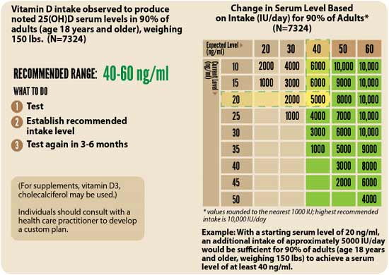
3.Retest in three to six months — Lastly, you’ll need to remeasure your vitamin D level in three to six months, to evaluate how your sun exposure and/or supplement dose is working for you.
Simple Way to Improve Your Vitamin D Absorption
Keep in mind that the specific dosage required to maintain an optimal vitamin D level can vary widely from person to person depending on a variety of factors, including skin pigmentation, age and weight. Your gut health can also play an important role in how well you absorb the vitamin D you take.25
When you have a healthy gut, beneficial bacteria produce butyrate by breaking down dietary fiber. Butyrate, in turn, helps increase vitamin D, so the more butyrate you have, the more vitamin D your body can absorb.
Another factor that can influence your vitamin D absorption is your magnesium level.26 Magnesium is required for the conversion of vitamin D into its active form.27,28,29,30 According to a scientific review31,32 published in 2018, about half of Americans taking vitamin D supplements may not get significant benefit as the vitamin D simply gets stored in its inactive form, and the reason for this is because they have insufficient magnesium levels.
Research by GrassrootsHealth33 shows you need 146% more vitamin D to achieve a blood level of 40 ng/ml (100 nmol/L) if you do not take supplemental magnesium, compared to taking your vitamin D with at least 400 mg of magnesium per day.
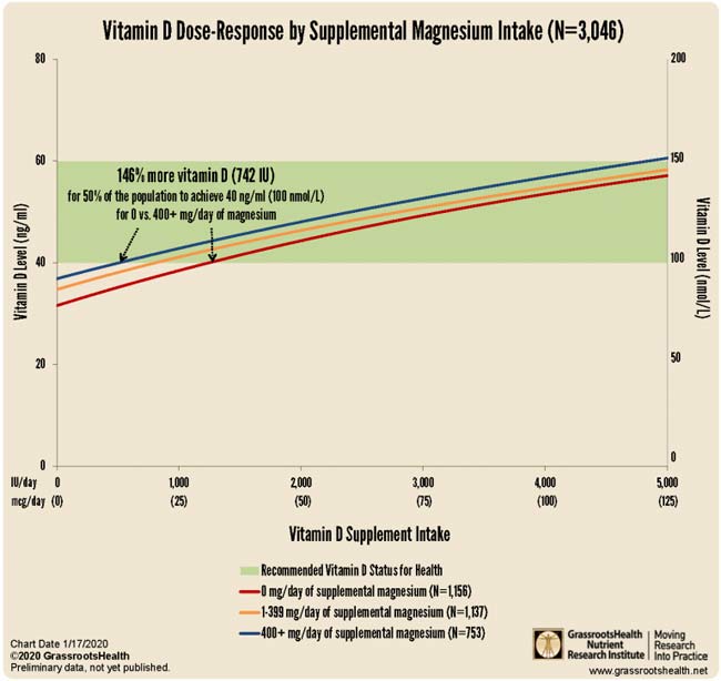
Your vitamin K2 intake can also affect your required vitamin D dosage. According to GrassrootsHealth,34 “combined intake of both supplemental magnesium and vitamin K2 has a greater effect on vitamin D levels than either individually,” and “those taking both supplemental magnesium and vitamin K2 have a higher vitamin D level for any given vitamin D intake amount than those taking either supplemental magnesium or vitamin K2 or neither.”
Data35 from nearly 3,000 individuals revealed 244% more oral vitamin D was required to get 50% of the population to achieve a vitamin D level of 40 ng/ml (100 nmol/L) if they weren’t concurrently also taking magnesium and vitamin K2. So, a simple way to optimize your vitamin D absorption is to take it in conjunction with magnesium and K2.
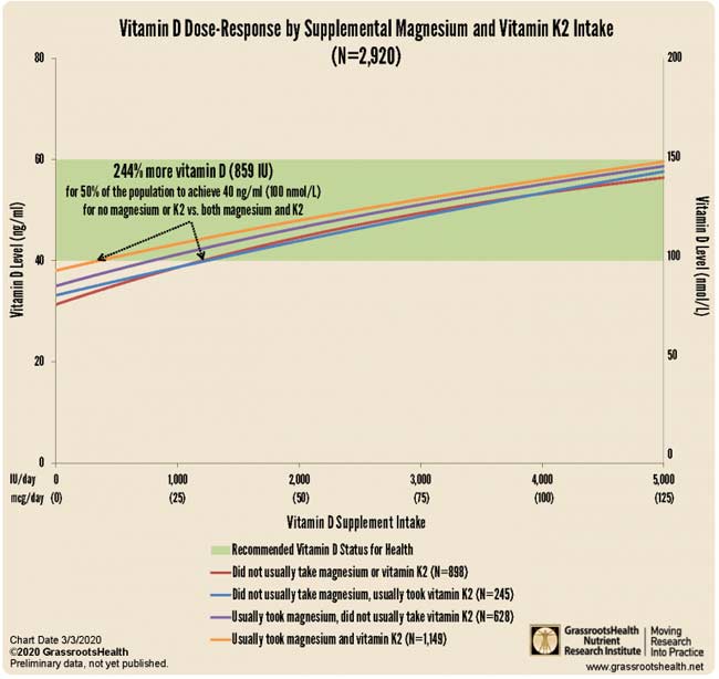
- 1, 5, 20 Nutrients October 31, 2020;12, 3361; doi:10.3390/nu12113361
- 2 Nutrients 2020; 12(11):3377
- 3 Nutrients October 31, 2020;12, 3361; doi:10.3390/nu12113361, Table 1
- 4 Nutrients October 31, 2020;12, 3361; doi:10.3390/nu12113361, Table 3
- 6 Nutrients, 2020;12:988
- 7 Advances in Pharmacological Sciences 2018; 2018: 8494816
- 8 ATS Journals October 5, 2010; 183(10)
- 9 Medicina 2019 Sep; 55(9): 541
- 10 Diabetes.co.uk January 15, 2019
- 11 The Lancet Diabetes & Endocrinology September 1, 2014; 2(9): 682-684
- 12 Current Treatment Options in Cardiovascular Medicine 2012 Aug; 14(4): 414–424
- 13 The Journal of Clinical Endocrinology & Metabolism October 27, 2020; dgaa733 [Epub ahead of print]
- 14 Endocrine.org October 27, 2020
- 15 Science Daily October 27, 2020
- 16 PLOS ONE September 17, 2020 DOI: 10.1371/journal.pone.0239252
- 17 Infectious Diseases April 8, 2020 DOI: 10.21203/rs.3.rs-21211/v1
- 18 Orthomolecular Medicine News Service June 22, 2020
- 19 Emerginnova.com June 4, 2020
- 21, 22 Alimentary Pharmacology & Therapeutics, 2020; doi.org/10.1111/apt.15777
- 23, 24 DMinder app
- 25 Nature Communications 2020; 11 Article number: 5997
- 26 BMC Medicine 2013; 11: 187
- 27 Live Science February 26, 2018
- 28 Medicalxpress.com February 27, 2018
- 29 News-Medical.net February 26, 2018
- 30 Wellandgood.com February 26, 2018
- 31 Journal of the American Osteopathic Association March 2018; 118: 181-189
- 32 Science Daily February 2018
- 33 GrassrootsHealth Is Supplemental Magnesium Important for Vitamin D Levels?
- 34, 35 GrassrootsHealth Magnesium and Vitamin K2 Combined Important for Vitamin D Levels
Dr. Mercola Files Lawsuit Against US Sen. Elizabeth Warren
Reproduced from original article:
https://articles.mercola.com/sites/articles/archive/2021/11/08/mercola-sues-sen-warren.aspx
The original Mercola article may not remain on the original site, but I will endeavor to keep it on www.leanmachine.com.au
Analysis by Dr. Joseph Mercola Fact Checked November 08, 2021

STORY AT-A-GLANCE
- In early September 2021, U.S. Sen. Elizabeth Warren sent a letter to Andy Jassy, chief executive officer of Amazon.com, demanding an “immediate review” of Amazon’s algorithms to weed out books peddling “COVID misinformation,” stressing that Amazon’s sale of such books was “potentially unlawful”
- Warren specifically singled out my book, “The Truth About COVID-19,” co-written with Ronnie Cummins, founder and director of the Organic Consumers Association (OCA), as a prime example of “highly-ranked and favorably-tagged books based on falsehoods about COVID-19 vaccines and cures” that she wants banned
- As a government official, it is illegal for Warren to violate the U.S. Constitution, and pressuring private businesses to do it for her is not a legal workaround
- Cummins and I, along with our publisher, Chelsea Green Publishing, and Robert F. Kennedy Jr., who wrote our foreword, are now suing Warren, both in her official and personal capacities, for violating our First Amendment rights and scaring book sellers into pulling and/or suppressing sales of our book
- Ironically, Warren’s claims of misinformation are themselves misinformation that puts lives at risk
In early September 2021, U.S. Sen. Elizabeth Warren sent a letter1 to Andy Jassy, chief executive officer of Amazon.com, demanding an “immediate review” of Amazon’s algorithms to weed out books peddling “COVID misinformation,” stressing that Amazon’s sale of such books was “potentially unlawful.”2,3,4
Warren specifically singled out my book, “The Truth About COVID-19,” co-written with Ronnie Cummins, founder and director of the Organic Consumers Association (OCA), as a prime example of “highly-ranked and favorably-tagged books based on falsehoods about COVID-19 vaccines and cures” that she wanted banned.
“Dr. Mercola has been described as ‘the most influential spreader of coronavirus misinformation online,” Warren wrote,5 adding: “Not only was this book the top result when searching either ‘COVID-19’ or ‘vaccine’ in the categories of ‘All Departments’ and ‘Books’; it was tagged as a ‘Best Seller’ by Amazon and the ‘#1 Best Seller’ in the ‘Political Freedom’ category.
The book perpetuates dangerous conspiracies about COVID-19 and false and misleading information about vaccines. It asserts that vitamin C, vitamin D and quercetin … can prevent COVID-19 infection … And the book contends that vaccines cannot be trusted …”
Warren Fancies Herself Above the Law
Warren should know that as a government official, it is illegal for her violate the U.S. Constitution, and pressuring private businesses to do it for her is not a legal workaround.
Since she willfully ignores the law, Cummins and I, along with our publisher, Chelsea Green Publishing, and Robert F. Kennedy Jr., who wrote our foreword, are suing Warren, both in her official and personal capacities, for violating our First Amendment rights. The federal lawsuit, in which Warren is listed as the sole defendant, was filed in the state of Washington. As noted in our complaint:
“Once upon a time, the First Amendment was understood to guarantee that books challenging governmental orthodoxy could be sold without fear of governmental intimidation or reprisal.
Almost sixty years ago, in Bantam Books v. Sullivan, 372 U.S. 58 (1963), the Supreme Court held that state officials violated the First Amendment by sending letters to booksellers warning that the sale of certain named books was potentially unlawful.
The ‘vice’ in such letters and in the ‘veiled threat’ of legal repercussions they communicated, explained the Court, is that they allow government to achieve censorship while doing an end-run around the judiciary, ‘provid[ing] no safeguards whatever against the suppression of … constitutionally protected’ speech, thus effecting an unconstitutional ‘prior restraint.’
It made no difference that the officials who sent the letter lacked the ‘power to apply formal legal sanctions’ — i.e., that the officials did not themselves have the power to sanction or prosecute the booksellers in any way. Indeed this fact made the unconstitutionality more apparent.
The officials ‘are not law enforcement officers; they do not pretend that they are qualified to give or that they attempt to give distributors only fair legal advice … [T]hey acted … not to advise but to suppress.’
It also made no difference, the Court expressly found, that the letters were framed as mere ‘exhort[ation]’ or that the booksellers were in theory ‘free’ to ignore the letters, because the officials had ‘deliberately set about to achieve the suppression of publications deemed ‘objectionable’,’ and ‘people do not lightly disregard public officers’ veiled threats.’
Today, certain members of the United States Congress have apparently forgotten, or think they are above, the law set forth in Bantam Books.”
Warren’s Attack on Constitutionally Protected Speech
There’s no doubt our book, “The Truth About COVID-19,” is constitutionally protected speech, and that Warren’s letter is calling on Amazon to suppress protected speech.
In our book, we share viewpoints, ideas, opinions, verifiable facts and factual hypotheses that our federal government just so happens to disfavor, as it counters their chosen narrative that SARS-CoV-2 emerged naturally, cannot be prevented by any means other than experimental gene therapy, and cannot be treated by any other means than certain experimental and exorbitantly costly drugs.
Since the start of the pandemic, government has systematically sought to suppress the kind of information shared in our book, using the same tactic as Warren used against us here — warning Internet-based companies that if they don’t censor these views, the full weight of the government’s wrath will be turned against them. As explained in our complaint:
“The term ‘vaccine misinformation’ as Warren uses it is propagandistic and false. As she uses it, ‘vaccine misinformation’ refers to any speech challenging the safety and efficacy of the COVID vaccines, even when that speech consists of factually accurate information or protected opinion …
On September 10, 2021, as a direct result of Warren’s letter, a major national bookseller chain, Barnes and Noble, notified the publisher of The Truth About COVID-19 by email that it would no longer sell the work as an e-book. Barnes and Noble has — for now — reversed that decision.
It is impossible for Plaintiffs to know with certainty whether, as a result of Warren’s letter, Amazon is now covertly demoting, downgrading, or otherwise suppressing The Truth About COVID-19 in numerous ways that would be hidden from view, but Plaintiffs believe that Amazon is in fact covertly taking such action.
Even if no bookseller in the country had yielded to Warren’s threats, her letter would still be actionable as a clear violation of the First Amendment.
In Backpage.com, LLC v. Dart, 807 F.3d 229 (7th Cir. 2015) (Posner, J.), relying on Bantam Books, the Court held that a governmental official ‘violates a plaintiff’s First Amendment rights’ if by ‘threat’ or ‘intimidation’ the official attempts to induce ‘a third party’ to stop ‘publishing or otherwise disseminating the plaintiff’s message,’ and emphasized that ‘such a threat is actionable and thus can be enjoined even if it turns out to be empty — the victim ignores it, and the threatener folds his tent.’
Such threats go ‘by the name of ‘prior restraint,’ and a prior restraint is the quintessential first-amendment violation.’ Accordingly, Plaintiffs ask this Court to vindicate clearly established law, to vindicate Plaintiffs’ constitutional rights, to vindicate the First Amendment itself, by declaring Warren’s conduct unconstitutional and by enjoining her from repeating such conduct in future.”
Warren Calls Out ‘Misinformation’ With Misinformation
In our complaint, we also emphasize the fact that Warren’s claims of misinformation are themselves misinformation. For example, Warren claims our book falsely “asserts that … vitamin D … can prevent COVID-19 infection.” According to Warren, this claim has no scientific basis. This is clearly and verifiably false as there are many studies, published in 2020 and 2021, supporting this claim.
For example, in May 2021, the National Institutes of Health’s website, PubMed.gov, published a Journal of Medical Virology article titled “Vitamin D Deficiency Is Associated With COVID-19 Positivity and Severity of the Disease.”6 Many other scientific articles have also linked vitamin D deficiency with a higher risk of COVID infection, more severe outcomes and increased rates of death.
Indeed, a recent systematic review7 of the literature, posted on the U.S. National Library of Medicine, which is another National Institutes of Health website, concluded that “blood vitamin D status can determine the risk of being infected with COVID-19, seriousness of COVID-19, and mortality from COVID-19.
Therefore, maintaining appropriate levels of Vitamin D through supplementation or natural methods … is recommended for the public to be able to cope with the pandemic.” As noted in our complaint:
“Thus while Warren professes to champion true COVID information to save lives, she is purveying false information that could lead to COVID deaths. Warren is telling people that vitamin D levels don’t matter for COVID, when in fact — as readers would learn from The Truth About COVID-19 — correcting vitamin D deficiencies could save their lives.
By her own logic and according to her own demands, every major social media platform should have banned Warren’s letter as ‘COVID misinformation.’ But officials like Warren only denounce ‘COVID misinformation,’ demand its censorship, and threaten legal repercussions when the statements in question challenge the COVID narrative they support — not when they themselves are misrepresenting the truth about COVID-19.
Warren’s letter further accuses The Truth About COVID-19 of disseminating ‘false and misleading information about vaccines,’ including by (in Warren’s words) ‘contend[ing] that vaccines cannot be trusted.’
The book’s stated thesis about the COVID vaccines is that their effectiveness ‘has been wildly exaggerated and major safety questions have gone unanswered.’ This statement is accurate and well within the bounds of constitutionally protected opinion …
Warren’s letter further cites a June, 2021, review of The Truth About COVID-19 that purports to list examples of the book’s ‘misinformation,’ the first of which is the following: ‘the authors argue that the SARS-CoV-2 coronavirus was engineered in a laboratory in Wuhan, China.’ It is true that The Truth About COVID-19 argues that that ‘the preponderance of evidence’ supports the lab-leak theory of the origins of the COVID virus.
But the claim that this position is ‘misinformation’ is, once again, itself misinformation. The lab-leak theory — long denounced as a ‘conspiracy theory’ by federal actors and suppressed on social media — is in fact supported by substantial and growing evidence. See, e.g., Wall St. Journal, ‘Science Closes In on Covid’s Origins: Four studies — including two from WHO — provide powerful evidence favoring the lab-leak theory,’ Oct. 5, 2021.8
The review’s next example of the supposed ‘misinformation’ in the The Truth About COVID-19 is this: the book ‘insists multiple times that the public health measures and restrictions will be permanent. Not true.
The CDC announced that fully vaccinated Americans could resume activities without wearing masks or physically distancing, resume domestic travel, and refrain from quarantine even when following a known exposure to the virus if they remain symptom-free.’
This CDC announcement obviously proved to be false, while the prediction made in The Truth About COVID-19 that health restrictions would continue after vaccination has proved more accurate.
Moreover, it is not the case that the Truth About COVID-19 ‘insists’ that these restrictions will be permanent — it says that certain restrictions on our liberty, beginning in the pandemic, will ‘probably’ be permanent, reflecting a humility about the certainty of one’s assertions that Warren might have profited from.”
This Is Only the Beginning
As noted in a press release by Cummins, this lawsuit is just the beginning. OCA and I are launching a campaign to fight back against the censorship that is taking root. This includes unraveling the threads that lead back to the fake fact checkers and disinformation agents in the media, but all of this will take time, so be patient.
It’s also a costly venture, so if you can afford it, consider making a donation to the OCA. As a thank you, I will match your donations dollar for dollar during the month of November.

As explained by Cummins:
“OCA’s federal lawsuit, filed jointly with Dr. Mercola, Robert F. Kennedy Jr. and Chelsea Green books is not just directed against Elizabeth Warren, but is intended to establish a legal precedent against the increasing censorship, slander, and intimidation coming from a wide variety of government, corporate, and media sources.
This Big Pharma/Big Media/Big Government Inquisition is fueled by disinformation and dark money coming from powerful international public relations firms such as the Publicis Groupe and front groups such as the so-called Center for Countering Digital Hate (CCDH).
We are under attack, not because we are purveyors of dangerous disinformation and hate, as Warren and her Establishment cohorts allege, but rather because, in the midst of an international health, economic, and political crisis, we are trying to expose the truth about the lab origins of this catastrophe, and explain how preventive and natural medicine and health, healthy organic food, natural supplements, low-cost generic drugs, strong immune systems, and a healthy environment are our best defenses against chronic disease and engineered pathogens.
We are not anti-vaccine, but rather pro-vaccine safety. We are not purveyors of disinformation, but rather firm defenders of free speech, unobstructed scientific inquiry, and freedom of choice …
We are castigated as ‘conspiracy theorists’ for publicizing the behind-the- scenes machinations of billionaires like Bill Gates, the World Economic Forum, and their ‘Hall of Shame’ collaborators9 in the military-industrial complex.
We are under siege for exposing the existential risks of genetic engineering and lab manipulation, a mad science not only contaminating our food, seeds and animals, but essentially weaponizing pathogenic viruses, bacteria, and insects, part of a catastrophic biological and medical arms race that threatens us all.
We are saddened and alarmed by the now routine attacks on free speech, free association, and medical freedom of choice. We are troubled by the extreme polarization and anger poisoning the body politic, and the debilitating impact of fear-mongering and shaming on our children and the public at large.
We are alarmed by the collateral damage to our health, our psyches, and the entire social fabric by government authoritarianism, virologists and gene engineers playing God, and Big Pharma greed …
America, and the once-hoped-for community of nations, are accelerating toward self-destruction. The body politic is sick, frightened, angry, and divided. People have apparently forgotten how to talk to one another when we disagree on politics, COVID responses, vaccine safety, and a range of other polarizing government dictates.
Former friends and co-workers have become enemies. Meanwhile the forests are burning. Water resources are diminishing … Our children and the most vulnerable are forced to struggle harder than ever, just to survive and preserve their sanity, making it harder and harder maintain a positive outlook, enjoy every day life, much less achieve true happiness.
If COVID-19, the product of mad science and insatiable greed, has taught us anything, it’s that we must transform our food and farming systems and take control of our health.
We must acknowledge, prevent, and resolve the dietary, environmental, and public health-related comorbidities of our ailing population, strengthen our immune systems to fight off chronic disease and pathogens, and provide special protection for the most vulnerable.
We must bring profit-at-any-cost corporations, captured media and regulatory agencies, indentured politicians, Silicon Valley surveillance capitalists, out-of-control genetic engineers, virologists, and bioweapons profiteers to heel.”
Stop the Madness
To this end, OCA has launched a Stop the Mad Science campaign. This global grassroots campaign aims to ban the engineering of viruses, bacteria and all potential pandemic pathogens (PPPs). Mounting evidence suggests COVID-19 was indeed the result of gain-of-function (GOF) research, paid for in part by U.S. taxpayers and carried out by U.S. and Chinese researchers.
Unless we put an end to this kind of dangerous research (and it goes on worldwide, not just in the U.S. and China), COVID won’t be the last manmade pandemic we’ll have to face. More than 65,000 people have already signed the petition in support of this effort. Please add your signature here if you haven’t done so already. As noted by Cummins:
“Current ongoing experiments, routinely funded with our tax dollars, that need to be stopped immediately include genetically engineering SARS-CoV-2 so that it can overcome or bypass natural immunity; combining the SARS-CoV-2 virus with deadly anthrax bacteria; engineering the bird flu and Ebola to be more transmissible; and other criminally insane experiments — hiding behind the excuse that lab and genetic engineering of pathogens are necessary for ‘biodefense’ and ‘biomedicine.’
Over the next six months we will begin to organize protests and picket lines outside the GoF labs and institutions where these dangerous experiments are being carried out. These street protests will be amplified by public education, petition gathering, litigation, and grassroots lobbying.”
- 1, 5 Warren’s letter to Andy Jassy September 7, 2021
- 2 National Interest September 12, 2021
- 3 The Guardian September 13, 2021
- 4 New York Times September 8, 2021
- 6 Journal of Medical Virology May 2021; 93(5):2992-2999
- 7 Risk Management Healthcare Policy 2021; 14: 31-38
- 8 Wall Street Journal October 5, 2021
- 9 OCA Gain of Function Hall of Shame
Is Niacin a Missing Piece of the COVID Puzzle?
Reproduced from original article:
https://articles.mercola.com/sites/articles/archive/2021/10/27/what-are-the-benefits-of-niacin.aspx
The original Mercola article may not remain on the original site, but I will en devour to keep it alive permantly on this site
Analysis by Dr. Joseph Mercola Fact Checked October 27, 2021

STORY AT-A-GLANCE
- B vitamins may play an important role in COVID-19 prevention and treatment, according to two recent papers
- Based on B vitamins’ effects on your immune system, immune-competence and red blood cells (which help fight infection), supplementation may be a useful adjunct to other prevention and treatment strategies
- B vitamins can influence several COVID-19-specific disease processes, including viral replication and invasion, cytokine storm induction, adaptive immunity and hypercoagulability
- Niacin appears particularly important. According to one published paper, there appears to be a causative link between low niacin status and SARS-CoV-2 infection
- SARS-CoV-2’s ability to invade your body is dependent on calcium signaling, which in turn is dependent on the presence of NAADP, which is formed from niacin. NAADP-dependent calcium signaling is responsible both for the inhibition of viral entry into cells and driving the virus out of already infected cells
This article was previously published January 20, 2021, and has been updated with new information.
While vitamins C and D have garnered much attention in the fight against COVID-19, B vitamins can also play an important role, according to two recent papers — niacin (B3) in particular.
The first, “Be Well: A Potential Role for Vitamin B in COVID-19,”1,2 was published in the February 2021 issue of the journal Maturitas. The paper is the result of a joint collaboration between researchers at the University of Oxford, United Arab Emirates University and the University of Melbourne, Australia.
While no studies using B vitamins have been performed on COVID-19 patients, the researchers stress that, based on B vitamins’ effects on your immune system, immune-competence and red blood cells (which help fight infection), supplementation may be a useful adjunct to other prevention and treatment strategies. As noted by the authors:3
“There is a need to highlight the importance of vitamin B because it plays a pivotal role in cell functioning, energy metabolism, and proper immune function.
Vitamin B assists in proper activation of both the innate and adaptive immune responses, reduces pro-inflammatory cytokine levels, improves respiratory function, maintains endothelial integrity, prevents hypercoagulability and can reduce the length of stay in hospital.
Therefore, vitamin B status should be assessed in COVID-19 patients and vitamin B could be used as a non-pharmaceutical adjunct to current treatments …
Vitamin B not only helps to build and maintain a healthy immune system, but it could potentially prevent or reduce COVID-19 symptoms or treat SARS-CoV-2 infection. Poor nutritional status predisposes people to infections more easily; therefore, a balanced diet is necessary for immuno-competence.”
B Vitamins Play Many Roles in COVID-19 Disease Process
Importantly, B vitamins can influence several COVID-19-specific disease processes, including:4
- Viral replication and invasion
- Cytokine storm induction
- Adaptive immunity
- Hypercoagulability
The paper goes on to detail how each of the B vitamins can help manage various COVID-19 symptoms:5
| Vitamin B1 (thiamine) — Thiamine improves immune system function, protects cardiovascular health, inhibits inflammation and aids in healthy antibody responses. Vitamin B1 deficiency can result in an inadequate antibody response, thereby leading to more severe symptoms. There’s also evidence suggesting B1 may limit hypoxia. |
| Vitamin B2 (riboflavin) — Riboflavin in combination with ultraviolet light has been shown to decrease the infectious titer of SARS-CoV-2 below the detectable limit in human blood, plasma and platelet products. |
| Vitamin B3 (niacin/nicotinamide) — Niacin is a building block of NAD and NADP, which are vital when combating inflammation. |
| Vitamin B5 (pantothenic acid) — Vitamin B5 aids in wound healing and reduces inflammation. |
| Vitamin B6 (pyridoxal 5′-phosphate/pyridoxine) — Pyridoxal 5′-phosphate (PLP), the active form of vitamin B6, is a cofactor in several inflammatory pathways. Vitamin B6 deficiency is associated with dysregulated immune function. Inflammation increases the need for PLP, which can result in depletion.
According to the authors, in COVID-19 patients with high levels of inflammation, B6 deficiency may be a contributing factor. What’s more, B6 may also play an important role in preventing the hypercoagulation seen in some COVID-19 patients. |
| Vitamin B9 (folate/folic acid) — Folate, the natural form of B9 found in food, is required for the synthesis of DNA and protein in your adaptive immune response.
Folic acid, the synthetic form typically found in supplements, was recently found6 to inhibit furin, an enzyme associated with viral infections, thereby preventing the SARS-CoV-2 spike protein from binding to and gaining entry into your cells. The research7 suggests folic acid may therefore be helpful during the early stages of COVID-19. Another recent paper8 found folic acid has a strong and stable binding affinity against SARS-CoV-2. This too suggests it may be a suitable therapeutic against COVID-19. |
| Vitamin B12 (cobalamin) — B12 is required for healthy synthesis of red blood cells and DNA. A deficiency in B12 increases inflammation and oxidative stress by raising homocysteine levels. Your body can eliminate homocysteine naturally, provided you’re getting enough B9 (folate), B6 and B12.9
Hyperhomocysteinemia — a condition characterized by abnormally high levels of homocysteine — causes endothelial dysfunction, activates platelet and coagulation cascades and decreases immune responses. B12 deficiency is also associated with certain respiratory disorders. Advancing age can diminish your body’s ability to absorb B12 from food,10 so the need for supplementation may increase as you get older. According to “Be Well: A Potential Role for Vitamin D in COVID-19”:11
|
Niacin — A Missing Piece of the COVID-19 Puzzle?
The second paper,12 “Sufficient Niacin Supply: The Missing Puzzle Piece to COVID-19 and Beyond?” (which is a preprint and has yet to undergo peer review), focuses specifically on niacin (B3), raising the question of whether this vitamin might actually be a crucial player in the COVID-19 disease process. As noted in the abstract:
“Definitive antiviral properties are evidenced for niacin, i.e., nicotinic acid (NA), as coronavirus disease 2019 (COVID-19) therapy for both disease recovery and prevention, to the level that reversal or progression of its pathology follows as an intrinsic function of NA supply.
This detailed investigation provides a thorough disentanglement of how the downstream inflammatory propagation of ensuing severe acute respiratory virus 2 (SARS-CoV-2) infection is entirely prohibited or reversed upstream out the body to expeditiously restore health with well-tolerated dynamic supplementation of sufficient NA (i.e., ~1-3 grams per day).”
As noted in this paper, a primary hallmark of COVID-19 pathology is the cytokine storm, which can lead to multiple organ failure and death. Marked elevations in proinflammatory cytokines are to blame for this chain of events, most notable of which are interleukin-6 (IL-6), interleukin-1β (IL-1β), tumor necrosis factor-α (TNF-α) and monocyte chemoattractant protein-1 (MCP-1).
If you can decrease and control these damaging cytokines, you stand a good chance of thwarting the cytokine storm and the downstream damage it causes. Nicotinamide adenine dinucleotide (NAD+) plays an important role in this, and niacin is a building block of NAD. As explained in “Be Well: A Potential Role for Vitamin D in COVID-19”:13
“NAD+ is released during the early stages of inflammation and has immunomodulatory properties, known to decrease the pro-inflammatory cytokines, IL-1β, IL-6 and TNF-α. Recent evidence indicates that targeting IL-6 could help control the inflammatory storm in patients with COVID-19.”
Aside from markedly decreasing proinflammatory cytokines, niacin has also been shown to:14
- Reduce the replication of a number of viruses, including vaccinia virus, human immunodeficiency virus, enteroviruses and hepatitis B virus
- Reduce neutrophil infiltration
- Have anti-inflammatory effect in patients with ventilator-induced lung injury
Niacin Modulates the Bradykinin Storm
COVID-19 also triggers bradykinin storms. Bradykinin is a chemical that helps regulate your blood pressure and is controlled by your renin-angiotensin system (RAS). The bradykinin hypothesis provides a model that helps explain some of the more unusual symptoms of COVID-19, including its bizarre effects on your cardiovascular system.
Researchers have discovered SARS-CoV-2 downregulates your body’s ability to degrade or break down bradykinin. The end result is a bradykinin storm, and this appears to be an important factor in many of COVID-19’s lethal effects, perhaps even more so than the cytokine storms associated with the disease. As bradykinin accumulates, the more serious COVID-19 symptoms appear.
Vitamin D has a significant impact on the RAS,15 and can therefore help prevent a bradykinin storm, but niacin also plays an important role. As noted in “Sufficient Niacin Supply: The Missing Puzzle Piece to COVID-19 and Beyond?”:16
“Immediate-release NA [niacin] administration has been reported as highly effective in preventing the lung tissue damage involved in this … pathology. As a matter of fact, authors of a March, 2020, paper17 in Nature for this very reason conclude with suggestion of niacin supplementation to COVID-19 patients as a ‘wise approach.'”
The paper also expounds on the role of NAD+, and why niacin is a useful strategy for boosting NAD+:18
“The major effects of COVID-19 are evidenced to involve tryptophan metabolism and the kynurenine pathway towards depletions of these precursors of NAD+ …
Exclusively sufficient dosage of immediate-release NA — through its processing in the mammalian body to form NAADP [nicotinic acid adenine dinucleotide phosphate, a calcium mobilizer] — leads to an inverse potential energy pump back upstream, from the core up and ultimately out the body, of the downstream ensuing propagation of such inflammatory disease that spreads into the cells.
This is made possible by the capability of NAADP to be readily formed by sufficient NA supply to induce Ca2+ [calcium] channeling back upstream out the body of built-up or ensuing inflammation, representing kinetic energy … that by electron gradient, moves downstream into the body.
Attempting to restore NAD+ with other NAD+-precursors aside from NA (e.g., nicotinamide, nicotinamide riboside, nicotinamide mononucleotide) only actually temporarily and in a sense, artificially, raises NAD+ levels, until they imminently deplete back down with further ensuing inflammation.
NA is in fact the only compound to readily produce NAADP if needed in acidic environments (as is characteristic to ensuing inflammatory disease pathology), which in turn provides a potential energy/H+ pump-out action of its inverse, downstream kinetic (heat) energy inflammation to ultimately restore NAD+ to normal, pre-inflammatory levels, as well as other inflammatorily-depleted cofactors and biochemical pathways towards a more thermodynamically homeostatic health status …
The ‘niacin red flush’ in fact is this thermodynamic exfoliation of ensuing disease, toxins, and (restoration of) free radical-damaged compounds being H+ (potential energy) pumped out the body.
It represents the anti-inflammatory or thermodynamic (i.e., energy transfer-like) therapy in action that only and exclusively sufficient oral intake of immediate-release NA is capable of (readily) accomplishing with potency.”
Recommended Use
The paper19 goes deep into the biochemical aspects of how niacin works in your body, so if you’re interested in that, you may want to read through it. In summary, as it pertains to COVID-19, the important thing to understand is that there appears to be a causative link between low niacin status and SARS-CoV-2 infection.
Nothing outside of sufficiently … supplied niacin is capable of readily leading to the NAADP supply needed … for therapeutic action that counteracts inflammatory disease progression.
According to the authors, SARS-CoV-2’s ability to invade your body is dependent on whether calcium signaling can properly proceed, which in turn is dependent on the presence of NAADP. And, as explained in the quoted section above, niacin forms NAADP in your body. NAADP-dependent calcium signaling is responsible both for the inhibition of viral entry into cells and driving the virus out of already infected cells.
And, again, the authors stress that “nothing outside of sufficiently, dynamically supplied niacin is capable of readily leading to the NAADP supply needed in these acidic environments for therapeutic action that counteracts inflammatory disease progression.”
They also point out that the flushing you get from niacin is part of how the niacin drives inflammatory free radicals out of the cells. As you continue to take the supplement at a consistent, sufficiently high dose, that flushing will gradually lessen, which is a sign that your body is reaching a healthy homeostasis.
“This represents perhaps the ideal state that should be worked up to and maintained thereafter — in terms of niacin dosing — to respectively reverse out and prevent inflammation,” the authors state.20
While the flushing can be uncomfortable, the authors stress that it is “indeed safe,” and actually “should be sought when needed for its anti-inflammatory properties.”
Suggested Dosing
As a “health restorative therapy” for those diagnosed with SARS-CoV-2, they recommend starting with a dose of 500 milligrams of immediate-release niacin, two to three times a day, ideally within the first 48 hours of symptom onset. As your flush response lessens, increase your dose to 1,000 mg, two to three times a day.21
“For the subgroup of patients still suffering with high cytokines profiles from deep, remnant damage of previously experienced SARS-CoV-2 infection — termed the ‘long-haulers’ — alleviation from ailment(s) towards complete health restoration to pre-infection state from initiating and maintaining the aforementioned dosage regimen has consistently been reported to assume within two days and to incrementally follow further over the course of weeks.”22
Although the authors suggest you can use niacin prophylactically, using that same dose, I disagree. According to the authors:23
“By readily providing sufficient NAADP, this same NA dosage regimen is capable of serving as prophylaxis, which can be interpreted as the physical/biochemical inability of sufficient progression of SARS-CoV-2 in order to enter into the body and/or thereafter induce replication, infection onset, or disease progression in a previously uninfected host.”
There may be some value to the high doses in acute COVID-19 infections but I am skeptical. I am a huge fan of NAD+ augmentation and have been using it for years. My research suggests you really only need about 25 mg per day of niacin, which will not cause flushing in nearly anyone. I believe most would benefit from taking 25 mg of niacin daily, preferably in a well-balanced B complex, which would have thiamine (B1) that has also been shown to be useful in COVID-19.
Other alternatives to high-dose niacin would be nicotinamide riboside (NR) and nicotinamide mononucleotide (NMN), which is my personal favorite. I believe that compounding these into rectal suppositories would avoid most of the methylation of the supplement and supply you with higher NAD+ tissue levels.
Another downside of high-dose niacin is that it breaks down to nicotinamide and in high doses, nicotinamide will inhibit Sirt1, which is an important longevity protein.
Personally, I believe a superior strategy to high-dose niacin in acute COVID-19 would be to use nebulized hydrogen peroxide at 0.1%. I have never seen or heard of this intervention failing in the treatment of COVID-19.
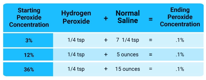
How to Improve Your Vitamin B Status
As a general rule, I recommend getting most if not all of your nutrition from real food. This will work well for most B vitamins, but not if you’re using niacin therapeutically, as described above. For that, you will need to take a supplement.
That said, the list below will show you which foods contain which B vitamins, as well as provide general guidance on dosage if you’re taking a supplement. If you’re trying to improve your vitamin B status, also consider limiting sugar and eating more fermented foods.
The reason for this is because the entire B group vitamin series is produced within your gut, assuming you have healthy gut flora. Eating real food, including plenty of leafy greens and fermented foods, will provide your microbiome with important fiber and beneficial bacteria to help optimize your internal vitamin B production.
| Nutrient | Dietary Sources | Supplement Recommendations |
|---|---|---|
| Vitamin B1 | Pork, fish, nuts and seeds, beans, green peas, brown rice, squash, asparagus and seafood.24 | The recommended daily allowance for B1 is 1.2 mg/day for men and 1.1 mg/day for women.25 |
| Vitamin B2 | Eggs, organ meats, lean meats, green vegetables such as asparagus, broccoli and spinach.26 | The RDA is 1.1 mg for adult women and 1.3 mg for men.
Your body cannot absorb more than about 27 mg at a time, and some multivitamins or B-complex supplements may contain unnecessarily high amounts.27 |
| Vitamin B3 | Liver, chicken, veal, peanuts, chili powder, bacon and sun-dried tomatoes have some of the highest amounts of niacin per gram.28
Other niacin-rich foods include baker’s yeast, paprika, espresso coffee, anchovies, spirulina, duck, shiitake mushrooms and soy sauce.29 |
The dietary reference intake established by the Food and Nutrition Board ranges from 14 to 18 mg per day for adults.
Higher amounts are recommended depending on your condition. For a list of recommended dosages, see the Mayo Clinic’s website.30 The dosage recommended as an anti-inflammatory, health-restorative therapy in “Sufficient Niacin Supply: The Missing Puzzle Piece to COVID-19 and Beyond?”31 is 500 mg two to three times a day, working your way up to 1,000 mg, two to three times a day as the flushing lessens. |
| Vitamin B5 | Beef, poultry, seafood, organ meats, eggs, milk, mushrooms, avocados, potatoes, broccoli, peanuts, sunflower seeds, chickpeas and brown rice.32 | The RDA is 5 mg for adults over the age of 19.
Pantothenic acid in dietary supplements is often in the form of calcium pantothenate or pantethine.33 |
| Vitamin B6 | Turkey, beef, chicken, wild-caught salmon, sweet potatoes, potatoes, sunflower seeds, pistachios, avocado, spinach and banana.34,35 | Nutritional yeast is an excellent source of B vitamins, especially B6.36
One serving (2 tablespoons) contains nearly 10 mg of vitamin B6. Not to be confused with Brewer’s yeast or other active yeasts, nutritional yeast is made from an organism grown on molasses, which is then harvested and dried to deactivate the yeast. It has a pleasant cheesy flavor and can be added to a number of different dishes. |
| Vitamin B9 | Fresh, raw, organic leafy green vegetables, especially broccoli, asparagus, spinach and turnip greens, and a wide variety of beans, especially lentils, but also pinto beans, garbanzo beans, kidney beans, navy and black beans.37 | Folic acid is a synthetic type of B vitamin used in supplements; folate is the natural form found in foods.
(Think: Folate comes from foliage, edible leafy plants.) For folic acid to be of use, it must first be activated into its biologically active form (L-5-MTHF). Nearly half the population has difficulty converting folic acid into the bioactive form due to a genetic reduction in enzyme activity. For this reason, if you take a B-vitamin supplement, make sure it contains natural folate rather than synthetic folic acid. Nutritional yeast is an excellent source.38 Research39 also shows your dietary fiber intake has an impact on your folate status. For each gram of fiber consumed, folate levels increased by nearly 2%. The researchers hypothesize that this boost in folate level is due to the fact that fiber nourishes bacteria that synthesize folate in your large intestine. |
| Vitamin B12 | Vitamin B12 is found almost exclusively in animal tissues, including foods like beef and beef liver, lamb, snapper, venison, salmon, shrimp, scallops, poultry, eggs and dairy products.
The few plant foods that are sources of B12 are actually B12 analogs that block the uptake of true B12. |
Nutritional yeast is high in B12, and is highly recommended for vegetarians and vegans.
One serving (2 tablespoons) provides nearly 8 mcg of natural vitamin B12.40 Sublingual (under-the-tongue) fine mist spray or vitamin B12 injections are also effective, as they allow the large B12 molecule to be absorbed directly into your bloodstream. |
– Sources and References
- 1 Maturitas 2021 Feb; 144: 108–111
- 2 Yahoo August 27, 2020
- 3, 4, 5, 13, 14 Maturitas August 15, 2020 DOI: 10.1016/j.maturitas.2020.08.007 [Epub ahead of print]
- 6, 7 ChemRxiv March 30, 2020 DOI: 10.26434/chemrxiv.12034980.v1 (PDF)
- 8 Research Square May 26, 2020 DOI: 10.21203/rs.3.rs-31775/v1
- 9 PLoS ONE 5(9): e12244
- 10 New York Times September 6, 2016
- 11 Maturitas August 15, 2020 DOI: 10.1016/j.maturitas.2020.08.007 [Epub ahead of print], 1.7. Vitamin B12
- 12, 16, 18, 19, 20, 21, 22, 23, 31 OSF Preprints Sufficient Niacin Supply: The Missing Puzzle Piece to COVID-19?
- 15 Vitamin D (Third Edition) 2011: 707-723
- 17 Cell Death and Differentiation 2020 May;27(5):1451-1454
- 24 My Food Data, Thiamine
- 25 Dietary Reference Intakes for B Vitamins
- 26, 27 NIH.gov Riboflavin
- 28 Healthaliciousness.com, Top 10 High Niacin Foods
- 29 Healthaliciousness.com, Niacin-Rich Foods
- 30 Mayo Clinic Niacin Dosing
- 32, 33 NIH.gov Pantothenic Acid
- 34 NIH Vitamin B6 Fact Sheet December 10, 2019
- 35 Healthalisciousness.com, Top 10 Foods High in Vitamin B6
- 36, 40 Self Nutrition Data, Nutritional Yeast
- 37 NIH Folate March 29, 2021
- 38 Chalkboard, Nutritional Yeast
- 39 American Journal of Clinical Nutrition 1997 Dec;66(6):1414-21
Protect Your Bones With 8 Natural Osteoprotectives
© GreenMedInfo LLC. This work is reproduced and distributed with the permission of GreenMedInfo LLC.
Want to learn more from GreenMedInfo? Sign up for the newsletter here:
www.greenmedinfo.com/greenmed/newsletter
Reproduced from original article:
www.greenmedinfo.com/blog/protect-your-bones-8-natural-osteoprotectives
Posted on: Friday, October 8th 2021 at 2:30 pm
Written By: Dr. Diane Fulton
This article is copyrighted by GreenMedInfo LLC, 2021

Your bones support your entire body. Isn’t it time to find natural alternatives to strengthen them?
Bones are literally the structure of your body. As you age, bone loss happens. But there are eight natural osteoprotectives — including puerarin, boswellia, citrus naringin, resveratrol, certain vitamins and whole foods like dried plums — that could help prevent the deterioration of your bones. When bones lose their density, this can lead to osteoporosis — a painful and debilitating condition.
1. Puerarin
Puerarin, an isoflavone from the kudzu plant, has osteoprotective properties. Ovariectomy-induced mice are often used for researching treatments for postmenopausal osteoporosis. In a systematic review of eight such animal studies with 203 subjects, puerarin significantly improved bone mass.[i]
Puerarin alleviates osteoclast-related loss of bone mass in ovariectomy-induced osteoporotic rats by inhibiting the tumor necrosis factor receptor–associated factor/reactive oxygen species or TRAF6/ROS-dependent MAPK/NF-κB signaling pathway within the bone tissue.[ii]
In an osteoporosis model of overiectomized rats, the anti-osteoporosis effects of puerarin were related to improvements in gut microbiota via regulating short chain fatty acid levels and repairing the intestinal mucosal integrity.[iii]
A high dose of puerarin and zinc together in an ovariectomized rat model worked better than either alone — reversing some bone loss and suppressing the adiposity of bone marrow, a marker of osteoporosis.[iv]
Type 1 and Type 2 diabetes are often associated with increased risk of bone fractures, osteopenia and osteoporosis. In a diabetes-induced study of mice, puerarin markedly attenuated bone loss and suppressed inflammatory markers associated with osteoporosis by inhibiting histone deacetylases (HDAC1/HDAC3) enzyme signaling.[v]
In another study of diabetes-induced rats, those given a puerarin injection of 100 milligrams (mg) per kilogram of body weight per day for six weeks had higher bone mineral density, improved osteoblast numbers, new bone formation and reduced caspase-3 expression — a marker for diabetic osteoporosis — compared to the control group.[vi]
2. Vitamin C
Many in vitro and animal model studies confirm the significant influence of vitamin C (ascorbic acid) on the skeletal system, and those who have a severe vitamin C deficiency would benefit from supplementation.
However, if you do not have such a deficiency, it is better to get your vitamin C by eating five servings of vegetables and fruits a day, rather than through vitamin supplementation, which remains controversial.[vii] In fact, some reviewers found vitamin C supplementation could actually increase your risk of fractures.[viii]
Greater dietary vitamin C intake — fruits and vegetables — was associated with a 33% lower risk of osteoporosis and a lower risk of hip fractures, as well as higher bone mineral density in a meta-analysis study.[ix] Similarly, in a study of 73 healthy people, those who added more plant foods to their diet increased their bone mineral density and had higher vitamin D levels.[x]
In a meta-analysis of six articles with 225,062 people, those who added at least one serving of fruits and vegetables per day to their diet decreased their bone fracture risk.[xi]
3. Vitamin D
Vitamin D deficiency has been linked to poor bone health, osteoporosis and higher risk of bone fractures. In a review of 66 patients aged 50 years or more with hip fractures, 74% had vitamin D deficiencies, with 62% diagnosed with osteoporosis and 18% with severe osteoporosis.[xii]
In a study of 100 postmenopausal women, 47% of the group was deficient in vitamin D and 31% had insufficient levels. Hip osteoporosis was 31.9% in the vitamin D deficient group compared to 18.2% in those with sufficient vitamin D levels. Vitamin D insufficiency is a risk factor for osteoporosis associated with increased bone remodeling and low bone mass.[xiii]
In an induced-vitamin D deficiency mouse model, vitamin D treatment reversed age-related osteoporosis symptoms while promoting proliferation, osteogenic differentiation and bone formation of bone marrow mesenchymal stem cells and osteoblasts — cells that build bones — while inhibiting bone aging and bone resorption — removal of old bones.
If bone resorption is higher than bone building, there are overall bone deficits.[xiv]
In research of induced-vitamin D deficiency mice, low vitamin D levels accelerated age-related bone loss, increased oxidative stress and DNA damage, were associated with higher bone cell aging, inhibited osteoblastic bone formation and stimulated osteoclastic bone resorption.[xv]
4. Vitamin K2
Vitamin K2 plays a significant role in the prevention and treatment of osteoporosis because it regulates bone remodeling. When the balance between bone resorption and bone formation shifts to a net bone loss, both men and women can develop osteoporosis.[xvi]
In a human vitro cell study, vitamin K2 promoted the osteogenic differentiation of mesenchymal stem cells by inhibiting miR-133a expression — an impactor of genes in muscle and skeletal development.[xvii] In a human studies review of vitamin K2, the commonly used dosage was 45 mg per day and highly benefitted bone health by regulating bone growth and bone loss mechanisms, particularly in osteoporotic postmenopausal women.[xviii]
Trial subjects including 311 men and postmenopausal women 50 to 75 years of age were randomly assigned to four groups — placebo, 50 micrograms per day or 90 micrograms per day of vitamin K2 or co-supplementation with calcium (500 mg per day) and vitamin D (10 micrograms per day) for one year.
The bone loss of femoral neck was significantly lower in postmenopausal women in the high dose vitamin K2 group compared with placebo, but had no effect in men. High dose supplementation of K2 significantly reduced bone loss in postmenopausal women with no benefit of adding calcium and vitamin D.[xix]
In a comprehensive review of vitamin K2, supplementation modulated necrosis factor kappa beta — NF-κB — signaling in the body, which ameliorated bone loss and promoted bone health.[xx]
One preclinical study showed that vitamin K2 administration in a Type 2 diabetic rat model increased serum osteocalcin, improved collagen cross-link profiles and increased bone strength, suggesting it as a preventative for bone deterioration and fractures resulting from Type 2 diabetes.[xxi]
5. Boswellia or Frankincense
Osteoarthritis is a joint disease involving articular cartilage degeneration causing patients pain, joint stiffness, physical disability and significantly reducing quality of life. Forty-nine patients who took a boswellia- and bromelain-based supplement for a period between one and six months found it significantly improved quality of life and various osteoarthritis symptoms.[xxii]
Based on a meta-analysis of seven trials including 545 osteoarthritis patients, boswellia effectively and safely ameliorated pain, stiffness and joint function with ideal treatment for four or more weeks.[xxiii] Boswellia — also called frankincense — has been found to play a significant role in eliminating inflammation and joint destruction in knee osteoarthritis, with no serious side effects.[xxiv]
The boswellic acids found in gum resin extract were strong anti-inflammatory agents and protectors of cartilage cells in a human in vitro study and improved pain and weight-bearing ability in osteoarthritis-induced rats, which received the supplement for 28 days.[xxv]
In an osteoarthritis-induced rat model, boswellia was orally administered once per day for three weeks and inhibited increases in osteoarthritis symptoms, synovial fluid cytokines, cartilage damage and expression levels of pro-inflammatory mediators/cytokines in the cartilage.[xxvi]
6. Citrus Naringin
Citrus naringin — a natural flavanone glycoside present in plants like grapefruits, tart cherries, tomatoes, and oregano — modulates different signaling pathways and interacts with numerous cell signaling molecules to treat inflammation, oxidative stress, metabolic syndrome, bone disorders and cancers.[xxvii]
Naringin enhanced osteogenic differentiation of human bone marrow mesenchymal stem cells seen through in vitro study by activating the extracellular signal-regulated kinase, or ERK signaling pathway.[xxviii]
Supplementation with naringin and rabbit bone marrow mesenchymal stem cells together was more efficient in repairing cartilage defects in rabbit knees than the use of either treatment alone. Citrus naringin activates and continuously regulates the growth factor beta, or TGF-β, signaling pathway, which encourages these stem cells to differentiate into chondrocytes, giving function and structure to cartilage.[xxix]
Naringin promoted bone repair and prevented bone loss in a drug-induced osteonecrosis rat model and through in vitro study impacted the Akt/Bad signal cascades.[xxx] Naringin also protected against bone loss in a steroid-treated inflammatory bowel disease rat model.[xxxi]
In a meta-analysis of naringin’s effect on postmenopausal osteoporosis in 10 ovariectomized rat studies, citrus naringin improved bone mineral density, bone volume and thickness.[xxxii]
7. Resveratrol
A comprehensive systematic review and meta-analysis of eight studies with 264 subjects showed improvements in two bone biomarkers after resveratrol supplementation over placebo treatment.
Compound medicine of both tanshinone and resveratrol showed the most improvement on peak bone mass compared to placebo or each alone in 40 growing rats.[xxxiii] In a bovine cartilage model, supplementation with both resveratrol and curcumin together effectively decelerated age-related and diabetes-induced osteoporosis.[xxxiv]
8. Dried Plums
Menopause drastically increases the risk of osteoporosis — ovarian hormone production stops, which causes accelerated bone loss. Fifty-eight postmenopausal women were randomly assigned to either 100 grams of dried plums or 75 grams of dried apples daily for three months. The dried plums significantly increased two indices of higher bone formation in postmenopausal women.[xxxv]
A follow up study — five years later — of postmenopausal women who previously consumed 100 grams of dried plums per day during a three-month trial showed long-lasting bone-protective effects.[xxxvi]
Osteoprotection
Using natural supplements such as pueraria, boswellia, certain vitamins and resveratrol or adding fruits and vegetables to your diet can help protect your bone health. Consult GreenMedInfo.com’s database for more detailed research on osteoprotectives, osteoporosis, osteoarthritis, bone fractures and bone diseases.
References
[i] Xiaobin Yang, Haishi Zheng, Yuan Liu, Dingjun Hao, Baorong He, Lingbo Kong. Puerarin for OVX-induced postmenopausal osteoporosis in murine model: systematic review and meta-analysis. Curr Stem Cell Res Ther. 2019 Jul 3. Epub 2019 Jul 3. PMID: 31269886
[ii] Long Xiao, Mengdan Zhong, Yu Huang, Jie Zhu, Wenkai Tang, Danyong Li, Jiandong Shi, Aiqing Lu, Huilin Yang, Dechun Geng, Hong Li, Zhirong Wang. Puerarin alleviates osteoporosis in the ovariectomy-induced mice by suppressing osteoclastogenesis via inhibition of TRAF6/ROS-dependent MAPK/NF-κB signaling pathways. Aging (Albany NY). 2020 11 7 ;12(21):21706-21729. Epub 2020 Nov 7. PMID: 33176281 [iii] Bo Li, Mingyan Liu, Yu Wang, Shiqiang Gong, Weifan Yao, Wenshuai Li, Hua Gao, Minjie Wei. Puerarin improves the bone micro-environment to inhibit OVX-induced osteoporosis via modulating SCFAs released by the gut microbiota and repairing intestinal mucosal integrity. Biomed Pharmacother. 2020 Dec ;132:110923. Epub 2020 Oct 28. PMID: 33125971 [iv] Hao Liu, Wei Li, Xiyuan Ge, Shengnan Jia, Binbin Li. Coadministration of puerarin (low dose) and zinc attenuates bone loss and suppresses bone marrow adiposity in ovariectomized rats. Life Sci. 2016 Dec 1 ;166:20-26. Epub 2016 Sep 30. PMID: 27697446 [v] Chang-Jun Guo, Jing-Jing Xie, Rong-Hua Hong, Han-Song Pan, Fu-Guo Zhang, Yi-Min Liang. Puerarin alleviates streptozotocin (STZ)-induced osteoporosis in rats through suppressing inflammation and apoptosis via HDAC1/HDAC3 signaling. Biomed Pharmacother. 2019 May 1 ;115:108570. Epub 2019 May 1. PMID: 31054509 [vi] Junbo Liang, Haixiao Chen, Weibo Pan, Chunli Xu. Puerarin inhibits caspase-3 expression in osteoblasts of diabetic rats. Mol Med Rep. 2012 Jun ;5(6):1419-22. Epub 2012 Apr 2. PMID: 22470138 [vii] Brzezińska O, Łukasik Z, Makowska J, Walczak K. Role of Vitamin C in Osteoporosis Development and Treatment-A Literature Review. Nutrients. 2020 Aug 10;12(8):2394. doi: 10.3390/nu12082394. PMID: 32785080; PMCID: PMC7469000 [viii] Heike A Bischoff-Ferrari, Bess Dawson-Hughes, John A Baron, Peter Burckhardt, Ruifeng Li, Donna Spiegelman, Bonny Specker, John E Orav, John B Wong, Hannes B Staehelin, Eilis O’Reilly, Douglas P Kiel, Walter C Willett. Calcium intake and hip fracture risk in men and women: a meta-analysis of prospective cohort studies and randomized controlled trials. Am J Clin Nutr. 2007 Dec;86(6):1780-90. PMID: 18065599 [ix] Hanieh Malmir, Sakineh Shab-Bidar, Kurosh Djafarian. Vitamin C intake in relation to bone mineral density and risk of hip fracture and osteoporosis: a systematic review and meta-analysis of observational studies. Br J Nutr. 2018 Apr ;119(8):847-858. PMID: 29644950 [x] J Berg, N Seyedsadjadi, R Grant. Increased Consumption of Plant Foods Is Associated with Increased Bone Mineral Density. J Nutr Health Aging. 2020 ;24(4):388-397. PMID: 32242206 [xi] Juliana E Brondani, Fabio V Comim, Liziane M Flores, Lígia Araújo Martini, Melissa O Premaor. Fruit and vegetable intake and bones: A systematic review and meta-analysis. PLoS One. 2019 ;14(5):e0217223. Epub 2019 May 31. PMID: 31150426 [xii] Ankit Dadra, Sameer Aggarwal, Prasoon Kumar, Vishal Kumar, Deba Prasad Dibar, Sanjay Kumar Bhadada. High prevalence of vitamin D deficiency and osteoporosis in patients with fragility fractures of hip: A pilot study. J Clin Orthop Trauma. 2019 Nov-Dec;10(6):1097-1100. Epub 2019 Mar 21. PMID: 31708635 [xiii] Vasundhara Kamineni, Akkenapally Prasanna Latha, K Ramathulasi. Association between serum 25-hydroxyvitamin D levels and bone mineral density in normal postmenopausal women. J Midlife Health. 2016 Oct-Dec;7(4):163-168. PMID: 28096639 [xiv] Renlei Yang, Jie Chen, Jiao Zhang, Ran Qin, Rong Wang, Yue Qiu, Zhiyuan Mao, David Goltzman, Dengshun Miao. 1,25-Dihydroxyvitamin D protects against age-related osteoporosis by a novel VDR-Ezh2-p16 signal axis. Aging Cell. 2020 02 ;19(2):e13095. Epub 2019 Dec 26. PMID: 31880094 [xv] Wanxin Qiao, Shuxiang Yu, Haijian Sun, Lulu Chen, Rong Wang, Xuan Wu, David Goltzman, Dengshun Miao. 1,25-Dihydroxyvitamin D insufficiency accelerates age-related bone loss by increasing oxidative stress and cell senescence. Am J Transl Res. 2020 ;12(2):507-518. Epub 2020 Feb 15. PMID: 32194899 [xvi] V D Myneni, E Mezey. Regulation of bone remodeling by vitamin K2. Oral Dis. 2017 Nov ;23(8):1021-1028. Epub 2017 Apr 5. PMID: 27976475 [xvii] Yuelei Zhang, Shiyang Weng, Junhui Yin, Hao Ding, Changqing Zhang, Youshui Gao. Vitamin K2 promotes mesenchymal stem cell differentiation by inhibiting miR-133a expression. Mol Med Rep. 2017 May ;15(5):2473-2480. Epub 2017 Mar 9. PMID: 28447758 [xviii] Julia Khéde Dourado Villa, Marisa Alves Nogueira Diaz, Virgínia Ramos Pizziolo, Hércia Stampini Duarte Martino. Effect of vitamin K in bone metabolism and vascular calcification: A review of mechanisms of action and evidences. Crit Rev Food Sci Nutr. 2017 Dec 12 ;57(18):3959-3970. PMID: 27437760 [xix] Yingfeng Zhang, Zhipeng Liu, Lili Duan, Yeyu Ji, Sen Yang, Yuan Zhang, Hongyin Li, Yu Wang, Peng Wang, Jiepeng Chen, Ying Li. Effect of Low-Dose Vitamin K2 Supplementation on Bone Mineral Density in Middle-Aged and Elderly Chinese: A Randomized Controlled Study. Calcif Tissue Int. 2020 Feb 14. Epub 2020 Feb 14. PMID: 32060566 [xx] Masayoshi Yamaguchi, M Neale Weitzmann. Vitamin K2 stimulates osteoblastogenesis and suppresses osteoclastogenesis by suppressing NF-κB activation. Int J Mol Med. 2011 Jan;27(1):3-14. Epub 2010 Nov 11. PMID: 21072493 [xxi] Jun Iwamoto, Yoshihiro Sato, Tsuyoshi Takeda, Hideo Matsumoto. Bone quality and vitamin K2 in type 2 diabetes: review of preclinical and clinical studies. Nutr Rev. 2011 Mar;69(3):162-7. Epub 2011 Feb 14. PMID: 21348880 [xxii] Giovanni Italiano, Mariangela Raimondo, Giovanna Giannetti, Anna Gargiulo. Benefits of a Food Supplement Containingand Bromelain for Improving the Quality of Life in Patients with Osteoarthritis: A Pilot Study. J Altern Complement Med. 2019 Nov 1. Epub 2019 Nov 1. PMID: 31674795 [xxiii] Giovanni Italiano, Mariangela Raimondo, Giovanna Giannetti, Anna Gargiulo. Benefits of a Food Supplement Containingand Bromelain for Improving the Quality of Life in Patients with Osteoarthritis: A Pilot Study. J Altern Complement Med. 2019 Nov 1. Epub 2019 Nov 1. PMID: 31674795 [xxiv] Raju Vaishya, Amit Kumar Agarwal, Amish Shah, Vipul Vijay, Abhishek Vaish. Current status of top 10 nutraceuticals used for Knee Osteoarthritis in India. J Clin Orthop Trauma. 2018 Oct-Dec;9(4):338-348. Epub 2018 Jul 20. PMID: 30449982 [xxv] Venkata Krishnaraju Alluri, Sreenath Kundimi, Krishanu Sengupta, Trimurtulu Golakoti, Eswar Kumar Kilari. An Anti-Inflammatory Composition ofResin Extracts Alleviates Pain and Protects Cartilage in Monoiodoacetate-Induced Osteoarthritis in Rats. Evid Based Complement Alternat Med. 2020 ;2020:7381625. Epub 2020 May 21. PMID: 32565872 [xxvi] Hae Lim Kim, Hae Jin Lee, Dong-Ryung Lee, Bong-Keun Choi, Seung Hwan Yang. Herbal Composition LI73014F2 Alleviates Articular Cartilage Damage and Inflammatory Response in Monosodium Iodoacetate-Induced Osteoarthritis in Rats. Molecules. 2020 Nov 23 ;25(22). Epub 2020 Nov 23. PMID: 33238379 [xxvii] Maryam Ghanbari-Movahed, Gloria Jackson, Mohammad Hosein Farzaei, Anupam Bishayee. A Systematic Review of the Preventive and Therapeutic Effects of Naringin Against Human Malignancies. Front Pharmacol. 2021 ;12:639840. Epub 2021 Mar 29. PMID: 33854437 [xxviii] Huichao Wang, Chunbo Li, Jianming Li, Yingjie Zhu, Yudong Jia, Ying Zhang, Xiaodong Zhang, Wenlong Li, Lei Cui, Wuyin Li, Youwen Liu. Naringin enhances osteogenic differentiation through the activation of ERK signaling in human bone marrow mesenchymal stem cells. Iran J Basic Med Sci. 2017 Apr ;20(4):408-414. PMID: 28804610 [xxix] Chao Ye, Jing Chen, Yi Qu, Hang Liu, Junxing Yan, Yingdong Lu, Zheng Yang, Fengxian Wang, Pengyang Li. Naringin and bone marrow mesenchymal stem cells repair articular cartilage defects in rabbit knees through the transforming growth factor-β superfamily signaling pathway. Exp Ther Med. 2020 Nov ;20(5):59. Epub 2020 Sep 4. PMID: 32952649 [xxx] Ming-Jie Kuang, Wei-Hao Zhang, Wei-Wei He, Lei Sun, Jian-Xiong Ma, Dachuan Wang, Xin-Long Ma. Naringin regulates bone metabolism in glucocorticoid-induced osteonecrosis of the femoral head via the Akt/Bad signal cascades. Chem Biol Interact. 2019 May 1 ;304:97-105. Epub 2019 Mar 14. PMID: 30878453 [xxxi] Chengli Li, Jun Zhang, Fang Lv, Xingtao Ge, Gang Li. Naringin protects against bone loss in steroid-treated inflammatory bowel disease in a rat model. Arch Biochem Biophys. 2018 Jul 15 ;650:22-29. Epub 2018 May 17. PMID: 29753722 [xxxii] Zhu Zhu, Wenjing Xie, Yanyan Li, Zaiou Zhu, Wei Zhang. Effect of Naringin Treatment on Postmenopausal Osteoporosis in Ovariectomized Rats: A Meta-Analysis and Systematic Review. Evid Based Complement Alternat Med. 2021 ;2021:6016874. Epub 2021 Feb 10. PMID: 33628301 [xxxiii] Fang-Fang Yang, Yu-Hai Gao, Hui-Rong Xi, Wen-Yuan Li, Hui-Ping Ma, Ke-Ming Chen. Effect of Compound Medicine of Tanshinone 2A and Resveratrol on Peak Bone Mass in Growing Rats. Zhongguo Yi Xue Ke Xue Yuan Xue Bao. 2018 Aug 30 ;40(4):456-462. PMID: 30193597 [xxxiv] Shikhar Mehta, Cameron C Young, Matthew R Warren, Sumayyah Akhtar, Sandra J Shefelbine, Justin D Crane, Ambika G Bajpayee. Resveratrol and Curcumin Attenuate Sugar-Induced Cartilage Glycation, Stiffening, Senescence, and Degeneration. Cartilage. 2021 Jan 20:1947603520988768. Epub 2021 Jan 20. PMID: 33472415 [xxxv] Bahram H Arjmandi, Dania A Khalil, Edralin A Lucas, Amanda Georgis, Barbara J Stoecker, Claudia Hardin, Mark E Payton, Robert A Wild. Dried plums improve indices of bone formation in postmenopausal women. Altern Ther Health Med. 2010 Mar-Apr;16(2):12-4. PMID: 11860726 [xxxvi] Bahram H Arjmandi, Sarah A Johnson, Shirin Pourafshar, Negin Navaei, Kelli S George, Shirin Hooshmand, Sheau C Chai, Neda S Akhavan. Bone-Protective Effects of Dried Plum in Postmenopausal Women: Efficacy and Possible Mechanisms. Nutrients. 2017 May 14 ;9(5). Epub 2017 May 14. PMID: 28505102
