Vitamins
now browsing by category
Bradykinin Hypothesis Explains COVID-19 Complexities
Reproduced from original article:
https://articles.mercola.com/sites/articles/archive/2020/09/14/bradykinin-hypothesis.aspx
Analysis by Dr. Joseph Mercola Fact Checked September 14, 2020
STORY AT-A-GLANCE
- Genetic analysis using the Oak Ridge National Lab supercomputer has revealed an interesting new hypothesis — the bradykinin hypothesis — that helps explain the disease progression of COVID-19
- The bradykinin hypothesis also strengthens the hypothesis that vitamin D plays a really important role in the disease
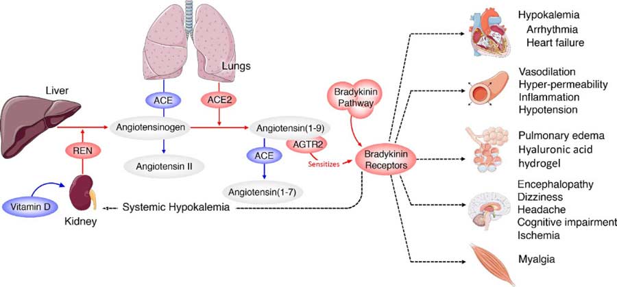
- SARS-CoV-2 not only infects cells that naturally have high numbers of ACE2 receptors but also tricks your body into upregulating ACE2 receptors in places where they’re usually expressed at lower levels, such as your lungs
- SARS-CoV-2 also downregulates your body’s ability to degrade or break down bradykinin, a chemical that helps regulate your blood pressure and is controlled by your renin-angiotensin system
- The end result is a bradykinin storm, which appears to be the primary cause behind many of COVID-19’s lethal effects, even more so than the cytokine storms associated with the disease
- The virus also increases production of hyaluronic acid (HLA) in your lungs. HLA has the ability to absorb more than 1,000 times its own weight in fluid, and when it combines with the built-up fluid in the lungs, it forms a thick hydrogel that makes breathing very difficult
Genetic analysis using the Oak Ridge National Lab supercomputer called the Summit has revealed an interesting new hypothesis that helps explain the disease progression of COVID-19. A September 1, 2020, Medium article1 by Thomas Smith reviewed the findings of what is now referred to as the bradykinin hypothesis.
As reported by Smith, the computer crunched data on more than 40,000 genes obtained from 17,000 genetic samples.
“Summit is the second-fastest computer in the world, but the process — which involved analyzing 2.5 billion genetic combinations — still took more than a week. When Summit was done, researchers analyzed the results. It was, in the words of Dr. Daniel Jacobson, lead researcher and chief scientist for computational systems biology at Oak Ridge, a ‘eureka moment.’”
The Bradykinin Hypothesis
Bradykinin is a chemical that helps regulate your blood pressure and is controlled by your renin-angiotensin system (RAS). As explained in the Academic Press’ book on vitamin D (which has a significant impact on the RAS):2
“The renin-angiotensin system (RAS) is a central regulator of renal and cardiovascular functions. Over-activation of the RAS leads to renal and cardiovascular disorders, such as hypertension and chronic kidney disease, the major risk factors for stroke, myocardial infarction, congestive heart failure, progressive atherosclerosis, and renal failure.”
The bradykinin hypothesis provides a model that helps explain some of the more unusual symptoms of COVID-19, including its bizarre effects on the cardiovascular system. It also strengthens the hypothesis that vitamin D plays a really important role in the disease.
The findings3 were published in the journal eLife July 7, 2020. Based on this new hypothesis, the researchers also suggest more than 10 potential treatments, most of which are readily available drugs already approved by the U.S. Food and Drug Administration. I’ll review those later on.
As detailed in previous articles, your ACE2 receptors are the primary gateways of the virus, as the virus’ spike protein binds to the ACE2 receptor. As explained by Smith:4
“… COVID-19 infection generally begins when the virus enters the body through ACE2 receptors in the nose … The virus then proceeds through the body, entering cells in other places where ACE2 is also present: the intestines, kidneys, and heart. This likely accounts for at least some of the disease’s cardiac and GI symptoms.
But once Covid-19 has established itself in the body, things start to get really interesting … The data Summit analyzed shows that COVID-19 isn’t content to simply infect cells that already express lots of ACE2 receptors. Instead, it actively hijacks the body’s own systems, tricking it into upregulating ACE2 receptors in places where they’re usually expressed at low or medium levels, including the lungs.
In this sense, COVID-19 is like a burglar who slips in your unlocked second-floor window and starts to ransack your house. Once inside, though, they don’t just take your stuff — they also throw open all your doors and windows so their accomplices can rush in and help pillage more efficiently.”

Bradykinin Storm Likely Responsible for Lethal Effects
In addition to upregulating ACE2 receptors throughout your body, the SARS-CoV-2 virus also downregulates your body’s ability to degrade or break down bradykinin.
The end result is a bradykinin storm, and according to the researchers, this appears to be an important factor in many of COVID-19’s lethal effects, even more so than the cytokine storms associated with the disease. As bradykinin accumulates, the more serious COVID-19 symptoms appear.
Mounting clinical data suggest COVID-19 is actually primarily a vascular disease rather than a respiratory one, and runaway bradykinin buildup can help explain this.
How SARS-CoV-2 Attacks the Lungs
That said, COVID-19 certainly has a respiratory component, and it appears the virus attacks the lungs in more ways than one. For starters, bradykinin increases vascular permeability, essentially causing your blood vessels to leak fluid. In the lungs, this leads to fluid buildup that can trigger inflammation when immune cells also leak out into the lungs.
But the Summit data also show the virus uses yet another pathway, which raises production of hyaluronic acid (HLA) in your lungs. HLA has the ability to absorb more than 1,000 times its own weight in fluid, and when it combines with the built-up fluid in the lungs, the effect is devastating, as it ends up forming a thick hydrogel that makes breathing near-impossible.
When this happens — in severe cases — even mechanical ventilation becomes ineffective, as the alveoli in the lungs are simply too clogged with this gel-like substance that prevents oxygen uptake.
Note that the HLA produced in your lungs does not mean that using supplemental HLA is a bad strategy. It is only when HLA is produced locally in high concentrations in pathologic conditions like COVID-19 that it becomes problematic. Otherwise it has important physiologic benefits.
How SARS-CoV-2 Attacks Your Heart and Brain
SARS-CoV-2 can also affect heart function, causing arrhythmias and low blood pressure. About 1 in 5 COVID-19 patients requiring hospitalizations have experienced damage to their heart. Your heart has ACE2 receptors, so SARS-CoV-2 has the ability to infect your heart directly. Arrhythmias and low blood pressure can also be the result of a bradykinin storm.
In some cases, COVID-19 has also been known to trigger neurological symptoms such as dizziness, seizures, delirium and stroke, and this too can be explained by bradykinin buildup.
At high levels, bradykinin can lead to a breakdown of your blood-brain barrier, thereby allowing harmful compounds to flood your brain. Bradykinin itself also causes blood vessel leakage. Together, these effects can trigger inflammation, brain damage and a variety of neurological symptoms.
SARS-CoV-2 Mimics ACE Inhibiting Drugs
Interestingly, as reported by Smith:5
“Increased bradykinin levels could also account for other common COVID-19 symptoms. ACE inhibitors — a class of drugs used to treat high blood pressure — have a similar effect on the RAS system as COVID-19, increasing bradykinin levels.
In fact, Jacobson and his team note in their paper that ‘the virus … acts pharmacologically as an ACE inhibitor’ — almost directly mirroring the actions of these drugs.
By acting like a natural ACE inhibitor, COVID-19 may be causing the same effects that hypertensive patients sometimes get when they take blood pressure-lowering drugs. ACE inhibitors are known to cause a dry cough and fatigue, two textbook symptoms of COVID-19.
And they can potentially increase blood potassium levels, which has also been observed in COVID-19 patients. The similarities between ACE inhibitor side effects and COVID-19 symptoms strengthen the bradykinin hypothesis, the researchers say.”
Another side effect associated with ACE inhibiting drugs is the loss of smell and taste. This is also an early sign associated with SARS-CoV-2 infection, and it’s a primary symptom of zinc deficiency too.
Zinc, as explained in “Swiss Protocol for COVID — Quercetin and Zinc,” plays a vital role in immunity as well as in blood clotting, cell division, thyroid health, smell and taste, vision and wound healing, and can effectively inhibit viral replication.
Your body does not store zinc, and it’s poorly absorbed, which appears to be why the combination of zinc and zinc ionophores such as quercetin and hydroxychloroquine are so effective when taken at first symptoms.
Bradykinin Hypothesis Explains Other COVID-19 Symptoms Too
The bradykinin storm also helps explain other odd COVID-19 symptoms such as “COVID toes,” a condition in which your toes become swollen and bruised. This may be due to leaky vasculature in your toes.
As explained by Smith, it can also shed helpful light on the gender differences seen in COVID-19. Women tend to have a lower mortality rate than men, and this may be due to the fact that women have twice the level of certain proteins involved in the RAS system.
Potential Treatments
The good news is that if bradykinin storms are to blame, there are a number of already existing drugs that can help prevent bradykinin storms, either reducing bradykinin or blocking its receptors. As noted in the study:6
“Several interventional points (most of them already FDA-approved pharmaceuticals) could be explored with the goal of increasing ACE, decreasing BK [bradykinin], or blocking BK2 receptors.
Icatibant is a BKB2R antagonist whereas Ecallantide acts to inhibit KLKB1, reducing levels of BK production. Androgens (danazol and stanasolol) increase SERPING1, although the side effects likely make these undesirable, but recombinant forms of SERPING1 could be administered to reduce BK levels.
It should be noted that any intervention may need to be timed correctly given that REN levels rise on a diurnal cycle, peaking at 4AM which corresponds with the commonly reported worsening of COVID-19 symptoms at night …
4-methylumbelliferone (Hymecromone) is a potent inhibitor of HAS1, HAS2, and HAS3 gene expression and results in the suppression of the production of hyaluronan in an ARDS model.
Hymecromone (4-methylumbelliferone) is approved for use in Asia and Europe for the treatment of biliary spasm. However, it can cause diarrhea with subsequent hypokalemia, so considerable caution should be used if this were to be tried with COVID-19 patients … Timbetasin may reduce COVID-19 related coagulopathies by increasing fibrinolysis.”
However, please understand that taking these drugs is absolutely not my recommendation. These are simply the conventional strategies that can be used if this hypothesis is correct.
Why on earth would we use dangerous drugs like these for a disease that they have not been tested on when we have so many other safe, inexpensive and highly effective interventions for COVID-19? Strategies like nebulized peroxide, ozone, molecular hydrogen, exogenous ketones, and quercetin with zinc.
Evidence for Vitamin D Strengthens
The researchers also highlight the usefulness of vitamin D, noting that “Another approach would be the modulation of REN levels via Vitamin D supplementation …” Vitamin D is involved in the RAS system,7,8,9 and can reduce a compound called renin (REN), thereby preventing a deadly bradykinin storm. Renin is an endopeptidase, the function of which is to generate angiotensin 1 from angiotensinogen in your plasma.
If you are vitamin D deficient, your renin expression is stimulated, and based on the latest data, that may render you more prone to bradykinin storm.
Several investigations have highlighted the apparent influence of vitamin D in COVID-19 incidence, severity and mortality, and its effects on RAS further strengthens the idea that vitamin D may be a crucial component in your COVID-19 defense arsenal.
As explained in the 2004 paper,10 “Vitamin D: A Negative Endocrine Regulator of the Renin-Angiotensin System and Blood Pressure,” when the RAS system is inappropriately activated, high blood pressure can result.
One factor that influences your RAS is your vitamin D level, as it suppresses renin biosynthesis. If you are vitamin D deficient, your renin expression is stimulated, and based on the latest data, that may render you more prone to bradykinin storm.
The Evidence for Vitamin D
In a November 1, 2020 commentary11 in the journal Metabolism Clinical and Experimental, JoAnn Manson and Shari Bassuk call for the elimination of vitamin D deficiency to effectively squelch the COVID-19 pandemic, noting that 23.3% of the total U.S. population have insufficient or deficient vitamin D levels, with people of color having disproportionately lower levels than non-Hispanic whites.
They list several types of studies showing vitamin D deficiency is “an important modifiable risk factor for COVID-19,” including:12
•Laboratory studies that demonstrate how vitamin D helps regulate immune function and the RAS, and modulate inflammatory responses to infection.
•Ecologic studies showing populations with lower vitamin D levels or lower UVB radiation exposure have higher COVID-19 mortality,13,14,15 and the fact that people identified as being at greatest risk for COVID-19 hospitalization and death (people of color, the elderly, nursing home residents and those with comorbidities such as obesity, vascular conditions and chronic kidney disease) also have a higher risk of vitamin D deficiency.
A pilot randomized clinical study16,17 published online August 29, 2020, found hospitalized COVID-19 patients in Spain who were given supplemental vitamin D (calcifediol) in addition to standard of care — which included the use of hydroxychloroquine and azithromycin — had significantly lower intensive care unit admissions.
Patients in the vitamin D arm received 532 micrograms of calcifediol on the day of admission (equivalent to 106,400 IUs of vitamin D18) followed by 266 mcg on Days 3 and 7 (equivalent to 53,200 IUs19). After that, they received 266 mcg once a week until discharge, ICU admission or death.
Of those receiving calcifediol, only 2% required ICU admission, compared to 50% of those who did not get calcifediol. None of those given vitamin D supplementation died, and all were discharged without complications.
•Observational studies showing low vitamin D levels are associated with a greater risk of testing positive for SARS-CoV-2 and contracting acute respiratory infections.
Most recently, a September 3, 2020 JAMA study20 reported that people who tested positive for SARS-CoV-2 were 1.77 times more likely to be deficient in vitamin D than those who tested negative for the virus.
•Randomized clinical trials showing vitamin D inhibits respiratory tract infections, especially in those with lower vitamin D levels at baseline.
REALLY IMPORTANT: Optimize Your Vitamin D Level Now
For years, I’ve stressed the importance of optimizing your vitamin D level, especially in anticipation of flu season, and it seems clear it can go a long way toward protecting yourself against COVID-19 as well.
Aside from what’s already been mentioned, vitamin D also helps Type II cells in your lungs produce surfactant that aids in fluid clearance. When you’re vitamin D deficient, your entire RAS is deranged or dysfunctional, thereby raising your risk of both bradykinin storm and cytokine storm.
In closing, experts have been warning that SARS-CoV-2 may reemerge in the fall when temperatures and humidity levels drop, thereby increasing the virus’ transmissibility.
Now is the time to check your vitamin D level and start taking action to raise it if you’re below 60 ng/mL. An easy and cost-effective way of measuring your vitamin D level is to order GrassrootsHealth’s vitamin D testing kit and learn more about vitamin D and its impact on your health.
Knowledge is empowerment, and that is particularly true during this pandemic. To learn more about the influence of vitamin D on your health in general and COVID-19 in particular, see my vitamin D report.
- 1, 4, 5 Medium September 1, 2020
- 2, 9 Vitamin D (Third Edition) 2011: 707-723
- 3, 6 eLife July 7, 2020; 9:e59177
- 7 Journal of Cellular Biochemistry February 1, 2003; 88(2):327-31
- 8 Journal of Nephropathology 2014; 3(2): 41–43
- 10 The Journal of Steroid Biochemistry and Molecular Biology May 2004; 89-90: 387-392
- 11 Metabolism Clinical and Experimental November 1, 2020; 112: 154322
- 12 etabolism Clinical and Experimental November 1, 2020; 112: 154322, Table 1
- 13 MedRxiv DOI: 10.1101/2020.04.08.20058578
- 14 Northwestern.edu May 7, 2020
- 15 Fox News May 7, 2020
- 16 The Journal of Steroid Biochemistry and Molecular Biology August 29, 2020 [Epub ahead of print]
- 17, 18, 19 Chrismasterjohnphd.com September 3, 2020
- 20 JAMA Network Open 2020;3(9):e2019722
Your lung function is an important predictor of health and longevity
Reproduced from original article:
www.naturalhealth365.com/lung-function-predicting-longevity-3550.html
by:
 (NaturalHealth365) Since none of us are immortal, the impulse to estimate how many years we might have left is perfectly natural. But, what is the most accurate predictor of lifespan? Some studies have focused on factors such as telomere length, while others have examined levels of important antioxidants like glutathione. But, it turns out that the biggest clue to longevity is your lung function.
In fact, a peer-reviewed study supports this point of view. And, with COVID on everyone’s mind, we thought this a very important topic to discuss. So, today, we’ll focus our attention on how lung health is directly connected to our ability to live a long, healthy life or not!
Health ALERT: Limited lung function can have severe consequences
Lung capacity is defined as the maximum amount of air the lungs can hold, while lung function involves the speed with which you can inhale and exhale. Lung function also involves how efficiently your lungs oxygenate the blood, while at the same time removing carbon dioxide.
Both lung function and lung capacity can be measured by a spirometry test. Also known as a pulmonary function test, spirometry measures the lungs’ forced vital capacity (FVC), which involves lung size and exhalation capability, and the FEV1 (forced expiratory volume) which measures how much air can be exhaled in one second.
When lung capacity and function are limited, less oxygen enters the bloodstream, cells and tissues – resulting in shortness of breath, reduced endurance and decreased cardiorespiratory fitness.
Because limited lung function causes the heart to work harder, this can lead over time to heart failure and heart attacks. Other adverse effects include impaired metabolic and digestive functions, problems with cognition and memory, increased inflammation and heightened susceptibility to respiratory infections.
Do NOT ignore the health dangers linked to toxic indoor air. These chemicals – the ‘off-gassing’ of paints, mattresses, carpets and other home/office building materials – increase your risk of headaches, dementia, heart disease and cancer.
Get the BEST indoor air purification system – at the LOWEST price, exclusively for NaturalHealth365 readers. I, personally use this system in my home AND office. Click HERE to order now – before the sale ends.
Study: Poor lung capacity can double your risk of premature death
In a 29-year study published in Chest, the peer-reviewed journal of the American College of Chest Physicians, researchers assessed the pulmonary function of 1,194 adults ranging in age from 20 to 89.
After adjusting for factors such as age, body mass, blood pressure, education and smoking, the team found that lung capacity was strongly related to all-cause mortality in both men and women.
Men with the poorest lung capacity were a shocking 2.24 times more likely to die from any cause than those with the highest capacity, while women were 1.81 times more likely to die.
Concluding that lung capacity and volume is a “strong and independent predictor of both all-cause and disease-specific mortality,” the researchers suggested that this could be used as an important tool for general health assessment.
By the way, this is not the only study linking lung capacity with lifespan. In an earlier investigation known as the Framingham study, researchers found that people with generous lung volume were healthier and lived longer than those with limited lung capacity.
Warning: Too many people experience poor lung health as early as age 30
As with so many other body functions, lung capacity declines with age. Lung tissue becomes less flexible, the diaphragm muscle becomes weaker, and the rib cage may contract, leaving less room for lungs to expand.
In fact, Dr. Adrian Draper, a respiratory consultant at Spire St. Anthony Hospital, reports that lung capacity at age 60 may be only two thirds of what it was at age 30. In addition, diseases such as COPD, asthma and pulmonary fibrosis (scarring) take a toll on lung capacity.
Conventionally speaking, lung function can’t be improved. However, the Lung Health Institute reports that lung capacity – the amount of air available to be used – can be.
Increasing lung capacity can provide a wealth of health benefits – including better immune defense against disease, accelerated wound healing, sharpened focus and concentration, improved digestion and more efficient elimination of waste.
Simple lifestyle choices can improve lung capacity
If you still smoke, quitting is the single most important thing you can do prevent and combat COPD and increase lung capacity. If you have tried to quit without success, don’t beat yourself up – but don’t give up. Many ex-smokers report that it took several attempts before they achieved success.
As excess fat can push on the chest and interfere with lung function, it can be helpful to shed pounds if you are overweight or obese. In addition, you can support healthy lung capacity by avoiding allergens, environmental toxins, secondhand smoke and dust. Remove dust and vacuum frequently – using a HEPA filter.
The Lung Health Institute recommends jettisoning household objects – such as drapes and tablecloths – that may act as “dust catchers,” and washing bed sheets regularly at high temperature.
Bypass chemical air fresheners in favor of scenting your home with essential oils, and substitute organic cleaning products for harsh cleaners. And, of course, a good indoor air filtration system is a powerful weapon against toxins and pollutants.
Breathing exercises and techniques – including coordinated breathing, deep breathing and diaphragmatic breathing – can also help restore lung capacity. You can find some suggestions here.
Vitamin D – which is antioxidant, anti-inflammatory and immune system-boosting – can be a boon to pulmonary function. Studies reveal that people with COPD who accompany standard rehabilitation measures with increased vitamin D intake show improvement in their ability to exercise. As always, check with your integrative doctor before supplementing.
Physical exercise can be highly beneficial for improving lung capacity. Experts recommend interspersing low-intensity activities with high-intensity exercise for maximum benefit. However, before beginning any exercise routine, consult your integrative doctor to work out a program that is safe and effective for you. (Note: This is especially important if you suffer from COPD).
Finally, you can promote relaxation and manage stress naturally with techniques such as biofeedback, acupuncture, guided meditation and yoga (which helps with both breathing and relaxation).
The key point is this: with the right lifestyle choices and techniques, you have the power to help your lungs tell the story of a longer, healthier life. Take action today and enjoy the results.
Sources for this article include:
FDA admits Sunscreens are Dangerous
Written by Brenton Wight, Health Researcher
Copyright [c] 1999-[y] Brenton Wight. All Rights Reserved.
Updated 18th September 2020
We have been told for decades:
– Sunscreen is safe
– Sunscreen protects us from Cancer
For the last 10 years, I have been advising everyone NOT to use sunscreen because of dangerous effects, while the media blindly follows standard medical advice – the “Slip, Slop, Slap” campaign that has been running for over 30 years promoting sunscreens. In that time, the incidence of relatively harmless cancers (squamous cell carcinomas and basal cell carcinomas) that rarely turn into anything serious, has reduced somewhat. However the incidence of potentially deadly melanomas has INCREASED, although now somewhat stabilised and over the last few years started to drop, not because of the stupid advertising for sunscreens, but for increasing knowledge that vitamins, especially vitamin D3, actually increase the ability to protect our skin from harmful radiation.
Governments and the Cancer institutions have been silent on the fact that more Office workers die from melanomas that Construction workers! And that many deadly melanomas start under armpits, soles of feet and other areas seldom reached by sunshine!
Dianne E. Godar, a chemist with the FDA ( Food and Drug Administration) wrote in an article: “Although outdoor workers get three to 10 times the annual UV dose that indoor workers get, they have similar or lower incidences of CMM.” CMM (Cutaneous Malignant Melanoma) is a dangerous form of melanoma that can spread if not treated.
The FDA Sunscreen Study:
-
- Avobenzone
- Oxybenzone
- Octocrylene
- Homosalate
- Octisalate
- Octinoxate
These cause hormone disruption and, ironically, skin cancer!
These six are not only toxic to our skin, but worse, the skin absorbs these chemicals into our blood, accumulating to dangerously high concentrations, far higher than the FDA’s own permitted maximum concentrations!
These ingredients have the following harmful effects:
- Mimic estrogen
- Cause hormonal imbalances
- Cause allergic reactions
- Cause skin irritations
- cause reproductive harm
- Attack body cells, causing premature aging
- Promote the onset of breast cancer
Enjoy the Sun
Natural Sunscreen with Zinc Oxide is a safer option where sunscreen is required.
Zinc oxide sits on top of the skin, reflecting and scattering UV rays without penetrating the skin’s deeper layers or entering the blood.
For more info, go here: www.leanmachine.com.au/vitamin-d3
You Don’t Have to Live in Florida to Grow a Lemon Tree
Reproduced from original article:
https://articles.mercola.com/sites/articles/archive/2020/09/11/growing-lemon-tree.aspx Analysis by Dr. Joseph Mercola Fact Checked September 11, 2020

STORY AT-A-GLANCE
- Lemons can be grown indoors or out and still produce a crop of healthy fruit for your table
- Lemons are high in vitamin C and contain the bioflavonoid rutin that helps strengthen the lining of your blood vessels
- Lemons may help reduce the pain of sunburn when applied topically. However, it makes your skin photosensitive, so don’t go in the sun after use
- Meyer lemon trees grow well indoors and outside when they’re given plenty of sunlight, fertilized once a month and not overwatered
They are bright yellow, the scent is refreshing and they remind you of summer. Lemons appeared millions of years ago, according to scientists writing in the journal Nature.1 However, the lemon we know today is a hybrid grown from some of the five ancient species of citrus that originated in the Himalayas.2
Each of these varieties has its own taste and consistency. They are easily crossbred, and the resulting hybrids can continue to replicate. Citrus trees flourish in semitropical climates, and there are many that make their home in Asia.
Nearly all lemons that are sold in North America are from two varieties, Eureka or Lisbon.3 They are so much alike that they are often grown and packed together. A smaller hybrid, the Meyer lemon, was introduced to the U.S. by Frank Meyer, an agricultural explorer, in 1908.4
Currently, this variety is planted extensively in the U.S. in the state of Texas as well as in Australia and New Zealand. It’s often grown for home use in California and Florida. Some lemon trees can produce up to 600 pounds of lemons every year and most produce fruit all year long. By growing your own trees, you’ll also have access to the leaves, which can be used to make tea.5
Lemons: Medicinal and Delicious
Lemons are a part of the Rutaceae family, having been cultivated for their alkaloids and medicinal properties.6 A lemon adds acid to foods, and can brighten flavors. One common way to use a lemon is to juice it.7
To get the most juice from your lemon, be sure the fruit is at room temperature. Roll it on the counter with the palm of your hand and wait a couple of minutes before cutting it in half and using a juicer. But, the juice isn’t the only part of this fruit that can be used. The whole fruit is edible.
You can zest the rind to add flavor to your baking, add juice to sauces and marinades and slice the peel to be added to soups and stews. Lemons are also a nontoxic, all-natural cleaner and hand deodorizer. If you have too many on hand from harvesting your own tree or purchasing them at the store, you’ll need to freeze them before they go bad.
While freezing whole lemons can cause the juice sacks to rupture, taking a few minutes to separate the juice and the rind improves your results. Juice your lemons and reserve the rind. The juice can be frozen first in ice cube trays and then popped out into freezer storage. The rinds can be sliced thinly and frozen separately on a tray before adding them to another freezer container. This prevents them from sticking together.
Lemons are high in vitamin C, low in calories and full of vitamins including calcium, iron, potassium and several of the B vitamins.8 Lemon peel also has a number of health benefits and can be used to produce essential lemon oil.
Health Benefits From Using Lemons
The health benefits of lemons come from using the juice or peel, not from making lemonade! While it is an acid outside the body, once eaten, it helps to alkalize your pH and counteract the acidifying effects of processed foods and sugar.9
The symptoms of too much acidity in your body include dry skin, sensitive teeth, digestive problems, headaches and brittle hair and nails.10 These clinical symptoms are a sign of a much deeper problem that researchers know contribute to diseases such as cancer, kidney stones, metabolic syndrome and osteoporosis.11
One study found “lemon juice has protective effects on alcohol-induced liver injury in mice.”12 It also may assist with passing gallstones.13 Lemon peels are known to be rich in polyphenols that contribute to digestion and insulin sensitivity.14
Rutin is a bioflavonoid found in lemons that helps vitamin C to work more efficiently.15 It also helps to reduce bleeding, treat hemorrhoids and reduce the potential for hemorrhagic stroke.16,17
Foods rich in polyphenols may also help with weight maintenance. In an animal study, researchers demonstrated that mice that were fed a high-fat diet as well as lemon polyphenols gained much less weight than those who did not receive the treatment.18 In addition, lemons have a molecule called nobiletin.19 The findings from animal studies suggest:20
“… that NOB improves adiposity, dyslipidemia, hyperglycemia and insulin resistance. These effects may be elicited by regulating the expression of lipid metabolism-related and adipokine genes, and by regulating the expression of inflammatory markers and activity of the insulin signaling pathway.”
Let’s Not Forget Essential Oil From the Sunny Lemon
Lemon juice has been commonly used by people with high blood pressure to help lower it. In one study published in 2012 in the Journal of Experimental and Clinical Medicine, researchers described how they engaged 98 people in a prospective study to test the effects of lemon juice on blood pressure.21
After two weeks, the authors did not record any beneficial effects directly from the juice, and they worried that lemons or other citrus fruits potentially could interfere with the metabolism of prescription drugs a patient may be taking. However, data from studies using aromatherapy have shown different results.
In a study from 2010, scientists discovered that the combination of essential oils of lemon, lavender and ylang ylang used in aromatherapy “… is effective in lowering systolic blood pressure and sympathetic nervous system activity.”22
In a second study, it was found that aromatherapy involving only lemon reduced systolic blood pressure after three or four days of intervention.23 The researchers gathered 100 people who had an acute myocardial infarction; they either received lemon inhalation aromatherapy or they were included as part of the control group.
Those who received the aromatherapy showed reduced anxiety and demonstrated greater regulation of their heart rate. The researchers suggested using this type of aromatherapy in coronary care units.
The pleasant citrus fragrance of the oil also makes it an effective air freshener. Lemon oil can also help remove oil, grease and other stains from clothing and other surfaces.24 It have demonstrated antimicrobial activity when tested against Staphylococcus aureus, Enterococcus faecalis, Escherichia coli, and Pseudomonas aeruginosa.25
Lemons Were a Luxury and Other Interesting News
In ancient Rome, lemons were a sign of wealth and privilege. This finding comes from a study by The Sonia & Marco Nadler Institute of Archaeology at Tel Aviv University in Israel. One discovery was that the lemon was relatively rare in ancient Rome and therefore was treated as a luxury only for the wealthy and elite.26
Lemon juice is an exfoliant, yet the acidity can change the pH in your skin and make it photosensitive in the sun.27 Even squeezing lemons outside on a sunny day can result in phytophotodermatitis and burns on the skin that are visible for months.
Another little-known fact is that when lemon juice has been left out in the air it can develop a distinct bitterness.28 Lemons contain a compound called limonin which is produced when the lemon is squeezed, and enzymes break down the precursor limonin glucoside. The reaction takes a little time and so the bitterness develops later.
Bring Summer Into Your Home All Year-Round
With the number of benefits from eating lemons each day, you may want to be able to just grab a few from your garden. Although they love semitropical climates, even if you live in northern climates, there is a lemon tree that can bear fruit indoors when cared for properly.
The easiest of the varieties to grow indoors are Meyer lemons, which are prized for a sweeter flavor and do not suffer from citrus greening disease, which is decimating the groves of citrus trees throughout Florida. Alternatives include Lisbon lemons and Bearss limes, which also can easily be grown indoors.29
Growing Meyer lemon trees is immensely rewarding as they are not only prolific producers, but the blossoms are fragrant and beautiful. They are self-pollinating, and when taken care of properly, a grafted Meyer lemon tree can produce fruit in as little as two years.30 If grown from seed, it takes between 4 and 7 years before you get your first lemon.
The lemon dwarf tree can grow up to 10 feet tall. To enjoy Meyer lemons year-round you’ll want to take a few simple steps to care for your tree. Christopher Satch is the head of plant education at The Sill. In an interview, he told Get Pocket:31
“The real key to success with lemons is giving them enough light and letting them thoroughly dry between waterings. The indoor, full direct sun will correct all their problems. As long as they are in a window that gets a few hours of direct sunlight, they will be fine.”
Your lemon tree will appreciate well-drained soil. Satch advises using potting mix and watering regularly in addition to making sure the plant gets enough light. The soil can be mixed with sand to help with drainage. Terracotta pots work best since they allow the soil to dry faster.
While the plants prefer growing outdoors, under the right conditions they will produce indoors. Plenty of light, high humidity and good air circulation encourages growth. If you plant them outside in a zone that gets weather below 50 degrees F, you need to bring them inside during Satch warns that you can expect a few leaves to drop as the tree will be getting less light and won’t be able to nutritionally support as many leaves as it does outside.
He advises to water the tree and then let the soil dry completely. If a lemon tree is not getting enough water the leaves will wilt. If the leaves are turning yellow and dropping it could be that the roots have gotten too wet or the plant has not been kept warm enough.
When they’re indoors it’s important they get as much sunlight as possible. The more direct sunlight they get, the higher their production. South-facing windows are usually best. He recommends fertilizing once monthly, or every two weeks when the tree is producing fruit.
- 1 Nature February 7, 2018
- 2, 28 Apeel, November 8, 2018
- 3, 7 Spruce Eats, February 14, 2020
- 4 Purdue University
- 5 That’s It, February 11, 2016
- 6, 8 International Journal of Ayurvedic and Herbal Medicine, 2013; 3(1)
- 9 NDTV Foods, March 26, 2018
- 10 The LifeCo
- 11 Integrative Medicine, 2015;14(1)
- 12 BioMed Research International, 2017;7463571
- 13 Alternative Medicine Review, 2009;14(3):258
- 14 Diabetes in Control, February 20, 2016
- 15 Nutrients, 2019;11(11)
- 16 SelfHacked, July 30, 2020
- 17 Saudi Pharmacological Journal, 2017;25(2):149
- 18 Clinical Biochemistry and Nutrition, 2008;43(3)
- 19 Nutra-Ingredients, March 22, 2017
- 20 Journal of Nutritional Biochemistry, 2013;24(1)
- 21 Journal of Experimental and Clinical Medicine, 2012;29:38
- 22 Journal of Korean Academy of Nursing, 2010;40(5)
- 23 Complementary Therapies in Clinical Practice, 2020;39
- 24 Stockpiling Moms, April 15, 2013
- 25 Pathogens, 2019;8(1)
- 26 From the Grapevine, August 17, 2017
- 27 Mashed, August 26, 2019
- 29 Grow Organic, March 12, 2018
- 30 Gardening Know How
- 31 Get Pocket
COVID-19 Pandemic Has Decimated Mental Health
Reproduced from original article:
https://articles.mercola.com/sites/articles/archive/2020/09/10/mental-health-coronavirus.aspx
Analysis by Dr. Joseph Mercola Fact Checked September 10, 2020
STORY AT-A-GLANCE
- Statistics show rates of depression, anxiety and suicide are currently at an all-time high due to COVID-19 pandemic responses
- According to poll results published in late August, 53% of American adults said their mental health “has been negatively impacted due to worry and stress over the coronavirus”
- 36% report having trouble sleeping, 32% have trouble eating, 12% report increased alcohol consumption and/or substance use and 12% report worsening of chronic health conditions due to worry and stress. Those faring the worst are people sheltering in place, 47% of whom report negative health effects, compared to 37% of those not sheltering in place
- A CDC study found 40.9% of respondents reported anxiety, depression or symptoms of trauma- and stressor-related disorder relating to the pandemic; 13.3% of American adults reported new or increased substance use as a way to manage stress, and 10.7% of adults said they’d seriously contemplated suicide in the past 30 days
- Unpaid caregivers for adults had the highest rate of suicidal ideation at 30.7%, followed by young adults, age 18 to 24 (25.5%) and essential workers (21.7%)
America has had an epidemic of poor health long before SARS-CoV-2 appeared earlier this year. This is largely a result of industry propaganda influencing their choices of large amounts of processed foods that are high in carbs and industrially processed vegetable oils.
However, over the past several months, several polls discovered more about the impact of the COVID-19 pandemic response on mental health, and it’s not good. In the featured video, Glenn Greenwald, host of System Update, discusses the “crumbling of the U.S. social fabric” and explores the rise in “deaths of despair” from drugs and suicide brought on by the pandemic.
Anxiety, Depression and Suicide Are at All-Time Highs
Indeed, statistics show rates of depression, anxiety and suicide are currently at an all-time high.1,2 The Kaiser Family Foundation has conducted polls at regular intervals since the beginning of the pandemic. Here’s a quick summary of findings over time:
•In the early-April poll,3 45% of respondents said the pandemic had disrupted their lives “a lot,” with women (49%) being more disproportionally affected than men (40%). At the time, 52% worried they would lose their job, and 85% worried local businesses would have to permanently close due to loss of revenue.
•The late-July poll4 found that 60% of American adults still felt that the worst effects of the pandemic were still to come (down from 74% in early April5), and 53% now said that “worry and stress related to coronavirus has had a negative impact on their mental health.” Of respondents, 4 in 10 reported having trouble affording basic necessities or falling behind on bills.
•Poll results6 published in late August revealed economic hardships have negatively affected many people’s mental health “and created new barriers for people already suffering from mental illness and substance use disorders.”
As in the previous month, 53% of American adults said their mental health “has been negatively impacted due to worry and stress over the coronavirus;” 36% report having trouble sleeping, 32% have trouble eating, 12% report increased alcohol consumption and/or substance use and 12% report worsening of chronic health conditions due to worry and stress.
Those faring the worst are people sheltering in place, 47% of whom report negative health effects, compared to 37% of those not sheltering in place. Women with children under the age of 18 also report higher rates of mental health problems than male counterparts. Kaiser Family Foundation reports:
“More than 1 in 3 adults in the U.S. have reported symptoms of anxiety or depressive disorder during the pandemic (weekly average for May: 34.5%; weekly average for June: 36.5%; weekly average for July: 40.1%).” For comparison, from January to June 2019, the rate of anxiety or depressive disorder was 11%.
Stress Is Taking a Toll, CDC Finds
Similarly, a study7 by the U.S. Centers for Disease Control and Prevention published August 14, 2020, found 40.9% of the 5,412 respondents reported struggling with anxiety, depression or symptoms of trauma- and stressor-related disorder (TSRD) relating to the pandemic; 13.3% of American adults reported new or increased substance use as a way to manage stress, and 10.7% of adults said they’d seriously contemplated suicide in the past 30 days.
Unpaid caregivers for adults had the highest rate of suicidal ideation at 30.7%, followed by young adults, age 18 to 24 (25.5%) and essential workers (21.7%). Symptoms of trauma- and stressor-related disorder caused by the pandemic, increased substance use and suicidal ideation were all also more prevalent among employed than unemployed respondents.
Now, as noted by Greenwald in the video above, depression and suicide rates were seeing significant increases even before the pandemic, especially among younger people. This, despite the explosion of social media, where you can stay connected to people across vast distances 24/7.
This paradoxical result has been taken as evidence that social media simply cannot replace actual face-to-face interactions. We need physical contact. We need to be able to interact face-to-face.
Between lockdowns and mandatory mask orders, not only has physical contact been extinguished, but we also cannot see each other’s facial expressions when wearing masks. Both significantly contribute to the feeling of alienation and loneliness.
Antidepressant Use Has Skyrocketed
According to an April 16, 2020, report8,9,10 by Express Scripts, an employer-based pharmacy benefit management company, prescriptions for antidepressants have also skyrocketed, rising 34.1% between mid-February and mid-March, by which time stay at home orders had been issued for many parts of the U.S.
Combined, drugs for anxiety, depression and insomnia rose by 21%. Mirroring poll results, far more women have turned to antidepressants than men, with women increasing use by 40% compared to men, who had a 22.7% rise in prescriptions.11
June 3, 2020, The Food and Drug Administration reported12 that the antidepressant Zoloft had been added to the FDA’s list of drugs experiencing shortages due to increased demand. Increased antidepressant use has also been reported in Queensland, Australia.13
All of this is regrettable, considering studies14,15,16,17,18,19 have repeatedly shown antidepressants work no better than placebo for mild to moderate depression and come with a long list of adverse health effects.
According to a 2017 systematic review20 that included meta-analysis and trial sequential analysis of 131 placebo-controlled studies, all trials were at high risk of bias, thereby rendering their clinical significance “questionable.” None of the trials, even when reporting a positive result, met the threshold for clinical significance of three points on the depression score.
The authors also pointed out that “SSRIs significantly increase the risk of both serious and non-serious adverse events,” and that “The potential small beneficial effects seem to be outweighed by harmful effects.”
Suicide Rates Are Spiking
As one would expect, we are also seeing a rise in suicides. In a July 2020 Buck Institute webinar reported by Townhall,21 CDC director Robert Redfield stated that lockdowns and lack of school attendance have had a disproportionally negative impact on children and teens’ mental health. Among high school students — who are at very low risk of dying from COVID-19 — suicides and drug overdoses have surpassed the death rate for COVID-19.
“This is why I keep coming back for the overall social being of individuals,” Redfield said. “Let’s all work together and find out how we can find common ground to get these schools open in a way that people are comfortable and they’re safe.”
Townhall pointed out that it’s unclear where Redfield got his data,22 but highlighted a May 21, 2020, report23 by ABC7 News, in which Dr. Mike deBoisblanc, at John Muir Medical Center in Walnut Creek, California, stated the hospital had “seen a year’s worth of suicide attempts” in just four weeks.
Trauma nurse Kacey Hansen was also quoted, saying, “What I have seen recently, I have never seen before. I have never seen so much intentional injury.” In March, FirstLink, a company that answers 211 helplines and the National Suicide Prevention Lifeline for North Dakota and parts of Minnesota, reported a 300% increase in call volume.24 Text conversations with the national Crisis Text Line were also double that of the normal volume during the week of March 15.25
Isolation and loneliness is bad for our health — both physical and mental … Lack of social connection heightens health risks as much as smoking three-quarters of a pack of cigarettes a day, every day. ~ Psycom
The week of April 20, which, looking back, coincided with the peak of COVID-19 deaths in the U.S., suicides exceeded COVID-19 deaths in Tennessee,26,27 and in Knox County (one of the few areas in Tennessee where nonessential businesses were ordered to close), eight suicides took place within a span of just 48 hours during the third week of March.28
Meanwhile, only six patients died from COVID-19 in the entire state that week. In a statement, Knoxville, Tennessee, Mayor Glenn Jacobs said:29
“That number is completely shocking and makes me wonder if what we are doing now is really the best approach. We have to determine how we can respond to COVID-19 in a way that keeps our economy intact, keeps people employed and empowers them with a feeling of hope and optimism — not desperation and despair.”
Isolation Worsens Already Existing Mental Health Issues
A report released in April by The Well Being Trust predicts30 “deaths of despair” due to the COVID-19 pandemic may reach as high as 75,000. Psycom, which reported the results, noted:31
“The research has been clear on this one for years: isolation and loneliness is bad for our health — both physical and mental. According to a meta-analysis co-authored by Julianne Holt-Lunstad, PhD, a professor of psychology and neuroscience at Brigham Young University, lack of social connection heightens health risks as much as smoking three-quarters of a pack of cigarettes a day, every day.
‘There is robust evidence that social isolation and loneliness significantly increase risk for premature mortality, and the magnitude of the risk exceeds that of many leading health indicators,’ Holt Lunstad says. The coronavirus has obviously exacerbated the problem.”
In a related article, Psycom notes that:32
“The effects of this crisis are more far-reaching than heightened anxiety, which pretty much everyone is feeling. There are also trickle-down effects to those with addiction and behavioral disorders like OCD, ADHD, and substance abuse.
Along with the good that social distancing can do for our own health and society in general, it also increases feelings of isolation, loneliness, stress, and fear — all of which can be triggers that make many mental illnesses infinitely worse.”
The article goes on to address what people struggling with nine of the most common mental health problems can do to cope better. This includes those with anxiety, depression, obsessive-compulsive disorder, substance abuse and eating disorders, ADHD, bipolar disorder, schizophrenia and post-traumatic stress disorder. For more details about each of those, please refer to the original Psycom article.33
Intentional and Unintentional Deaths Reported by CDC
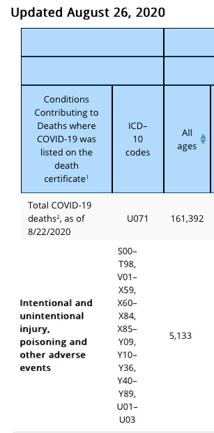
According to data34 released by the CDC August 26, 2020, only 6% of the total COVID-19-related death toll had COVID-19 listed as the sole cause of death on the death certificate. Six percent of 161,392 is 9,683. “For deaths with conditions or causes in addition to COVID-19, on average, there were 2.6 additional conditions or causes per death,” the CDC states.
Now, we’ve heard reports of accident victims being listed as COVID-19 deaths, and Table 3 in this August 26 report is granular enough that we can see that a total of 5,133 “COVID-19-related deaths” also had “intentional or unintentional injury, poisoning or other adverse events” listed on the death certificate.
This number likely also contains suicides where the individual was found to test positive for SARS-CoV-2, or was suspected of testing positive. (Please note, these data are accurate as of this writing, as shown in the partial screenshots below. The CDC does not notate when data are altered as new death certificates come in, so the numbers may therefore be different from what is reported here, depending on when you’re looking at it. For the most up-to-date figures, see the CDC’s website.35)
The Difference Between Being Scared and Being Fearful
Fear is the primary emotion that the technocrats are using to manipulate and coerce their tyrannical controls. It is the most powerful tool at their disposal and highly effective which is why it is being deployed.
As explained in “You Can Control Fear,” there’s a difference between being scared and being fearful. Right now, the whole world is holding its proverbial breath in anticipation for what might come next. It’s easy to feel fearful, considering the daily barrage of bad news.
However, understanding the difference between being scared and living in fear can be very helpful, as fear has a paralyzing effect while being scared primarily heightens alertness. Novel threats raise a person’s level of anxiety to a greater degree than familiar threats, even when they have the same or similar consequences. This is thought to be related to activity in your amygdala, which processes emotions.36
Ryan Holiday, media strategist and author of 10 books, including “The Daily Stoic” and “The Obstacle Is the Way,” writes:37
“Being afraid? That’s not fight or flight. That’s paralysis. That only makes things worse. Especially right now. Especially in a world that requires solutions to the many problems we face. They’re certainly not going to solve themselves. And inaction (or the wrong action) may make them worse, it might put you in even more danger. An inability to learn, adapt, to embrace change will too.”
While fleeting feelings of concern are expected when faced with new experiences, when such feelings are allowed to continuously dominate, paralysis can set in. To thrive in times of great uncertainty and fear, Holiday stresses the importance of training, education and preparation, which are the foundations of courage.38
The difference between being fearful and being scared is that fear paralyzes your ability to evaluate what’s happening and to make decisions. But preparation and information help you to make decisions and act, even when you’re scared. This is the definition of courage — taking action despite being scared.
How to Reduce Fear and Find Courage
There are several strategies you can use to reduce fear and find courage. It is important to begin with the understanding that your thoughts engender feelings. Your emotions do not have a life of their own, but rather are dependent on your thinking.
So, one of the strategies you can use to reduce fear is to change the way you think about things. Yes, we’re dealing with many unknowns right now, but focusing on planning and preparation rather than getting locked into panic mode can go a long way toward safeguarding your mental health. Take the safety precautions you can take, and limit your exposure to the news.
Psychology Today39 recommends reducing anxiety by consuming positive news stories while keeping up with what’s going in the world. Also do your best to apply some critical thinking when reading the news.
It’s important to pay careful attention to “vague or loaded terms, cited statistics, and unstated assumptions.” In other words, don’t accept at face value what’s in the news but, rather, consider the information and ask questions about what you’re being told. Other stress- and depression-reducing techniques include:40
- Getting enough exercise
- Eating whole foods
- Limiting sugar
- Getting quality sleep
- Practicing meditation and mindfulness training
When you’re tired and your body doesn’t have adequate nutrition to function, you’re more apt to fall into the trap of fear-based thinking. Additional nondrug treatment strategies for depression can be found in “What Does the Best Evidence Say About Antidepressants?”
The Emotional Freedom Techniques
A strategy that can provide more immediate results is the use of Emotional Freedom Techniques, or EFT. In the video above, Julie Schiffman demonstrates a tapping strategy to relieve anxiety and other challenging emotions brought on by news and uncertainty about this pandemic and/or self-quarantining. If you aren’t familiar with EFT you’ll find a library of demonstrations at “Basic Steps to Your Emotional Freedom.”
The NET FAST Technique
Another alternative is the Neuro-Emotional Technique’s First Aid Stress Tool, or NET FAST, demonstrated in the video above. Firstaidstresstool.com also provides an excellent printable summary with visuals of the technique,41 which even a young child can do. Here is a summary of the FAST procedure:
- While thinking about an issue that is bothering you, place your right wrist, palm up, into your left hand. Place three fingers of your left hand onto the area of your right wrist where you can feel your pulse
- Place your open right hand on your forehead. Gently breathe in and out several times while concentrating on feeling the issue that bothers you
- Switch hands and repeat Steps 1 and 2
Suicide Prevention Resources
If you feel depressed, anxious or creeping despair, please do not hesitate to reach out to family, friends or any of the available suicide prevention services:
- The National Suicide Prevention Lifeline (U.S.) — Call 1-800-273-TALK (8255)
- Crisis Text Line — Text HOME to 741741 to connect with a crisis counselor
- Lifeline Crisis Chat — Chat online with a specialist who can provide emotional support, crisis intervention and suicide prevention services at suicidepreventionlifeline.org
- Alternatively, call 911, or simply go to your nearest Hospital Emergency Department
- 1 News-Medical.net August 25, 2020
- 2 PR Newswire May 1, 2020
- 3, 5 KFF.org April 2, 2020
- 4 KFF.org July 27, 2020
- 6 KFF.org August 21, 2020
- 7 CDC.gov MMWR August 14, 2020; 69(32): 1049-1057
- 8 Express Scripts, America’s State of Mind Report April 16, 2020
- 9, 11 CNN Health April 16, 2020
- 10 Ecowatch April 16, 2020
- 12 US FDA June 3, 2020
- 13 MSN May 7, 2020
- 14 PLOS Medicine February 26, 2008
- 15 Science Translational Medicine January 8, 2014: 6(218):218ra5
- 16 Prevention & Treatment 1998; 1(2), article ID 2a
- 17 JAMA 2010 Jan 6;303(1):47-53
- 18 J Clin Psychiatry. 2011 Apr;72(4):464-7
- 19 Zeitschrift fur Psychologie 2014; 222(3): 128–134
- 20 BMC Psychiatry February 8, 2017; 17(1): 58
- 21, 22 Townhall July 28, 2020
- 23 ABC7 News May 21, 2020
- 24 The Sacramento Bee March 25, 2020
- 25 Boston Globe March 24, 2020 (Archived)
- 26 The Federalist March 27, 2020
- 27 Red State March 29, 2020
- 28, 29 WBIR.com March 27, 2020
- 30 Well Being Trust Projected deaths of despair during covid-19
- 31 Psycom Impact of Covid-19 on suicide rates
- 32, 33 Psycom How coronavirus is affecting mental health of millions of Americans
- 34, 35 CDC.gov August 26, 2020
- 36 PLOS|ONE, 2013;8(5):e63220 Abstract
- 37, 38 Ryan Holiday Blog, The Important Thing Is to Not Be Afraid
- 39, 40 Psychology Today, March 13, 2020
- 41 Firstaidstresstool.com
131 Ways For An Infant to Die
© 8th September 2020 GreenMedInfo LLC. This work is reproduced and distributed with the permission of GreenMedInfo LLC. Want to learn more from GreenMedInfo? Sign up for the newsletter here www.greenmedinfo.com/greenmed/newsletter
Reproduced from original article:
www.greenmedinfo.com/blog/131-ways-infant-die
Posted on: Tuesday, September 8th 2020 at 12:00 pm
Written By: Neil Z. Miller
This article is copyrighted by GreenMedInfo LLC, 2020

There are 130 official ways for an infant to die (as categorized in the ICD), and one unofficial way for an infant to die: following an adverse reaction to one or more vaccines.
There are 130 official ways for an infant to die. These official categories of death, sanctioned by the Centers for Disease Control and Prevention (CDC) and the World Health Organization (WHO), are published in the International Classification of Diseases (ICD).1-3 When a baby dies, coroners must choose from among these 130 categories.
The official causes of death listed in the ICD include nearly every imaginable — and tragic — possibility. However, there is NO category for infant deaths caused by vaccines.4 This is odd because the federal government is aware that vaccines permanently disable and kill some babies — the very reason Congress established a “death and disability” tax on childhood vaccines more than 25 years ago when the National Childhood Vaccine Injury Act of 1986 (Public Law 99-660) created the National Vaccine Injury Compensation Program (VICP).
Many parents don’t realize that when they purchase vaccines for their babies, the cost is taxed and the money goes into a special fund to compensate them if and when those vaccines seriously injure or kill their babies. As of June 2014, $2.7 billion was granted for thousands of injuries and deaths caused by vaccines. Numerous cases are still pending. Awards were issued for permanent injuries such as learning disabilities, seizure disorders, mental retardation, paralysis, and numerous deaths, including many that were initially misclassified as sudden infant death syndrome (SIDS).5
Since vaccine-related deaths are officially recognized by the federal government but there is NO official classification for vaccine-related deaths in the ICD, two important questions must be asked:
- Are some deaths that are listed within the 130 infant mortality death categories really deaths that are associated with vaccination?
- Are some vaccine-related deaths hidden within the death tables?
Sudden Infant Death Syndrome (SIDS)
Prior to the introduction of organized vaccination programs, “crib death” was so rare that it was not mentioned in infant mortality statistics. In the United States, national immunization campaigns were initiated in the 1960s when several new vaccines were introduced and promoted. For the first time in history, most U.S. infants were required to receive several doses of DPT (diphtheria, pertussis, tetanus), polio, and measles vaccines.6 By 1969, an alarming epidemic of sudden unexplained infant deaths impelled researchers to create a new medical term — sudden infant death syndrome (SIDS).7 By 1972, SIDS had become the leading cause of post-neonatal mortality (deaths of infants from 28 days to one year old) in the United States.8 In 1973, the National Center for Health Statistics, operated by the CDC, created a new cause-of-death category to document deaths due to SIDS.9,10
SIDS is defined as the sudden and unexpected death of an infant which remains unexplained after a thorough investigation, including performance of an autopsy and review of the clinical history.11 Although there are no specific symptoms associated with SIDS, an autopsy often reveals congestion and edema of the lungs and inflammatory changes in the respiratory system.9,12
In 1984, Congress held a hearing on vaccine safety. The suspected link between vaccines and sudden infant deaths was addressed. The following excerpt is from a statement made by a distraught grandmother testifying before the Congressional Committee on Labor and Human Resources:
My name is Donna Gary. I am a constituent of Senator Kennedy’s from Massachusetts. Our family should have celebrated our very first granddaughter’s first birthday last month. Instead, we will commemorate the anniversary of her death at the end of this month.
Our granddaughter, Lee Ann, was just 8 weeks old when her mother took her to the doctor for her routine checkup. That included, of course, her first DPT inoculation and oral polio vaccine. In all her entire 8 weeks of life this lovable, extremely alert baby had never produced such a blood-curdling scream as she did at the moment the shot was given. Neither had her mother ever before seen her back arch as it did while she screamed. She was inconsolable. Even her daddy could not understand Lee Ann’s uncharacteristic screaming and crying.
Four hours later, Lee Ann was dead. “Crib death,” the doctor said — “SIDS.”
“Could it be connected to the shot?” her parents implored.
“No.”
“But she just had her first DPT shot this afternoon. Could there possibly be any connection to it?”
“No, no connection at all,” the emergency room doctor said definitely.
My husband and I hurried to the hospital the following morning after Lee Ann’s death to talk with the pathologist before the autopsy. We wanted to make sure he was alerted to her DPT inoculation such a short time before her death — just in case there was something else he could look for to make the connection. He was unavailable to talk with us. We waited two-and-a-half hours. Finally, we got to talk to another doctor after the autopsy had been completed. He said it was SIDS.
In the months before Lee Ann was born, I regularly checked with a friend as to the state of her grandchild’s condition. He is nearly a year-and-a-half older than Lee Ann. On his first DPT shot he passed out cold for 15 minutes, right in the pediatrician’s office.
“Normal reaction for some children,” the pediatrician reassured. The parents were scared, but they knew what a fine doctor they had. They trusted his judgment. When it was time for the second shot, they asked “Are you sure it’s alright? Is it really necessary?” Their pediatrician again reassured them. He told them how awful it was to experience, as he had, one of his infant patient’s bout with whooping cough. That baby had died. They gave him his second DPT shot that day. He became brain-damaged.
This past week I had an opportunity to read through printed copies of the hearings of this committee. I am dismayed to learn that this same talk has been going on for years, and nothing has seemed to progress to incorporate what seems so obvious and necessary to keep from destroying any more babies, and to compensate financially those who have already been damaged for life. How accurate are our statistics on adverse reactions to vaccines when parents have been told, are still being told, “No connection to the shot, no connection at all.”?
What about the mother I have recently talked with who has a 4-year-old brain-damaged son? On all three of his DPT shots he had a convulsion in the presence of the pediatrician. “No connection,” the pediatrician assured.
I talked with a father in a town adjoining ours whose son died at the age of 9 weeks, several months before our own granddaughter’s death. It was the day after his DPT inoculation. “SIDS” is the statement on the death certificate.
Are the statistics that the medical world loves to quote to say, “There is no connection,” really accurate, or are they based on poor diagnoses, poor record keeping? What is being done to provide a safer vaccine? Who is overseeing? Will it be the same scientists and doctors who have been overseeing in the past? How much longer does the public have to wait? How are physicians and clinics going to be held accountable to see that parents are informed of the possible reactions? And how are those children who should not receive the vaccine to be identified before they are damaged — or dead?
Today is the National Day of Prayer. My prayer is that this committee be instrumental in doing what needs to be done — and soon. May there not be yet another year pass by with more children afflicted, and some dead, because those who can do so refuse to make the right connection.13
Throughout the 1980s, sudden infant deaths continued to skyrocket. Parental concerns about an apparent link between childhood vaccines and SIDS reached a fever pitch. Many parents were afraid to vaccinate their babies. Authorities sought to reassure parents that vaccines are safe and claimed that sudden unexplained infant deaths following vaccines were merely coincidental.
Back to Sleep
In 1992, the American Academy of Pediatrics (AAP) came up with a plan to reduce the unacceptable SIDS rate while reassuring concerned moms and dads that sudden unexplained infant deaths were not related to vaccines. The AAP initiated a national “Back to Sleep” campaign, telling parents to place their infants supine, rather than prone, during sleep. From 1992 through 2001, the post-neonatal SIDS rate dropped by an average annual rate of 8.6%. It seemed as though the “Back to Sleep” campaign was successful and that the real cause of SIDS was due not to vaccinations but from babies sleeping on their bellies.
However, a closer inspection of the ICD — the 130 official ways for an infant to die — revealed a loophole. Medical certifiers, such as coroners, could choose from among several categories of death when a baby suddenly expired. They didn’t have to list the death as SIDS. Although the post-neonatal SIDS rate dropped by an average annual rate of 8.6% from 1992 through 2001 following the AAP’s seemingly successful “Back to Sleep” campaign, the post-neonatal mortality rate from “suffocation in bed” (ICD-9 code E913.0) increased during this same period at an average annual rate of 11.2%. Sudden, unexplained infant deaths that were classified as SIDS prior to the “Back to Sleep” campaign, were now being classified as deaths due to suffocation in bed!
The post-neonatal mortality rate from “suffocation other” (ICD-9 code E913.1-E913.9), from “unknown and unspecified causes” (ICD-9 code 799.9), and from “intent unknown” (ICD-9 code E980-E989), all increased during this period as well.10 In Australia, a similar subterfuge seemed to occur. Researchers observed that when the SIDS rate decreased, deaths attributed to asphyxia increased.14-16
From 1999 through 2001, the number of U.S. deaths that were attributed to “suffocation in bed” and “unknown causes” increased significantly. Although the post-neonatal SIDS rate continued to decline, there was no significant change in the total post-neonatal mortality rate. In a recent paper (Malloy and MacDorman) published in Pediatrics, SIDS researchers made the following observation:
If death-certifier preference has shifted such that previously classified SIDS deaths are now classified as “suffocation,” the inclusion of these suffocation deaths and unknown or unspecified deaths with SIDS deaths then accounts for about 90 percent of the decline in the SIDS rate observed between 1999 and 2001 and results in a non-significant decline in SIDS.10 (Figure 1)
Reclassification of SIDS Deaths to “Suffocation in Bed” and “Unknown Causes”

Figure 1. The postneonatal SIDS rate appears to have declined from 61.6 deaths (per 100,000 live births) in 1999 to 50.9 in 2001. However, during this period there was a significant increase in post-neonatal deaths attributed to “suffocation in bed” and due to “unknown causes.” When these sudden unexpected causes of infant deaths are combined with SIDS deaths, the total SIDS rate remains relatively stable, resulting in a non-significant decline.
Other Evidence Linking SIDS to Vaccines
Although some studies were unable to find positive correlations between SIDS and vaccines,17-19 there is other evidence that a subset of infants may be more susceptible to SIDS shortly after being vaccinated. For example, as early as 1933 the Journal of the American Medical Association (JAMA) published a paper by Madsen documenting the sudden deaths of two infants soon after pertussis vaccination. The first child developed cyanosis and convulsions 30 minutes after vaccination and died suddenly a few minutes later. The second child developed cyanosis 2 hours after vaccination and then died suddenly.20
In 1946, Werne and Garrow published a paper in JAMA documenting the sudden deaths of identical twins 24 hours after pertussis vaccination. The babies had symptoms of shock throughout the night prior to their fatal reactions.21
In the 1960s and 1970s Aborigine infants began to mysteriously die at astonishing rates. In some regions of Australia, 1 of every 2 babies succumbed to an unexplained death — a fatality rate of 50 percent! Kalokerinos solved the riddle when he realized that the deaths were occurring shortly after the babies were vaccinated. Health officials had recently initiated a mass vaccination campaign to “protect” Aborigine babies; their deaths corresponded with the vaccination program. Kalokerinos realized that these babies were severely malnourished, including a vitamin C deficiency. Their undeveloped immune systems couldn’t handle the additional stress of vaccination. Kalokerinos was able to save other babies from the same fate by administering small quantities of vitamin C (100mg per month of age) prior to their vaccines.22
In Japan, from 1970 through 1974, there were 37 documented sudden infant deaths following pertussis vaccinations, inciting parents and doctors to reject the shot. In 1975, Japanese authorities reacted to these events by raising the age of vaccination from three months to two years. As a result, the number of vaccine injury compensation claims that were paid out for sudden deaths following vaccination dropped from 37 cases during a 5-year period to just 3 cases during the next 6-and-a-half years (from 1975 through August of 1981). The sudden death rate following vaccination dropped from 1.47 to 0.15 deaths per million doses — a 90% improvement.23,24 In addition, from the early 1970s (a period when 3-month-old infants were vaccinated) to the mid-1980s (ten years after the age of vaccination was raised to 2 years) the Japanese infant mortality rate (infant deaths per 1,000 live births) dramatically declined from 12.4 to 5.0 — a 60 percent drop!25
According to a special task force (Cherry et al) that investigated the Japanese data and published their summary in Pediatrics:
The category of “sudden death” is instructive in that the entity disappeared following both whole-cell and acellular vaccines when immunization was delayed until a child was 24 months of age.24
Cherry et al also made the following observation:
It is clear that delaying the initial vaccination until a child is 24 months, regardless of the type of vaccine, reduces most of the temporally associated severe adverse reactions.24
There is other more recent evidence that delaying vaccinations until a later age could save babies from severe vaccine-related adverse reactions, including sudden deaths. For example, Human and Experimental Toxicology published a study by Goldman and Miller that investigated more than 38,000 infant reports filed with the Vaccine Adverse Event Reporting System (VAERS). (This federally mandated vaccine safety surveillance program collects information about possible adverse reactions from vaccines.) Cases that listed either “hospitalization” or “death” were evaluated relative to all infant reports, including those that were non-serious. The hospitalization rate for infants that were vaccinated shortly after birth was an astonishing 20.1% but decreased in a statistically significant linear fashion to 10.7% for infants that were vaccinated just prior to their first birthday. This study also revealed a statistically significant lower mortality rate for infants that were vaccinated between 6 months and 1 year of age when compared to infants vaccinated between birth and 6 months of age.26
In 1982, William Torch, MD, director of Child Neurology, Department of Pediatrics, University of Nevada School of Medicine, presented a study at the 34th Annual Meeting of the American Academy of Pediatrics showing that two-thirds of babies who had died from SIDS had been vaccinated against DPT prior to death. Of these, 6.5% died within 12 hours of vaccination; 13% within 24 hours; 26% within 3 days; and 37%, 61%, and 70% within 1, 2, and 3 weeks, respectively. (Figure 2)
The Pertussis Vaccine and Sudden Infant Death Syndrome


Figure 2. In a study of 103 children who died of SIDS, more than two-thirds had been vaccinated with pertussis prior to death. Of these, 6.5 percent died within 12 hours of vaccination; 13 percent within 24 hours; 26 percent within three days; and 37, 61, and 70 percent within one, two, and three weeks, respectively.
Torch also found that unvaccinated babies who died from SIDS did so most often in the fall or winter while vaccinated babies died most often at 2 and 4 months — the same ages when initial doses of DPT were given to infants. He concluded that:
DPT may be a generally unrecognized major cause of sudden infant and early childhood death, and that the risks of immunization may outweigh its potential benefits. A need for re-evaluation and possible modification of current vaccination procedures is indicated by this study.27
In 1983, Pediatric Infectious Diseases published a study by Baraff et al that analyzed 17 infants that had been vaccinated within 28 days prior to their sudden deaths, which were classified as SIDS. They calculated the expected frequency of SIDS deaths per day and compared that with the actual number of sudden deaths in each of the 28 days after vaccination. A statistically significant number of excess deaths happened in the first week following vaccination (6.75 sudden deaths were expected and 17 actually occurred) — a 250% increase. The greatest number of excess deaths happened within 24 hours after vaccination (0.96 sudden deaths were expected and 6 actually occurred) — a 625% increase over statistical expectations.28
In 1987, the American Journal of Public Health published a paper by Walker et al, once again confirming an apparent link between vaccination and sudden deaths. Babies died at a rate more than seven times greater than normal within 3 days after getting a DPT vaccination.29
In 1991, Scheibner and Karlsson presented strong evidence of an association between DPT injections and cot death (SIDS) at the Second National Immunisation Conference in Canberra, Australia. They were able to develop a sophisticated microprocessor that was placed under infants’ mattresses to precisely measure their breathing patterns before and after vaccination. The microprocessor generated computer printouts in integrals of a weighted apnea (cessation of breathing) hypopnea (abnormally shallow breathing) density (WAHD). The data clearly revealed that pertussis vaccination caused an inordinate increase in episodes where breathing either nearly ceased or stopped completely.30-32 (Figure 3)
The Pertussis Vaccine and Stress-Induced Breathing Patterns: A Link to SIDS

Figure 3. This chart represents a 17-day record of one child’s breathing patterns before and after receiving the pertussis vaccine. Values above 1000 indicate acute stress-induced breathing. The author of this study concluded that “vaccination is the single most prevalent and most preventable cause of infant deaths.”
These episodes continued for months following DPT vaccinations. The lead author of the paper concluded that “vaccination is the single most prevalent and most preventable cause of infant deaths.”31
In 2006, Ottaviani et al published a paper in Virchows Archiv (European Journal of Pathology) documenting the case of a 3-month-old infant who died suddenly and unexpectedly shortly after being given six vaccines in a single injection. After dissecting the brainstem and examining the cardiac conduction system, authors of the study made the following observation: “This case offers a unique insight into the possible role of hexavalent vaccine in triggering a lethal outcome in a vulnerable baby.” They also noted that “any case of sudden unexpected death occurring … in infancy, especially soon after a vaccination, should always undergo a full necropsy study,” otherwise a true association between vaccination and death may escape detection.33
That same year, another team of scientists (Zinka et al) published a paper in Vaccine documenting six cases of SIDS that occurred within 48 hours following the administration of a hexavalent vaccine. At postmortal examination, these cases showed “unusual findings in the brain” that appeared compatible with an association between hexavalent vaccination and sudden infant death syndrome.34
In 2011, Statistics in Medicine published a paper by Kuhnert et al that examined an association between multi-dose vaccinations and death. Authors of the paper demonstrated a 16-fold increase in sudden unexpected death after the fourth dose of a pentavalent shot (five different vaccines in one injection) or hexavalent shot (six different vaccines in one injection).35
Today, unsuspecting parents continue to experience the heartbreak of losing healthy children after vaccinations. Here is another unnecessary death labeled as SIDS, as reported by a distraught mother:
Our beautiful 2-month-old daughter recently died. What was unusual was that earlier on the day that she died, I had taken her to the military base hospital for her two-month checkup. The doctor told me that she was just perfect. Then he said that she needed four shots. I replied, “Four!”? She assured me that it was completely normal.
That evening after feeding our daughter, we laid her down to sleep. We checked on her 45 minutes later and discovered that she was dead. I told the police, coroner, and investigators that I thought it was from the shots because she was perfectly fine that day and before the shots. But after three weeks we finally got an answer from the autopsy that it was SIDS. To this day, I believe that her death was caused by the shots. No one can convince me otherwise.36
More Vaccine Fatalities – Hidden in the Death Tables
“SIDS,” “suffocation in bed,” and death due to “unknown and unspecified causes,” are just three of the 130 official cause-of-death categories that might be concealing fatalities that were really caused by vaccination. Several other ICD categories are possible candidates for incorrect infant death classifications: unspecified viral diseases, diseases of the blood, diseases of the nervous system, unspecified diseases of the respiratory system, and shaken baby syndrome. All of these official categories may be repositories of vaccine-related infant deaths reclassified as common fatalities.
For example, a vaccine against rotavirus-induced diarrhea (Rotarix) was licensed by the Food and Drug Administration (FDA) in 2008. However, in a clinical study that evaluated the safety of this vaccine, vaccinated babies died at a significantly higher rate than non-vaccinated babies — mainly due to a statistical increase in pneumonia fatalities.37 (One biologically plausible explanation is that natural rotavirus infection might have a protective effect against respiratory infection.)38 Although these deaths appear to be vaccine related, coroners are likely to have misclassified them as pneumonia.
Some infant fatalities that occur shortly after vaccinations are incorrectly classified as shaken baby syndrome. Retinal and subdural bleeding can result from an adult that shook the baby or from vaccine damage. Expert testimony by medical practitioners has exonerated innocent parents of all charges against them.39 This is just another example of how the true cause of death can be reclassified or hidden within the death tables.
The practice of reclassifying ICD data greatly concerns the CDC “because inaccurate or inconsistent cause-of-death determination and reporting hamper the ability to monitor national trends, ascertain risk factors, and design and evaluate programs to prevent these deaths.”40 Thus, medical certification practices need to be monitored to determine how often vaccine-related infant deaths are being reclassified as ordinary mortality in the ICD. More importantly, parents need to be warned that vaccine safety is grossly overestimated when vaccine-related deaths are not being accurately documented.
Vaccine Safety, Informed Consent and Human Rights
There are 130 official ways for an infant to die (as categorized in the ICD), and one unofficial way for an infant to die: following an adverse reaction to one or more vaccines. When vaccine-related deaths are hidden within the death tables, parents are denied the ability to ascertain honest vaccine risk-to-benefit ratios, and true informed consent to vaccinations is not possible. When families are urged to vaccinate their children without access to accurate data on vaccine-related deaths, their human rights have been violated. Medical health authorities, pediatricians, and the vaccine industry then become criminal accomplices to each infant death caused by vaccines — even when vaccines are not officially acknowledged as the cause of death. Finding ways to increase vaccine safety, providing families with true informed consent, and preserving human rights must be the top priorities.
Notes
1. WHO. International Classification of Diseases, 9th Revision. Geneva, Switzerland: World Health Organization, 1979.
2. WHO. International Statistical Classification of Diseases and Related Health Problems, 10th Revision. Geneva, Switzerland: World Health Organization, 1992.
3. CDC. Table 31. Number of infant deaths and infant mortality rates for 130 selected causes, by race: United States, 2006. Natl Vital Stat Rep 2009; 57: 110–112.
4. ICD-6, issued in 1948, and ICD-7, issued in 1955, included “Prophylactic inoculation and vaccination” as a separate cause of death category with subcategories (codes Y40-Y49) for death due to “vaccination against smallpox” (code Y40), “inoculation against whooping cough” (code Y42), “inoculation against other infectious disease” (code Y49), etc. The ICD-8, issued in 1965, deleted the subcategories for death due to inoculation against individual diseases while maintaining “Prophylactic inoculation and vaccination” (code Y42) as a separate cause of death category. When the ICD-9 was issued in 1979, authorities removed all cause of death classifications associated with vaccination.
5. Health Resources and Services Administration. National Vaccine Injury Compensation Program. U.S. Department of Health and Human Services. (Data as of June 2, 2014).
6. The measles vaccine was administered at 9 months of age from 1963 to 1965. ACIP. Measles prevention: recommendations of the Immunization Practices Advisory Committee (ACIP). MMWR (December 29, 1989) / 38(S-9): 1-18.
7. Bergman AB, Beckwith JB, Ray CG, eds. Sudden Infant Death Syndrome. Proceedings of the Second International Conference on Causes of Sudden Death in Infants, Seattle and London: University of Washington Press, 1970:18.
8. Bergman AB. The “Discovery” of Sudden Infant Death Syndrome. New York, NY, USA: Praeger Publishers, 1986: 209 (Appendix III).
9. MacDorman MF and Rosenberg HM. Trends in infant mortality by cause of death and other characteristics, 1960-88 (vital and health statistics), Volume 20. Hyattsville,MD, USA: National Center for Health Statistics, U.S. Government Printing, 1993.
10. Malloy MH and MacDorman M. Changes in the classification of sudden unexpected infant deaths: United States, 1992-2001. Pediatrics 2005; 115: 1247–1253.
11. Willinger M, James LS, Catz C. Defining the sudden infant death syndrome (SIDS): deliberations of an expert panel convened by the National Institute of Child Health and Human Development. Pediatr Pathol 1991; 11: 677–84.
12. Goldwater PN. Sudden infant death syndrome: a critical review of approaches to research. Arch Dis Child 2003; 88: 1095–100.
13. Vaccine Injury Compensation. Hearing Before the Committee on Labor and Human Resources; 98th Congress, 2nd Session, (May 3, 1984): 63-67.
14. Mitchell E, Krous HF, Donald T, and Byard RW. Changing trends in the diagnosis of sudden infant death. Am J Forensic Med Pathol 2000; 21: 311–314.
15. Overpeck MD, Brenner RA, Cosgrove C, Trumble AC, Kochanek K, and MacDorman M. National under ascertainment of sudden unexpected infant deaths associated with deaths of unknown cause. Pediatrics 2002; 109: 274–283.
16. Byard RW and Beal SM. Has changing diagnostic preference been responsible for the recent fall in incidence of sudden infant death syndrome in South Australia? J Pediatr Child Health 1995; 31: 197–199.
17. Vennemann MM, Butterfass-Bahloul T, Jorch G, Brinkmann B, Findeisen M, Sauerland C, et al. Sudden infant death syndrome: no increased risk after immunisation. Vaccine 2007; 25: 336–340.
18. Stratton K, Almario DA, Wizemann TM, and McCormick MC. Immunization safety review: vaccinations and sudden unexpected death in infancy. Washington DC, USA: National Academies Press, 2003.
19. Essery SD, Raza MW, Zorgani A, MacKenzie DA, et al. The protective effect of immunisation against diphtheria, pertussis and tetanus (DPT) in relation to sudden infant death syndrome. FEMS Immunology and Microbiology 1999 August 1; 25(1-2): 183-92.
20. Madsen T. Vaccination against whooping cough. JAMA 1933; 101: 187-88.
21. Werne J and Garrow I. Fetal anaphylactic shock: occurrence in identical twins following second injection of diphtheria toxoid and pertussis antigen. JAMA 1946; 131: 730-35.
22. Kalokerinos A. Every Second Child Was Doomed to Death — Unless One Dedicated Doctor Could Open His Colleagues’ Eyes and Minds. New Canaan, CT: Keats Publishing, Inc., 1974.
23. Noble GR., et al. Acellular and whole-cell pertussis vaccines in Japan: report of a visit by U.S. scientists. JAMA 1987; 257: 1351-56.
24. Cherry JD., et al. Report of the task force on pertussis and pertussis immunization. Pediatr (Jun 1988); 81(6): 933-84.
25. Congressional Budget Office. Factors contributing to the infant mortality ranking of the United States. CBO Staff Memorandum (February 1992): Table 2, International Infant Mortality Rates by Ranking.
26. Goldman GS and Miller NZ. Relative trends in hospitalizations and mortality among infants by the number of vaccine doses and age, based on the Vaccine Adverse Event Reporting System (VAERS), 1990–2010. Human and Experimental Toxicology 2012; 31(10): 1012-1021.
27. Torch WC. DPT immunization: A potential cause of the sudden infant death syndrome (SIDS). Amer Acad of Neur, 34th Annual Meet, Apr 25-May 1, 1982. Neur 32(4): pt. 2.
28. Baraff LJ, Ablon WJ, Weiss RC. Possible temporal association between diphtheria-tetanus toxoid-pertussis vaccination and sudden infant syndrome. Pediatric Infectious Disease Journal 1983; 2: 7-11.
29. Walker AM., et al. Diphtheria-tetanus-pertussis immunization and sudden infant death syndrome. Am J Public Health (August 1987); 77(8): 945-51.
30. Scheibner V. and Karlsson LG. Evidence of the association between non-specific stress syndrome, DPT injections and cot death. Proceedings of the 2nd National Immunisation Conference in Canberra (May 27-29, 1991).
31. Scheibner V. Vaccination: 100 Years of Orthodox Research Shows that Vaccines Represent a Medical Assault on the Immune System. Blackheath, NSW, Australia: Scheibner Publications, 1993: 59-70; 225-235; 262.
32. Scheibner V. Dynamics of critical days as part of the dynamics of non-specific stress syndrome discovered during monitoring with Cotwatch breathing monitor. Journal of the Australasian College of Nutritional & Environmental Medicine (December 2004); 23(3): 1-5.
33. Ottaviani G, Lavezze AM, and Matturri L. Sudden infant death syndrome (SIDS) shortly after hexavalent vaccination: another pathology in suspected SIDS? Virchows Archiv 2006; 448: 100–104.
34. Zinka B, Rauch E, Buettner A, Rueff F and Penning R. Unexplained cases of sudden infant death shortly after hexavalent vaccination. Vaccine 2006; 24(31-32): 5779–5780.
35. Kuhnert R, Hecker H, Poethko-Muller C, Schlaud M, Vennemann M, Whitaker HJ, et al. A modified self-controlled case series method to examine association between multidose vaccinations and death. Stat Med 2011; 30(6): 666–677.
36. Unsolicited correspondence received by the Thinktwice Global Vaccine Institute.
37. GlaxoSmithKline. Rotarix1 (Rotavirus Vaccine, Live, Oral) Oral Suspension. Product insert from the manufacturer (April 2008): 6.
38. FDA. Center for biologics evaluation and research, vaccines and related biological products advisory committee meeting (February 20, 2008): 127–128.
39. Buttram H and England C. Shaken Baby Syndrome or Vaccine-Induced Encephalitis: Are Parents Being Falsely Accused? Bloomington, Indiana: Author House, 2011.
40. CDC. About the sudden unexpected infant death investigation (SUIDI) reporting form. Department of Health and Human Services (accessed May 13, 2013).

Low Vitamin K in Elderly Linked to Increased Risk of Death
Reproduced from original article:
https://articles.mercola.com/sites/articles/archive/2020/09/02/vitamin-k-and-elderly.aspx
Analysis by Dr. Joseph Mercola Fact Checked September 02, 2020

STORY AT-A-GLANCE
- People with higher levels of vitamin K have a lower risk of death from all-cause mortality
- There are two types of vitamin K; the body’s not very good at storing them so they must be consumed every day for optimal health
- Vitamin K is important in the workings of vitamin D, as it helps prevent osteoporosis and arterial calcification and contributes to blood clotting
Vitamin K is a fat-soluble vitamin that can be found in some foods. The body uses it for certain functions, such as those required for bone metabolism, blood clotting and a variety of other cellular functions.1
Your body has a limited ability to store vitamin K, so it must be absorbed from your food or a supplement on a daily basis. It’s also recycled within your system, optimizing its benefits.2 Several different medications, such as antibiotics, weight loss drugs, cholesterol medications and the blood thinner warfarin, may interfere with its absorption, and therefore affect blood clotting.
Vitamin K is distributed throughout the body, including in your brain, heart, bones and liver.3 Since it’s broken down and excreted quickly it rarely reaches toxic levels in a way that happens with other fat-soluble vitamins.
Since the vitamin is crucial to several bodily functions, it’s not surprising to learn that if you don’t get enough every day, on a daily basis, you could be at risk for an earlier death.
Vitamin K Lowers Risk of Death in Older Adults
Recently, a paper was published in the American Journal of Clinical Nutrition by a team that investigated the vitamin K-dependent proteins that may affect atherosclerosis, which is the stiffness and calcification of the arterial vessels.4
The aim of the investigation was to determine if there was an association between circulating vitamin K, cardiovascular disease and all-cause mortality. The study was carried out at Tufts University and led by Kyla Shea, who commented:5
“The possibility that vitamin K is linked to heart disease and mortality is based on our knowledge about proteins in vascular tissue that require vitamin K to function. These proteins help prevent calcium from building up in artery walls, and without enough vitamin K, they are less functional.”
The researchers gathered data from three science databases, including the Framingham Offspring Study. The participants had circulating vitamin K1 (phylloquinone) measured, and each person either had some type of heart event or they died. There were 3,891 men and women with 858 heart events and 1,209 deaths over 13 years.
Interestingly, although vitamin K is associated with vascular health, the data did not show an association between low vitamin K and cardiovascular disease (CVD). However, there was a link between low levels and all-cause mortality. They found a 19% increased risk of all-cause mortality in those with the lowest serum levels of vitamin K.6
Since the study was observational, the scientists could not establish a causal link, yet the data fit with what we know about the function of vitamin K in the body. One-third of the participants in the study were not white, which allowed the researchers to theorize that the association is multi-ethnic.7 Dr. Daniel Weiner, nephrologist from Tufts Medical center, explained the association between stiff arteries and CVD:8
“Similar to when a rubber band dries out and loses its elasticity, when veins and arteries are calcified, blood pumps less efficiently, causing a variety of complications. That is why measuring risk of death, in a study such as this, may better capture the spectrum of events associated with worsening vascular health.”

Vitamin K: The Basics
There are two basic types of vitamin K.9 Vitamin K1 is found in green leafy vegetables and works through the liver to help maintain a healthy clotting system. This is the type researchers measured in the study. Vitamin K1 also helps reduce calcification in the blood vessels and assists your bones in retaining calcium.
Vitamin K2 (menaquinone) is produced by bacteria in your gut and can be found in some animal products and fermented foods, particularly cheese and natto. This is the one that helps activate vitamin D and move calcium out of the arterial system and into your bones and teeth.10
There are also several different forms of vitamin K2. They are named for the length of the chemical chain, beginning with the letter M for menaquinone. The two most important types are found in animal products and fermented foods. Most Western diets are deficient in both forms of vitamin K2.11
In one study, researchers sought to compare the effectiveness of giving supplements of these two types, MK-4 and MK-7, to healthy women. They “demonstrated that a nutritional dose of MK-7 is well absorbed in humans, and significantly increases serum MK-7 levels, whereas MK-4 had no effect on serum MK-4 levels.”12
More Benefits of Vitamin K
Vitamin K is integral to the body’s ability to coagulate, or clot, blood. These clotting factors are involved in a cascade of events that stop bleeding and are produced in the liver. Therefore, individuals with severe liver disease can have difficulty with vitamin K-dependent clotting and are at an increased risk for hemorrhage.13
As discussed previously, Vitamin K is also integral to moving calcium out of the arterial system and to preventing soft tissue calcification. This function is central to preventing osteoporosis and maintaining strong bone health.
Another benefit is a vitamin K-dependent protein, growth arrest specific gene 6 protein (Gas6), which was identified in 1993 and is found in the heart, kidneys, lungs and cartilage. It contributes to many cellular functions that may play a role in protecting an aging nervous system, and it appears to regulate vascular homeostasis.
Separate studies using vitamin K2 on cancer cells in the lab have demonstrated an apoptotic effect. In one study, researchers found vitamin K2 induced apoptosis in bladder cancer cells and in another, against triple negative breast cancer.14,15
Vitamin K2 and Magnesium Optimize Vitamin D Supplementation
The evidence that your serum vitamin D level is protective against COVID-19 and reduces the severity of the disease continues to grow. While your body produces vitamin D from the reaction to sunlight, many don’t get enough sun to raise their vitamin D levels above 30 ng/mL.
To reach this minimal goal, you may need a supplement. Yet, taking vitamin D3 supplements without magnesium and vitamin K2 may offer little effective protection and could increase your risk of atherosclerosis. Before you can determine how much supplementation is needed, you’ll want to test your current level. You can do this using a home kit through GrassrootsHealth.16
The kit is delivered to your home where you use a finger stick and send the kit back to the lab. Only you get the results of the test, which you can then input into the GrassrootsHealth calculator to determine the amount of supplement you’ll need to achieve your desired result.17
Vitamin D is important in the absorption and utilization of vitamin K2. In fact, a vitamin K deficiency may contribute to symptoms of “vitamin D toxicity.” In my interview with Dr. Kate Rheaume-Bleue, natural health physician and author of Vitamin K2 and the Calcium Paradox, she talked about this challenge:18
“We don’t see symptoms of D toxicity very often but when we do those symptoms are inappropriate calcification. That’s a symptom of D toxicity and that is actually a lack of vitamin K2 that will cause that. K2 protects you against that toxicity quite simply.”
Added to this, I’ve written about the importance of vitamin K2 when you’re taking supplemental vitamin D3 to avoid excessive calcification in your arteries. Magnesium is also a crucial part of raising your vitamin D level as it is necessary for the activation of the vitamin.
Without sufficient amounts your body can’t properly use the vitamin you’re taking. Mohammed Razzaque is a professor of pathology at Lake Erie College of Osteopathic Medicine in Pennsylvania. He notes that without magnesium, vitamin D supplements may not be useful.19
To lower the risk of vitamin D deficiency it’s necessary to have optimal amounts of magnesium.20 According to one scientific review, as many as 50% of Americans who are taking vitamin D supplementation may not benefit, as it’s stored in the inactive form without sufficient levels of magnesium.21,22
The relationship is a two-way street. While vitamin D improves the absorption of magnesium, large doses of vitamin D may deplete magnesium.23,24 This is because large doses of vitamin D will require large doses of magnesium to convert it to the active form.
GrassrootsHealth published research that was based on data from nearly 3,000 people showing you must take 244% more vitamin D if you aren’t also taking a magnesium and vitamin K2 supplement.25 Practically speaking, this means when you take all three in combination you’ll need far less vitamin D to achieve a healthy level.
According to researchers, “those taking both supplemental magnesium and vitamin K2 have a higher vitamin D level for any given vitamin D intake amount than those taking either supplemental magnesium or vitamin K2 or neither.”26
Consider Supplementing With Magnesium and Vitamin K2
There are several forms of magnesium. In each form, the magnesium is bound to another molecule. Some forms have compounds that serve other functions in the body, as you’ll find in “NYC Doctor Highlights Importance of Vitamin D Optimization”:
- Magnesium glycinate,27 a powder with low solubility. Glycine is an important amino acid and precursor for glutathione.
- Ionic magnesium found in molecular hydrogen tablets. Each water-soluble tablet has about 80 mg of highly bioavailable unbound magnesium ions, which is about 20% of the recommended daily allowance.
- Magnesium threonate28 is another excellent choice as it seems it can efficiently penetrate the blood-brain barrier.
- Magnesium malate,29 which dissolves well in water. Malate is an intermediary in the Krebs cycle, so it likely contributes to ATP production.
- Magnesium citrate30 also dissolves well and has a pleasant citric acid taste.
Since most people eating a Western diet are deficient in vitamin K2 and the vitamin is quickly used and excreted, it’s important to either change the foods you eat, or consider supplementation. As noted in the featured study, those with higher levels of vitamin K have a lower risk of death.
It can be difficult to tell if you’re getting enough vitamin K, as there’s no easy way to screen or test for vitamin K2 sufficiency. For more information about getting enough vitamin K, see “Spinach, Eggs and Gouda Cheese to Combat COVID?”
- 1 National Institutes of Health
- 2, 9, 13 Linus Pauling Institute
- 3 Harvard T.H. Chan School of Public Health
- 4 The American Journal of Clinical Nutrition, 2020; doi.org/10.1093/ajcn/nqaa082
- 5, 7, 8 Science Daily, June 15, 2020
- 6 Pharmacy Times, August 3, 2020
- 10, 11 Integrative Medicine: A Clinician’s Journal, 2015;14(1)
- 12 Nutrition Journal, 2012;11(93)
- 14 PLOS|One, 2016; doi.org/10.1371/journal.pone.0161886
- 15 Science Direct, 2015; dx.doi.org/10.1016/j.nutres.2015.05.014
- 16 GrassrootsHealth, Daction
- 17 GrassrootsHealth, Dcalculator
- 18 YouTube, January 15, 2013
- 19 EurekAlert! February 27, 2018
- 20 Live Science February 26, 2018
- 21 Science Daily, February 2018
- 22 Journal of the American Osteopathic Association March 2018;118:181
- 23 Magnesium Research 1995;8(1):19
- 24 American Journal of Therapeutics 2019;26(1):e124
- 25, 26 GrassrootsHealth, Vitamin D levels
- 27 Journal of Innovative Optical Health Sciences Vol. 10, No. 3 (2017) 1650052
- 28 Magnesium Research 2016
- 29, 30 Biological Trace Element Research December 27, 2017
IV High-Dose Vitamin C Saves Critically Ill COVID-19 Patient
Reproduced from original article:
https://articles.mercola.com/sites/articles/archive/2020/08/28/iv-vitamin-c-coronavirus.aspx
Analysis by Dr. Joseph Mercola Fact Checked August 28, 2020

STORY AT-A-GLANCE
- Vitamin C is an essential component of immune cell function, it has potent anti-inflammatory effects and at high doses even works as an antiviral
- Vitamin C is a foundational component of two distinct and highly effective treatment protocols developed by Dr. Paul Marik — one for sepsis and another, called MATH+, specifically for COVID-19
- According to a recent case history, high-dose IV vitamin C was successfully used to save the life of an elderly patient with critical COVID-19 infection, sepsis and acute respiratory distress syndrome
- According to the doctors who reported the case, she was the first patient to successfully discontinue mechanical ventilation early at that hospital. The median duration of mechanical ventilation for COVID-19 patients is 10 days, whereas this patient was able to get off it in five
- Previous research found IV vitamin C at a dose of 50 mg per kilo every six hours for 96 hours significantly reduced mortality and shortened ICU stays among patients with sepsis and ARDS
While U.S. media and self-appointed “fact checkers” claim the use of vitamin C against COVID-19 is nothing but fake news and dangerous nonsense, the Chinese have been hard at work looking at vitamin C against this pandemic illness.
One source where you can find pertinent research material is the Orthomolecular Medicine News Service website,1 which has published more than a dozen reports on this topic.
Earlier this year I interviewed Dr. Andrew Saul, editor-in-chief of the Orthomolecular Medicine News Service, about vitamin C against viral illnesses such as COVID-19, in which he reviewed some of its established benefits. Importantly, vitamin C is an essential component of immune cell function, it has potent anti-inflammatory effects and at high doses even works as an antiviral.
Vitamin C is a foundational component of two distinct and highly effective treatment protocols developed by Dr. Paul Marik — one for sepsis and another, called MATH+, specifically for COVID-19. I’ve reviewed both protocols in previous articles.
COVID-19 Patient Brought From Brink of Death by Vitamin C
An August 10, 2020, MedPage Today article2 highlights a recent case3 history in which high-dose IV vitamin C was successfully used to save the life of an elderly patient.
A 74-year-old woman was admitted to a Flint, Michigan, hospital after suffering with low-grade fever, dry cough and shortness of breath for two days. She’d recently undergone a knee replacement procedure, and her knee is now red, swollen and painful, suggesting infection has set in.
Over the next 24 hours, her condition continued to worsen, despite treatment with oxygen, antibiotics and other drugs. Her RT-PCR test also came back positive for SARS-CoV-2 infection. As reported by MedPage Today:4
“Clinicians start treatment with oral hydroxychloroquine 400 mg once and then 200 mg twice a day, along with intravenous azithromycin 500 mg once a day, zinc sulfate 220 mg three times a day, and oral vitamin C 1 g twice a day.
When blood and sputum cultures are negative for any organisms, broad-spectrum antibiotics are discontinued. The patient’s dyspnea rapidly worsens, and oxygen requirements increase to 15 liters. She is drowsy, in moderate distress, and her airways remain unprotected …
By day 6 … Bilateral alveolar infiltrates are evident on chest x-ray, due to pneumonia and interstitial edema, consistent with acute respiratory distress syndrome (ARDS). Given her rapid deterioration, the patient is intubated on an emergent basis and started on pressure-regulated volume-controlled mechanical ventilation.”
By day 7, the woman’s family requested she be placed on high-dose IV vitamin C, and over the next 24 hours, she received 11 grams total. Two days later, her condition began to gradually improve, and on day 10, a second chest x-ray revealed considerable improvement of both the pneumonia and interstitial edema, at which point she was extubated. The patient’s breathing continued improving over the following five days.
“On day 16, she shows significant recovery, with oxygen saturation of 92% while breathing ambient air, and chest radiography reveals almost complete resolution of the infiltrates,” MedPage Today reports.5
“During her hospitalization, the patient received a total of 5 days of treatment with hydroxychloroquine and azithromycin along with 4 days of colchicine. Treatment with high-dose vitamin C infusion and oral zinc sulfate were continued for a total of 10 days.”
Take-Home Message: Fight for the Treatment You Want
Remarkably, while suffering from severe COVID-19 infection, septic shock and ARDS — and being on mechanical ventilation — this elderly woman made a full recovery.
According to the doctors who reported the case, she was the first patient to successfully discontinue mechanical ventilation early at that hospital. The median duration of mechanical ventilation for COVID-19 patients is 10 days, whereas this patient was able to get off it in five.
Vitamin C has been recognized as an essential component of immune cell function with a critical role in numerous immune system mechanisms. Severe vitamin C deficiency is also known to increase the risk for … pneumonia and other infections.
A key take-home message from this story is that the IV infusion of vitamin C was done at the insistence of her family. It’s an excellent example demonstrating that when things are going sideways, you need to step in and push for the care you want. You have that right.
It’s also an example of why it’s so important to have an advocate to speak up for you when you’re admitted to the hospital. This woman was placed on mechanical ventilation, which means she was sedated and was in no position to make any of her own decisions. Fortunately, her family knew about the potential benefits of vitamin C and insisted it be tried. In doing so, they saved her life. As noted by MedPage Today:6
“Vitamin C infusion is not approved as a standard treatment for SARS-CoV-2 and thus was not part of the hospital’s COVID-19 treatment regimen.
Nevertheless, the case authors point out that for decades, vitamin C has been recognized as an essential component of immune cell function with a critical role in numerous immune system mechanisms. Severe vitamin C deficiency is also known to increase the risk for … pneumonia and other infections.
Vitamin C enhances neutrophil motility, phagocytosis, microbial killing by activating reactive oxygen species, and apoptosis, and prevents oxidative damage by its antioxidant properties.
It also promotes the proliferation of B and T lymphocytes and antibody production. More recent research suggests that vitamin C also prevents the production of pro-inflammatory cytokines, including interleukin-6, a component of the cytokine release syndrome associated with severe COVID-19 that results in lung injury and leads to ARDS.”
Yes, Doctors Are Using Vitamin C Against COVID-19
While the U.S. media continues to censor claims that vitamin C can be useful against COVID-19, many practicing physicians and medical researchers are taking it seriously, primarily based on previous studies showing benefit against sepsis, viral illnesses and ARDS.
For example, in one 2019 trial7 involving 167 ICU patients with sepsis and ARDS, IV vitamin C at a dose of 50 mg per kilo every six hours for 96 hours significantly reduced mortality and shortened ICU stays.
At day 28, mortality was 46.3% in the placebo group compared to 29.8% in the vitamin C group, and the number of ICU-free days was 7.7 in the placebo group compared to 10.7 in the vitamin C group. The mean difference in ICU stay was 3.2 days.
Back in March 2020, Northwell Health, the largest hospital system in New York, also reported that vitamin C was being “widely used” against COVID-19 within its 23 hospitals, in conjunction with hydroxychloroquine and azithromycin (an antibiotic). As reported by the New York Post, March 24, 2020:8
“Seriously sick coronavirus patients in New York state’s largest hospital system are being given massive doses of vitamin C … Dr. Andrew G. Weber, a pulmonologist and critical-care specialist affiliated with two Northwell Health facilities on Long Island, said his intensive-care patients with the coronavirus immediately receive 1,500 milligrams of intravenous vitamin C.
Identical amounts of the powerful antioxidant are then re-administered three or four times a day, he said … The regimen is based on experimental treatments administered to people with the coronavirus in Shanghai, China …
‘The patients who received vitamin C did significantly better than those who did not get vitamin C,’ he said. ‘It helps a tremendous amount, but it is not highlighted because it’s not a sexy drug’ …
Weber … said vitamin C levels in coronavirus patients drop dramatically when they suffer sepsis, an inflammatory response that occurs when their bodies overreact to the infection. ‘It makes all the sense in the world to try and maintain this level of vitamin C,’ he said.”
COVID-19 Studies Underway
In China, improvements among COVID-19 patients receiving vitamin C have been so consistently observed that they’re actually looking into it as a stand-alone treatment.9 The trial,10 which began in February 2020, is expected to have results by the end of September 2020. As detailed in the clinical trial description:
“Vitamin C … has antioxidant properties. When sepsis happens, the cytokine surge caused by sepsis is activated, and neutrophils in the lungs accumulate in the lungs, destroying alveolar capillaries. Early clinical studies have shown that vitamin C can effectively prevent this process.
In addition, vitamin C can help to eliminate alveolar fluid by preventing the activation and accumulation of neutrophils, and reducing alveolar epithelial water channel damage. At the same time, vitamin C can prevent the formation of neutrophil extracellular traps, which is a biological event of vascular injury caused by neutrophil activation.”
Research is also underway at the Cleveland Clinic in Florida.11 Here, they will investigate whether newly diagnosed COVID-19 patients are less likely to require hospitalization when given vitamin C and zinc. They’ll also assess whether the combo might reduce disease severity and duration. The goal is to enroll 520 patients and start them on the supplements within two days of diagnosis.
Vitamin C Recommendations and Guidance
For the actual treatment of sepsis and/or COVID-19, the dosages needed generally require IV administration. That said, Dr. Robert Rowen, whom I’ve interviewed about the use of vitamin C and ozone therapy for COVID-19, suggests taking upward of 6 grams (6,000 mg) per hour for acute illness, to simulate intravenous administration levels.
While doses higher than 20 grams per day of oral non-liposomal vitamin C typically result in loose stools, you can take up to 100 grams (100,000 mg) of liposomal or IV vitamin C without encountering such problems.
Prophylactically, it is not recommended to take such high doses, however. In fact, I discourage people from taking mega doses of vitamin C on a regular basis if they’re not actually sick, because in high doses it is essentially a drug — or at least it works like one — and doing so could result in nutritional imbalances.
For example, taking large doses of vitamin C on a regular basis lowers your level of copper, so if you are already deficient in copper and take high doses of vitamin C, you can actually compromise your immune system. So, instead of taking it all the time, simply start mega-dosing at the first sign of symptoms of illness, and continue until symptoms recede.
Yet another alternative that I’m starting to think might be even better than vitamin C is nebulized hydrogen peroxide at 0.1% dilution. You can find more information about this in “Could Hydrogen Peroxide Treat Coronavirus?” I’ve recently come across examples of people with moderate to severe COVID-19 illness who rapidly recovered after this simple treatment. I’ll be discussing that in a future article.
Contraindications
The only contraindication to high-dose vitamin C treatment is if you are glucose-6-phosphate dehydrogenase (G6PD) deficient, which is a genetic disorder.12 G6PD is required for your body to produce NADPH, which is necessary to transfer reductive potential to keep antioxidants, such as vitamin C, functional.
Because your red blood cells do not contain any mitochondria, the only way it can provide reduced glutathione is through NADPH, and since G6PD eliminates this, it causes red blood cells to rupture due to inability to compensate for oxidative stress.
Fortunately, G6PD deficiency is relatively uncommon, and can be tested for. People of Mediterranean and African decent are at greater risk of being G6PD deficient. Worldwide, G6PD deficiency is thought to affect 400 million individuals, and in the U.S., an estimated 1 in 10 African-American males has it.13
- 1 Orthomolecular Medicine News Service
- 2, 4, 5, 6 MedPage Today August 10, 2020 (Archived)
- 3 American Journal of Case Reports July 25, 2020; 21:e925521
- 7 JAMA 2019 Oct 1; 322(13): 1261–1270
- 8 New York Post March 24, 2020
- 9 California Institute for Human Science March 20, 2020
- 10 Clinicaltrials.gov Identifier: NCT04264533
- 11 MSN May 23, 2020
- 12, 13 NIH.gov Glucose-6-Phosphate Dehydrogenase Deficiency
Quercetin and Vitamin C: Synergistic Therapy for COVID-19
Reproduced from original article:
https://articles.mercola.com/sites/articles/archive/2020/08/24/quercetin-and-vitamin-c-synergistic-effect.aspx
Analysis by Dr. Joseph Mercola Fact Checked August 24, 2020
STORY AT-A-GLANCE
- Vitamin C and quercetin have synergistic effects that make them useful in the prevention and early at-home treatment of COVID-19. Both are part of the MATH+ protocol developed by the Front Line COVID-19 Critical Care Working Group (FLCCC)
- For COVID-19 prophylaxis, the FLCCC recommends vitamin C, quercetin, zinc, melatonin and vitamin D3
- The at-home treatment for mildly symptomatic patients is very similar to the prophylactic regimen, but adds several optional drugs, including aspirin, famotidine (an antacid) and ivermectin (a heartworm medication that has been shown to inhibit SARS-CoV-2 replication in vitro)
- The in-hospital MATH+ protocol calls for intravenous methylprednisone, high-dose ascorbic acid (vitamin C), thiamine and heparin. Optional additions include melatonin, zinc, vitamin D3, atorvastatin, famotidine and magnesium
- There are two distinct phases or stages of COVID-19 — the viral replication stage and the immune dysfunction stage — and the treatment must be appropriate for the stage you’re in. Equally crucial is starting aggressive treatment as early as possible
Quercetin was initially found to provide broad-spectrum protection against SARS coronavirus in the aftermath of the SARS epidemic that broke out across 26 countries in 2003.1,2,3 Now, some doctors are advocating its use against SARS-CoV-2, in combination with vitamin C, noting that the two have synergistic effects.
Incidentally, ascorbic acid (vitamin C) and the bioflavonoid quercetin (originally labeled vitamin P) were both discovered by the same scientist — Nobel prize winner Albert Szent-Györgyi.4,5 Quercetin’s antiviral capacity has been attributed to five main mechanisms of action:
- Inhibiting the virus’ ability to infect cells by transporting zinc across cellular membranes
- Inhibiting replication of already infected cells
- Reducing infected cells’ resistance to treatment with antiviral medication
- Inhibiting platelet aggregation — and many COVID-19 patients suffer abnormal blood clotting
- Promoting SIRT2, thereby inhibiting the NLRP3 inflammasome assembly involved with COVID-19 infection
Similarly, vitamin C at extremely high doses also acts as an antiviral drug, effectively inactivating viruses. During the 2003 SARS pandemic, a Finnish researcher called6 for an investigation into the use of vitamin C after research showed it not only protected broiler chicks against avian coronavirus, but also cut the duration and severity of common cold in humans and significantly lowered susceptibility to pneumonia.
The MATH+ Protocol
While high-dose vitamin C is new for COVID-19 treatment, it’s been used as a treatment for sepsis since about 2017. The vitamin C-based sepsis treatment was developed by Dr. Paul Marik, a critical care doctor at Sentara Norfolk General Hospital in East Virginia, which has since adopted it as standard of care for sepsis.
In the interview above, Marik explains how the COVID-19 critical care protocol grew out of his sepsis treatment, as he and other doctors noticed there were many similarities between sepsis and severe COVID-19 infection, in particular the out-of-control inflammatory cascade.
To address the differences between the two conditions, a group of doctors, including Marik, founded the Front Line COVID-19 Critical Care Working Group7 (FLCCC), and began developing a modified protocol specifically for COVID-19.
The original protocol for COVID-19 is detailed in “COVID-19 Critical Care.” Known as the MATH+ protocol,8,9 it involves the use of three key medicines, all of which need to be started within six hours of hospital admission:
- Intravenous methylprednisolone, to suppress the immune system and prevent organ damage from cytokine storms
- Intravenous ascorbic acid (vitamin C), to control inflammation and prevent the development of leaky blood vessels in the lungs
- Subcutaneous heparin (enoxaparin), to thin the blood and prevent blood clots
MATH+ Prophylactic and At-Home Treatment Protocol
The initial MATH+ protocol10 was released in April 2020. In early July and August, it was updated11,12 to include quercetin and a number of optional nutrients and drugs, not only for critical care but also for prophylaxis and mild disease being treated at home.
There is evidence that vitamin C and quercetin co-administration exerts a synergistic antiviral action due to overlapping antiviral and immunomodulatory properties and the capacity of ascorbate to recycle quercetin, increasing its efficacy.
For prophylaxis, the FLCCC recommends:13
- Vitamin C — 500 mg
- Quercetin — 250 mg to 500 mg
- Zinc — 75-100 mg/day (acetate, gluconate or picolinate). Zinc lozenges are preferred. After one month, reduce the dose to 30 mg to 50 mg per day
- Melatonin (slow release) — Begin with 0.3 mg and increase as tolerated to 2 mg at night
- Vitamin D3 — 1,000 to 4,000 IUs per day
The at-home treatment for mildly symptomatic patients is very similar, but adds several optional drugs, including aspirin (ASA), famotidine (an antacid), ivermectin (a heartworm medication that has been shown to inhibit SARS-CoV-2 replication in vitro14). (For dosages, see the Critical Care Management Protocol15 summary, available on the Easter Virginia Medical School’s site.)
They also recommend monitoring your oxygen saturation with a pulse oximeter and to go to the hospital if you get below 94%. The medical evidence to support each drug and nutrient can be found under “Medical Evidence”16 on the FLCCC’s website.
MATH+ Critical Care In-Hospital Protocol
In July, the in-hospital protocol was revised17 again to include thiamine (which is also a key ingredient in Marik’s sepsis protocol). As of the last revision, the in-hospital MATH+ protocol18 calls for:
- IV methylprednisone
- High-dose ascorbic acid (vitamin C)
- Thiamine
- Heparin
- Optional: melatonin, zinc, vitamin D3, atorvastatin, famotidine and magnesium
According to the FLCCC, “By initiating the protocol soon after a patient meets criteria for oxygen supplementation, the need for mechanical ventilators and ICU beds will decrease dramatically.” While heparin is an important part of the protocol due to the clotting complications in the microvasculature of the lung, it is likely that N-acetyl cysteine (NAC) is likely a far better choice as it is far safer and likely as effective.
The Two Phases of Disease Require Different Treatments
This is a key point: There are two distinct phases or stages of COVID-19 — the viral replication stage and the immune dysfunction stage — and the treatment must be appropriate for the stage you’re in. Equally crucial is starting aggressive treatment as early as possible. The graphic below details the two stages of disease, and the FLCCC’s suggested treatment focus for each.
Peak viral replication takes place at the earliest signs of symptoms, which include cold/flu-like symptoms, loss of taste and smell, myalgia (muscle pain) and general malaise.
From the time of symptom onset to the time that immune dysregulation starts to set in (accompanied by worsening symptoms) is about five or six days. During this time, you need to aggressively treat, whether you’re at home (see the at-home treatment for symptomatic patients) or in the hospital.
The key remedies in this phase are antivirals (which is what vitamin C, quercetin and zinc are). Anti-inflammatories should be avoided in this phase, Marik warns. Again, if treating at home, be sure to monitor your oxygen saturation with a pulse oximeter. If your oxygen drops to 94% or below when sitting or walking, it’s time to go to the hospital.
If your immune system is unable to successfully combat the virus, then five to six days after first symptoms, early pulmonary dysfunction can set in. At this point, anti-inflammatories — i.e., corticosteroids — and immune suppressive therapeutics are required.
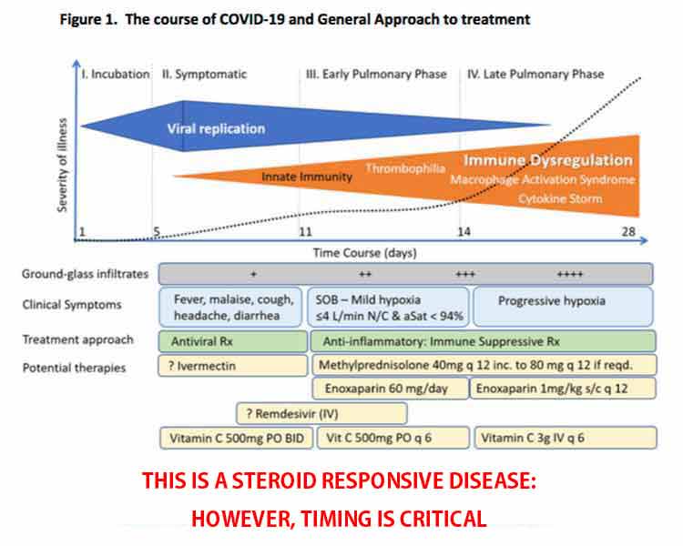
Viral Load Declines as Inflammation Rises
Another important concept explained by Marik in the featured interview is that the inflammatory response rises as the viral load decreases. They do not rise together.
“So, it’s really not the virus that is causing cytopathic effects,” he says. By the time you enter the pulmonary phase of the disease, your viral load has actually significantly decreased, but for some reason the inflammatory response then starts to run amok.
Your oxygen saturation is “the key indicator of pulmonary involvement,” Marik says. Once your oxygen saturation starts to decline, you are entering the early pulmonary phase where inflammation is rapidly increasing.
This is why it’s so important to make sure you’re measuring your oxygen saturation. Do not try to treat at home if your oxygen is dropping. Go to the hospital. Again, early treatment is crucial. Hopefully, your doctor will be willing to implement the MATH+ protocol.
Corticosteroids Are a Crucial Component
In a short essay19 co-written by the entire FLCCC team, they express their conviction that the MATH+ protocol is one of the best, most effective, critical care protocols for COVID-19 to date.
“Months ago, early on in COVID19, the FLCCC created the MATH+ protocol based on our physicians’ insights into COVID19 as a steroid-responsive disease.
This treatment recommendation went against all the major national and international health care societies that had misinterpreted the medical literature, a body of published evidence which, upon careful and deep review, actually supported the use of corticosteroids in prior pandemics …
Thousands of patients who became critically ill with Covid19 and who were suffering from massive inflammation may have been saved if this safe and powerful anti-inflammatory medicine had been provided.”
The essay20 stresses the importance of corticosteroids in the treatment of COVID-19, and cites results from the RECOVERY trial,21,22,23 a large, randomized controlled COVID-19 study by the University of Oxford, that validates their recommendation to use corticosteroids as soon as the patient is hospitalized.
In that study, the corticosteroid dexamethasone improved survival by one-third in ventilated patients and one-fifth in those requiring oxygen. However, the FLCCC believes another type of corticosteroid, methylprednisolone, is a better, more effective, choice.
First, because it reaches higher concentrations in lung tissue, and secondly, because it most closely matches the inflammatory gene activation patterns induced by SARS-CoV-2. They also believe the dexamethasone dose used in the RECOVERY trial was too low, especially for severe cases.
“In the hospitals of two of our FLCCC physicians — each having treated over 100 hospitalized patients with MATH+ often early on in the hospitalization, the hospital mortality rate to date is 7% in one hospital (Dr. Paul Marik, Norfolk, Va.) and less than 1% in the other (Dr. Joseph Varon, Houston, Texas),” the essay states.24
Vitamin C and Quercetin Work Synergistically
June 19, 2020, Marik published the paper,25 “Quercetin and Vitamin C: An Experimental, Synergistic Therapy for the Prevention and Treatment of SARS-CoV-2 Related Disease (COVID-19)” in the journal Frontiers in Immunology, which notes:
“Ascorbic acid is a crucial vitamin necessary for the correct functioning of the immune system. It plays a role in stress response and has shown promising results when administered to the critically ill. Quercetin is a well-known flavonoid whose antiviral properties have been investigated in numerous studies.
There is evidence that vitamin C and quercetin co-administration exerts a synergistic antiviral action due to overlapping antiviral and immunomodulatory properties and the capacity of ascorbate to recycle quercetin, increasing its efficacy.
Safe, cheap interventions which have a sound biological rationale should be prioritized for experimental use in the current context of a global health pandemic.”
The paper presents evidence for the use of vitamin C and quercetin — based on their biological actions and pharmacokinetics profiles — both as prophylaxis in high-risk populations, and as an adjunct to drugs such as Remdesivir or convalescent plasma in the treatment of hospitalized COVID-19 patients.
Post-COVID Syndrome
In the interview, Marik also addresses the issue of “post-COVID syndrome,” which he says is very similar to that of post-sepsis syndrome. In some cases, COVID-19 patients have recovered from the infection, only to die due to pulmonary embolism (blood clots in the lungs) or other organ dysfunction.
Marik suspects this is because the inflammatory response is still overactive. Many sepsis patients will have very high cytokine levels even a year after their recovery. He believes steroids are one key to downregulating the inflammatory response, which would prevent this problem.
A good way to screen for this, Marik says, is to measure CRP, which appear to be a good marker for ongoing inflammation. If CRP is high after recovering from COVID-19, Marik suggests doing a short course of corticosteroids to downregulate the inflammatory response. Aspirin may also be helpful if D-dimer is high. These should be used under medical supervision.
I believe this information needs to be shared far and wide, if we are to prevent more people from dying unnecessarily. More and more, as doctors are starting to speak openly about their clinical findings, we see there are quite a few different ways to tackle this illness without novel antivirals or vaccines, using older, inexpensive and readily available medications and nutrients that are already known to be safe.
- 1 Journal of Virology Sep 2004, 78 (20) 11334-11339, Antiviral activity of an analog of luteolin
- 2 Bioorg Med Chem. 2006 Dec 15;14(24):8295-306
- 3 Maclean’s February 24, 2020
- 4 ACS.org Albert Szent-Gyorgyi’s discovery of vitamin C
- 5, 25 Frontiers in Immunology June 19, 2020 DOI: 10.3389/fimmu.2020.01451
- 6 Journal of Antimicrobial Chemotherapy December 2003; 52(6): 1049-1050
- 7 covid19criticalcare.com
- 8, 10 MATH+ Protocol (PDF)
- 9 COVID-19 Management Protocol (MATH+) (PDF)
- 11 COVID-19 Management Protocol (MATH+) Updated July 9, 2020 (PDF)
- 12, 13 EVMS Critical Care COVID-19 Management Protocol August 1, 2020
- 14 Antiviral Research June 2020; 178: 104787
- 15 EVMS Critical Care COVID-19 Management Protocol June 17, 2020
- 16 FLCCC.net Medical Evidence
- 17, 18 MATH+ Hospital Treatment Protocol for COVID-19 July 14, 2020 (PDF)
- 19, 20, 24 Medium June 16, 2020
- 21 Recovery Trial
- 22 BMJ 2020;369:m1626
- 23 Science Translational Medicine June 5, 2020
12 Targeted Nutritional Strategies for Chronic Obstructive Pulmonary Disease
Reproduced from original article:
www.naturalhealth365.com/chronic-obstructive-pulmonary-disease-3525.htmlby: Lori Alton, staff writer | August 21, 2020
 (NaturalHealth365) The U.S. Centers for Disease Control and Prevention (CDC) reports that over 16 million Americans are currently living with chronic obstructive pulmonary disease (COPD), a debilitating condition characterized by difficulty breathing and blocked airflow to the lungs. While Western medicine may attempt to treat COPD with drugs such as bronchodilators and corticosteroids, these can feature toxic side effects that can include osteoporosis, pneumonia and even premature death.Fortunately, natural solutions do exist! Scientific research has shown that certain foods and supplements can markedly improve lung function and help alleviate COPD. Let’s take a look at twelve of the most promising nutritional strategies.
Inflammation and oxidative stress are the twin engines driving chronic obstructive pulmonary disease
Chronic obstructive pulmonary disease encompasses two primary conditions: emphysema (damage to the tiny air sacs in the lungs that allow for oxygen exchange) and chronic bronchitis due to inflamed airways. As the disease develops, narrowing and scarring of the airway may occur.
Common COPD symptoms include shortness of breath, persistent cough, wheezing and the production of sputum. Oxidative stress, in which harmful free radicals lead to cell and tissue damage, is a major factor in COPD. Inflammation – the presence in the lungs of inflammatory cells such as eosinophils -also plays a role.
Cigarette smoking, which accounts for 75 percent of cases, is the primary cause of COPD. Exposure to other irritants and fumes – such as coal and woodsmoke – can also play a role.
The following nutrients have been shown to help alleviate COPD. Of course, you should consult your experienced integrative doctor before adding any to your daily health routine.
Turn to N-acetylcysteine (NAC) for natural COPD relief
NAC, a supplement derived from the amino acid cysteine, has been credited by researchers with the ability to dissolve mucous, quell coughs and counteract oxidative stress.
Extensive studies have shown that NAC improves symptoms and helps slow declines in lung function. In one influential study, people with moderate-to-severe COPD who were given 1,200 mg a day of NAC improved their performance on post-exercise lung function tests.
But, remember: If you have an organ transplant, stomach ulcer or take any medication like, nitroglycerin – talk to your doctor (and pharmacist) before taking NAC.
Go with glutathione for improved respiratory function
Glutathione, a natural chemical produced in the liver, plays a vital role in boosting the immune system and neutralizing environmental toxins. Plus, its extraordinary antioxidant powers can help it combat COPD.
Glutathione is found in high concentrations in lung epithelial lining fluid and can help improve lung function and reduce inflammation. You can ramp up your dietary intake of glutathione with fresh avocados, asparagus, cucumbers and tomatoes. Eating cruciferous vegetables, such as spinach and cauliflower, also spurs production of glutathione.
Natural healers typically advise glutathione in dosages of 500 to 1,000 mg a day. For maximum benefit, opt for a liposomal formulation – and take it with vitamin C.
Good news: Studies suggest CoQ10 improves chronic obstructive pulmonary disease
A vitamin-like nutrient that is needed for muscle growth and repair, coenzyme Q10 can play an important role in reversing damage to the smooth muscle cells of the airway.
One influential clinical study showed that CoQ10 had favorable effects on muscular energy metabolism in patients with COPD. Good dietary sources include grass-fed beef liver, poultry, cold-water fatty fish, spinach and broccoli.
Don’t forget fish oil
Fish oil is rich in EPA and DHA, a pair of fatty acids hailed for their anti-inflammatory effects.
Peer-reviewed clinical studies have shown that these beneficial compounds reduce shortness of breath and inflammatory markers in COPD patients. Natural health experts typically advise dosages of 1,000 to 1,200 mg a day.
Research supports the value of resveratrol
Resveratrol, a flavonoid found in grapes, peanuts, cocoa and blueberries, has piqued researchers’ interest for its potent anti-inflammatory and antioxidant effects.
In a just-published review appearing in Current Opinion in Clinical Nutrition and Metabolic Care, the authors noted that resveratrol decreases inflammation and oxidative stress in the lung – and called it a “promising candidate” for decreasing lung injury from COPD.
Utilize the disease-fighting power of zinc
Zinc, an essential trace mineral, is believed to protect the health and function of respiratory cells during lung inflammation or injury.
In a study published in Respiratory Medicine, researchers measured the effect of zinc picolinate on volunteers with chronic obstructive pulmonary disease. The team concluded that zinc supplementation could have favorable effects on the antioxidant capacity of COPD patients.
Significantly, researchers report that zinc deficiencies are linked with increased risk of both cystic fibrosis and COPD. Good sources of zinc include oysters, grass-fed beef, poultry and pumpkin seeds.
Boswellia serrata – an ancient herbal remedy earns scientific credibility
Also known as Indian frankincense, boswellia has been utilized by herbalists for centuries to treat respiratory problems.
Animal studies have shown that boswellic acids from frankincense help to block the production of pro-inflammatory chemicals, alleviate airway dysfunction and prevent the progression of experimentally induced lung fibrosis (scarring).
These encouraging COPD-fighting results have been reinforced by human studies. One clinical study showed that boswellia caused substantial improvement in COPD by specifically inhibiting human leukocyte elastase (an enzyme which triggers emphysema and chronic bronchitis).
Boswellia may be recommended in dosages of 300 to 500 mg, two to three times a day.
Quercetin helps put the brakes on inflammation
Quercetin, an antioxidant flavonoid found in fruits and vegetables, has been shown to significantly decrease lung inflammation. And, researchers found in one preclinical trial that it helped to prevent COPD from progressing.
You can increase dietary intake of quercetin with apples, onions, grapes and green tea.
Vitamin D – the “sunshine vitamin” – benefits lung function
Vitamin D, which is produced in the skin in response to sunlight, has been shown to help regulate airway contraction.
Studies have revealed that having low levels of vitamin D raises the risk of COPD – while high-dose vitamin D supplementation has been shown to improve muscle strength and exercise capacity in COPD patients.
Natural health experts advise getting 20 minutes of direct sunlight several times a week to ensure sufficient vitamin D levels. You can also boost dietary intake with cold-water fatty fish, cage-free eggs, mushrooms and almonds.
While individual needs can vary, integrative doctors typically recommend between 5,000 and 8,000 IU of vitamin D a day. Supplementation is particularly important if you’re deficient. (below 30 ng/mL)
Address COPD with antioxidant vitamin C
Vitamin C, also known as ascorbic acid, has potent antioxidant, antihistamine and anti-inflammatory effects that make it useful in treating COPD. In fact, studies have shown that people with low levels of vitamin C have an increased risk of COPD.
In a 2016 review, the authors noted that dietary vitamin C can lower oxidative stress and restore the proliferation of alveolar cells in the lungs. Good sources include citrus fruits, red bell peppers, broccoli and strawberries.
Many integrative doctors recommend supplementation, with liposomal formulations of vitamin C considered the most bioavailable.
Enlist vitamin E to help prevent COPD
Like vitamin C, vitamin E can help reduce the odds of developing COPD.
In a ten-year population-based trial of over 38,000 women, the scientists concluded that taking 600 IU of vitamin E a day significantly reduced the risk of lung disease. You can ramp up your intake of vitamin E with sunflower seeds, almonds, spinach and wheat germ oil.
Scientific review: Proper nutrition linked with milder COPD symptoms
Overall, a healthy diet is associated with lower risk of COPD – along with better outcomes for those who develop the disease. In research published in European Respiratory Review, the authors noted an association between fruit intake and significantly improved pulmonary function.
Antioxidant-rich fruits (along with vegetables, nuts and seeds) are a virtual treasure trove of micronutrients and flavonoids that are particularly helpful to COPD – including the aforementioned quercetin, zinc and vitamins A, C and E.
And, cruciferous vegetables like Brussels sprouts, arugula and broccoli not only trigger production of beneficial glutathione – but also contain sulforaphane, an anti-inflammatory plant compound that may help enhance the therapeutic effects of COPD medications.
If you are living with COPD, you may find that the twelve natural strategies listed above really can help you to “breathe easier.”
Sources for this article:
CDC.gov
LifeExtension.com
LungInstitute.com
NIH.gov
NIH.gov
Chiroeco.com
ScienceDirect.com