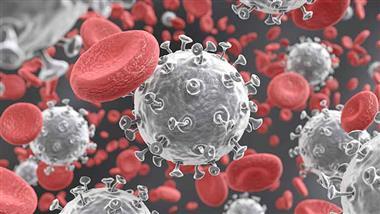
Endothelium
now browsing by category
Can This Unique Raspberry Protect Your Heart?
Reproduced from original article:
https://articles.mercola.com/sites/articles/archive/2024/02/01/endothelial-health.aspx
The original Mercola article may not remain on the original site, but I will endeavor to keep it on this site as long as I deem it to be appropriate.
Analysis by Dr. Joseph Mercola February 01, 2024

STORY AT-A-GLANCE
- Your endothelium is the collective group of cells that line your blood vessels, representing about 1% of your body mass and a surface area of 5,000 square meters
- The endothelium is directly involved in a number of diseases, including heart disease, diabetes and chronic kidney failure
- Black raspberry decreases markers of inflammation and improves endothelial function, reducing risk factors of cardiovascular disease
- Drinking green tea is associated with an increase in flow-mediated dilation (FMD) of the brachial artery, a measure of endothelium function
- Pomegranate, grape seed extract and garlic are other natural compounds that support endothelial health
Your endothelium is the collective group of cells that line your blood vessels. Representing about 1% of your body mass and a surface area of 5,000 square meters, the endothelium has been described as a “multifunctional endocrine organ strategically placed between the vessel wall and the circulating blood.”1
To put this into perspective, a single layer of endothelial cells lines the inner surface of your entire vascular system, separating your blood from the vessel wall. This “tissue-blood barrier” is semipermeable and plays a key role in regulating the transfer of molecules as well as vascular homeostasis.2
While endothelial dysfunction — an early sign of atherosclerosis3 — is a hallmark of many diseases, including cardiovascular disease, there are a host of natural options to protect your endothelium and keep it healthy.
The Role of Your Endothelium on Overall Health
While once regarded as a simple barrier, the endothelium is now regarded as a dynamic endocrine organ that has a major influence on human health. In the International Journal of Biological Sciences, it’s noted:4
“The endothelium was once thought of as the ‘cellophane wrapper’ of the vascular tree, with no other specific functions than affording selective permeability to water and electrolytes. However, enormous advances since the 1980’s have led to an understanding of the complex functions of this large endocrine organ. Vascular endothelial cells line the entire circulatory system, from the heart to the smallest capillaries.
These cells have very distinct and unique functions that are paramount to vascular biology. These functions include fluid filtration, such as in the glomeruli of the kidneys, blood vessel tone, hemostasis, neutrophil recruitment, and hormone trafficking.”
In addition to serving as a physical barrier, endothelial cells metabolize, synthesize and release vasoactive and other compounds that affect vascular tone, blood pressure, blood flow, coagulation, fibrinolysis, inflammation, immunological reactions and more.
“Any perturbation affecting the capacity and equilibrium of the endothelium as a physical barrier and to metabolize, synthesize and release these substances will cause endothelial dysfunction, which contributes to the development and progression of cardiovascular diseases,” according to a review in the World Journal of Cardiology.5 Further, the endothelium is directly involved in a number of diseases, including:6
| Peripheral vascular disease | Stroke | Heart disease |
| Diabetes | Insulin resistance | Chronic kidney failure |
| Tumor growth and metastasis | Venous thrombosis | Viral infectious diseases |
Black Raspberry and Other Natural Options for Endothelial Health
A healthy lifestyle, including diet and exercise, supports endothelial function,7 but so, too, do a variety of substances from nature.
| Black raspberry — Anthocyanins, the most abundant flavonoids in black raspberries, have anti-inflammatory and anti-angiogenic effects, meaning they help prevent angiogenesis, which may promote cancer. In human intestinal microvascular endothelial cells and human esophageal microvascular endothelial cells, black raspberry extract had both anti-angiogenic and anti-inflammatory effects.8
In a study on patients with metabolic syndrome, black raspberry also increased circulating endothelial progenitor cells (EPCs) and improved cardiovascular risks after 12 weeks.9 Separate research, also on people with metabolic syndrome, similarly revealed that black raspberry significantly decreased inflammatory cytokines, improving vascular endothelial function.10 Animal studies also suggest that black raspberry decreases markers of inflammation and improves endothelial function, reducing risk factors of cardiovascular disease.11 Flavonoids like those in black raspberries even alleviate the vascular endothelial barrier dysfunction that’s induced by advanced glycation end products.12 It’s believed that metabolism of flavonoids is involved in their beneficial role in cardiovascular health, as metabolism increases flavonoids’ vascular efficacy, “resulting in a diversity of structures of varying bioactivity in human endothelial cells.”13 |
| Green tea — Drinking green tea is associated with an increase in flow-mediated dilation (FMD) of the brachial artery, a measure of endothelium function. “The beneficial effect of green tea on endothelial function may be attributed to its high flavonoid content.
As has been shown, epigallocatechin gallate, a major catechin in tea, acutely improves endothelial function in humans with coronary artery disease,” researchers explained in the European Journal of Preventive Cardiology.14 Green tea also increases nitric oxide production by endothelial cells and may boost vascular function via anti-inflammatory pathways. According to the European Journal of Preventive Cardiology scientists:15
Green tea is also a rich source of quercetin. In one study of 30 men with coronary heart disease, consuming quercetin-rich polyphenol extract led to an increase in flow-mediated dilation of arteries, signaling improved endothelial health.16 It also inhibits platelet aggregation and has vasorelaxant properties that help lower blood pressure and prevent cardiac hypertrophy, in which the heart muscles thickens. |
| Black chokeberry — Black chokeberry (Aronia melanocarpa), known for its astringent berries, is another concentrated source of beneficial phenolic compounds, including proanthocyanidins, flavanols, anthocyanins, flavonoids and chlorogenic and caffeic acids.17
Black chokeberry extracts significantly induce endothelial cells nitric oxide (NO) production, even at relatively low concentrations. NO plays an important role in vascular function and endothelial cell dysfunction may impair NO production, increasing the risk of cardiovascular diseases. Writing in the Journal of Food Biochemistry, researchers explained:18
|
Grape seed extract — Grape seed extract contains antioxidant proanthocyanidins, a type of polyphenol that may also benefit endothelial function, thereby protecting heart health. Writing in the journal Nutrients, scientists from Tokyo Medical and Dental University noted:19
In the study of middle-aged adults with prehypertension, participants received either low-dose or high-dose grape seed proanthocyanidin extract or a placebo for 12 weeks. Grape seed extract improved vascular elasticity, while high-dose grape seed extract also decreased blood pressure.20 |
| Pomegranate — Due to its high polyphenol content, pomegranate acts against oxidative stress and is useful for endothelial dysfunction. Pomegranate contains antihypertensive, antiatherogenic, antihyperglycemic and anti-inflammatory compounds that protect heart health by improving endothelial function.
In describing its protective role in endothelial dysfunction, researchers with Federal University of Espirito Santo in Vitoria, Brazil, explained:21
Many of pomegranate’s — known as “the jewel of autumn” — beneficial polyphenols are stored in the peel, which is why pomegranate peel powder is one of my favorite supplements. Research shows pomegranate peel contains more than twice the amounts of antioxidants — specifically phenolics, flavonoids and proanthocyanidins — than the pulp, for instance, and has been shown to protect low-density lipoprotein against oxidation to a far greater degree than pulp.22,23 Other research found polyphenol-rich pomegranate peel extract improved endothelial dysfunction in mice by modulating gut microbiota.24 |
| Garlic — Garlic is a powerful antioxidant that may help fight reactive oxygen species (ROS) in your body. In one study, aged garlic extract reduced ROS, helping to prevent endothelial dysfunction.25 Garlic’s therapeutic properties may act synergistically to support heart health and protect against heart-related events like heart attack.
Aged garlic extract supplementation may increase microcirculation, helping to prevent the atherosclerotic process, while a garlic-herb preparation blocked atherosclerosis progression 1.5-fold in postmenopausal women, with the benefit lasting for 12 months.26 Further, according to a review published in the journal Antioxidants:
Among garlic’s protective effects is the ability to lower blood pressure levels. Aged black garlic (ABG), which contains more antioxidants than raw garlic,27 may be particularly beneficial. While it’s unknown exactly how garlic may lower blood pressure, scientists noted that ABG extract helps improve vasodilation and levels of endothelial nitric oxide synthase (eNOS), among other benefits.28 |
| Fibrinolytic enzymes — The connection between enzymes and the endothelium was highlighted during the COVID-19 pandemic. Studies suggested damage to the endothelium contributed to the development of blood clots, or thrombosis, in the blood vessels of severely ill COVID-19 patients.29
When physicians at the Yale School of Medicine began running clotting tests on their patients,30 levels of Von Willebrand factor (VWF), a clotting protein released by endothelial cells, were found to be significantly elevated, which suggested that damaged endothelial cells may be releasing large quantities of VWF, leading to clots.31 “Our findings show that endotheliopathy is present in COVID-19 and is likely to be associated with critical illness and death. Early identification of endotheliopathy and strategies to mitigate its progression might improve outcomes in COVID-19,” the researchers concluded.32 Under healthy conditions, blood cells can pass through the endothelium lining blood vessels, but when exposed to viral infections and other inflammatory agents, the endothelium becomes sticky and releases VWF. The end result is a cascade of clotting and inflammation, both characteristics of severe COVID-19. In the European Heart Journal it’s stated, “COVID-19, particularly in the later complicated stages, represents an endothelial disease.”33 This is where enzymes come in. One study reported three case studies of patients with severe COVID‐19 respiratory failure who were treated with tissue plasminogen activator (tPA), a serine protease enzyme found on endothelial cells that’s involved in fibrinolysis, or the breakdown of blood clots.34 All three patients benefitted from the treatment. Beyond COVID-19, another study involved 1,062 people with mild hyperlipidemia and/or mild atherosclerosis. They took the fibrinolytic enzyme nattokinase, which “effectively managed the progression of atherosclerosis and hyperlipidemia with a significant improvement in the lipid profile.”35 Significant reduction in carotid artery intima-media thickness, a measure of the extent of arterial thickening related to endothelial dysfunction, was noted, with improvement rates ranging from 66.5% to 95.4%. When using these enzymes for fibrinolytic therapy they need to be taken on an empty stomach, at least one hour before or two hours after meals. Lumbrokinase, which is about 300 times stronger than serrapeptase and nearly 30 times stronger than nattokinase,36 is my strong personal preference and recommendation if you are using a fibrinolytic enzyme. |

Download this Article Before it Disappears
Endothelial Health Depends on a Healthy Lifestyle
Protecting endothelial health is much like protecting heart health — it involves a comprehensive, healthy lifestyle. A sedentary lifestyle, smoking, excess alcohol37 and ultraprocessed foods can all negatively affect endothelial health, while exercise38 and fresh, whole foods, including citrus fruits and dark green vegetables, are protective.39
In addition to black raspberry and the other natural substances above, coenzyme Q10 (CoQ10), a fat-soluble antioxidant, also acts directly on your endothelium, dilating your blood vessels and lowering blood pressure.40,41 CoQ10 is associated with significant improvements in endothelial dysfunction.42 By the age of 65, your body typically produces only about half the amount of CoQ10 it did at 2543 so supplementation with CoQ10 or its reduced form ubiquinol is helpful in some cases.
- 1, 2 Pathophysiology June 2008, Volume 15, Issue 1, Pages 49-67
- 3 Open Cardiovasc Med J. 2010; 4: 302–312
- 4, 6 Int J Biol Sci. 2013; 9(10): 1057–1069
- 5 World J Cardiol. 2015 Nov 26; 7(11): 719–741
- 7 Metabolism December 2011, Volume 60, Issue 12, Pages 1736-1740
- 8 Microvascular Research January 2015, Volume 97, Pages 167-180
- 9 J Med Food. 2016 Apr;19(4):346-52. doi: 10.1089/jmf.2015.3563. Epub 2016 Feb 18
- 10 Phytother Res. 2014 October;28(10);1492-8
- 11 Journal of Food Biochemistry September 12, 2021
- 12 Nutrients. 2022 Mar; 14(5): 1026
- 13 The Journal of Nutrition February 3, 2016
- 14 European Journal of Cardiovascular Prevention and Rehabilitation, Volume 15, Issue 3, 1 June 2008, Pages 300–305
- 15 European Journal of Cardiovascular Prevention and Rehabilitation, Volume 15, Issue 3, 1 June 2008, Pages 300–305, Clinical implications
- 16 Pharmacogn Rev. 2016 Jul-Dec; 10(20): 84–89., Cardiovascular disease prevention
- 17, 18 J Food Biochem. 2016 Aug; 40(4): 404–410
- 19, 20 Nutrients. 2019 Dec; 11(12): 2844
- 21 Curr Pharm Des. 2020;26(30):3684-3699. doi: 10.2174/1381612826666200406152147
- 22 Food Chemistry May 2006; 96(2): 254-260
- 23 Foodnavigator.com July 19, 2008
- 24 Scientific Reports volume 9, Article number: 14150 (2019)
- 25 Antioxidants (Basel). 2020 Jul; 9(7): 619., 5.1 Garlic Properties
- 26 Antioxidants (Basel). 2020 Jul; 9(7): 619., 5.2.2. Cardiovascular Diseases
- 27 Food Chem. 2016 May 15:199:135-9. doi: 10.1016/j.foodchem.2015.11.128. Epub 2015 Nov 30
- 28 Nutrients, 2022; 14(3), Discussion
- 29, 30, 31 The Scientist November 3, 2020
- 32 The Lancet Haematology June 30, 2020
- 33 European Heart Journal, Volume 41, Issue 32, 21 August 2020, Pages 3038–3044, doi: 10.1093/eurheartj/ehaa623
- 34 Diapharma, Tissue Plasminogen Activator
- 35 Front. Cardiovasc. Med., 22 August 2022
- 36 Townsend Letter May 2018
- 37 European Journal of Physiology June 4, 2023
- 38 J Cardiovasc Transl Res. 2022; 15(3): 604–620
- 39 Cleveland Clinic, Endothelium
- 40 Molecular Aspects of Medicine. 1994;15(1):s257-s263
- 41 Circ Res. 1989;65(1):1-21
- 42 Atherosclerosis April 2012, Volume 221, Issue 2, Pages 311-316
- 43 Int J Mol Sci. 2020 Sep; 21(18): 6695., Intro
Are Enzymes a Key to COVID Endothelial Injury?
Reproduced from original article:
https://articles.mercola.com/sites/articles/archive/2023/02/10/coronavirus-enzymes.aspx
The original Mercola article may not remain on the original site, but I will endeavor to keep it on this site as long as I deem it to be appropriate.
Analysis by Dr. Joseph Mercola Fact Checked February 10, 2023
STORY AT-A-GLANCE
- Studies suggest damage to the endothelium, which are cells covering blood vessels, is contributing to the development of blood clots, or thrombosis, in the blood vessels of severely ill COVID-19 patients
- Enzymes may turn out to be the missing link in helping to break up clusters of clotting proteins involved in this dangerous thrombosis, which is linked to increased mortality in COVID-19
- Levels of Von Willebrand factor (VWF), a clotting protein released by endothelial cells, were found to be significantly elevated in COVID-19 patients in advanced stages of the disease
- Proteolytic enzymes such as lumbrokinase, serrapeptase and nattokinase also act as natural anticoagulants by breaking down the fibrin that forms blood clots
From Dr. Joseph Mercola
Since COVID-19 first entered the scene, exchange of ideas has basically been outlawed. By sharing my views and those from various experts throughout the pandemic on COVID treatments and the experimental COVID jabs, I became a main target of the White House, the political establishment and the global cabal.
Propaganda and pervasive censorship have been deployed to seize control over every part of your life, including your health, finances and food supply. The major media are key players and have been instrumental in creating and fueling fear.
I am republishing this article in its original form so that you can see how the progression unfolded.
Originally published: November 30, 2020
Enzymes catalyze many biological reactions in your body. They regulate the rate of these chemical reactions, speeding them up so necessary functions like digestion, muscle contractions and other aspects of cellular metabolism can occur.1
Enzymes are also emerging as key players in COVID-19, as studies suggest damage to the endothelium, which are cells covering blood vessels, is contributing to the development of blood clots, or thrombosis, in the blood vessels of severely ill COVID-19 patients.2 Enzymes may turn out to be the missing link in helping to break up clusters of clotting proteins involved in this dangerous thrombosis.
Endothelium Damage Found in Critically Ill COVID-19 Cases
After noticing blackened fingers and toes — signs of what appeared to be microvascular thrombosis, or tiny blood clots in small blood vessels — in COVID-19 patients in advanced stages of the disease, physicians at the Yale School of Medicine began running clotting tests on their patients.3
Levels of Von Willebrand factor (VWF), a clotting protein released by endothelial cells, were found to be significantly elevated, which suggested to hematologist Alfred Lee that damaged endothelial cells may be releasing large quantities of VWF, leading to clots.4 This prompted the team to screen for additional markers of endothelial cell and platelet activation in critically and noncritically ill COVID-19 patients.
The study, which was conducted in April 2020, included 68 hospitalized patients with COVID-19 and 13 asymptomatic controls. VWF antigen was significantly elevated in COVID-19 patients admitted to the intensive care unit (ICU) compared to non-ICU COVID-19 patients,5 as was soluble platelet selectin (sP-selectin), which is sometimes used as a biomarker for infection and mortality.6
Specifically, mean VWF was 565% among ICU patients and 278% among non-ICU patients while soluble P-selectin was 15.9 ng/mL compared to 11.2 ng/mL.7 “Our findings show that endotheliopathy is present in COVID-19 and is likely to be associated with critical illness and death. Early identification of endotheliopathy and strategies to mitigate its progression might improve outcomes in COVID-19,” the researchers concluded.8
Likely not coincidentally, endothelial dysfunction is also associated with insulin resistance and plays a role in the vascular complications of diabetes,9 as well as being involved in obesity and high blood pressure,10 conditions that raise the risk of severe COVID-19.
Even mild obesity may raise the risk of COVID-19 severity — COVID-19 patients with mild obesity had a 2.5 times greater risk of respiratory failure and a five times greater risk of being admitted to an ICU compared to nonobese patients. Those with a BMI of 35 and over were also 12 times more likely to die from COVID-19.11
Another study looking into the impact of coexisting health conditions like high blood pressure, heart disease and diabetes on COVID-19 outcomes found they’re linked to “poorer clinical outcomes,” such as admission to an intensive care unit, a need for invasive ventilation or death.12
It’s possible that the endothelial damage in all of these conditions plays a role in worsening COVID-19 outcomes, but it’s unclear which comes first — endothelial damage or COVID-19.
Endothelial Cells Are the ‘Main Target’ of SARS-CoV-2
Imperial College London cardiologist Thomas Lüscher told The Scientist that the endothelium is the main target of SARS-CoV-2, the virus that causes COVID-19.13 Under healthy conditions, blood cells can pass through the endothelium lining blood vessels, but when exposed to viral infections and other inflammatory agents, the endothelium becomes sticky and releases VWF.
The end result is a cascade of clotting and inflammation, both characteristics of severe COVID-19. According to a case report published April 8, 2020, “A hallmark of severe COVID-19 is coagulopathy, with 71.4% of patients who die of COVID-19 meeting … criteria for disseminated intravascular coagulation (DIC) while only 0.6% of patients who survive meet these criteria.”14
Writing in the European Heart Journal, Lüscher argues, “COVID-19, particularly in the later complicated stages, represents an endothelial disease,”15 which may help explain why multiple organ systems, including the lungs, heart, brain, kidney and vasculature, may be affected.
An additional study by Canadian researchers, published in Critical Care Explorations in September 2020, also revealed elevated VWF and soluble P-selectin levels in COVID-19 patients, along with higher glycocalyx-degradation products,16 a sign of damage to the glycocalyx, which envelops the endothelium.17 This can also be a sign of sepsis. Taken together, the research suggests that therapies targeting the endothelium may be useful for COVID-19, which is where enzymes come in.
Download this Article Before it Disappears
Enzymes Used to Treat COVID-19
With the role of coagulopathy in severe COVID-19 becoming clearer, researchers have experimented with enzymes in the treatment of the disease. Fibrinolytic therapy, which uses drugs or enzymes to break up blood clots, has been used in a Phase 1 clinical trial that showed the treatment reduced mortality and led to improvements in oxygenation.18 Further, researchers wrote in the Journal of Thrombosis and Haemostasis:19
“There is evidence in both animals and humans that fibrinolytic therapy in acute lung injury and acute respiratory distress syndrome (ARDS) improves survival, which also points to fibrin deposition in the pulmonary microvasculature as a contributory cause of ARDS.
This would be expected to be seen in patients with ARDS and concomitant diagnoses of DIC on their laboratory values such as what is observed in more than 70% of those who die of COVID‐19.”
The researchers reported three case studies of patients with severe COVID‐19 respiratory failure who were treated with tissue plasminogen activator (TPA), a serine protease enzyme found on endothelial cells that’s involved in fibrinolysis, or the breakdown of blood clots.20
All three patients benefited from the treatment, with partial pressure of oxygen/FiO2 (P/F) ratios, a measure of lung function, improving from 38% to 100%.21 While it should be noted that several of the authors have patents pending related to both coagulation/fibrinolysis diagnostics and therapeutics, the results suggest such treatments deserve further evaluation in certain COVID-19 patients.
An evaluation of organ tissues from people who died from COVID-19 also revealed extensive lung damage, including clotting, and long-term persistence of virus cells in pneumocytes and endothelial cells.22
The findings indicate that virus-infected cells may persist for long periods inside the lungs, contributing to scar tissue. In an interview with Reuters, study co-author Mauro Giacca, a professor at King’s College London, described “really vast destruction of the architecture of the lungs,” with healthy tissue “almost completely substituted by scar tissue,”23 which could be responsible for cases of “long COVID,” in which symptoms persist for months.
“It could very well be envisaged that one of the reasons why there are cases of long COVID is because there is vast destruction of lung (tissue),” he told Reuters. “Even if someone recovers from COVID, the damage that is done could be massive.”24 Dissolving scar tissue is another area where enzymes, particularly proteolytic enzymes, may be useful.
Three Top Enzymes Act as Natural Anticoagulants
Holistic prophylactic alternatives that might be beneficial against blood clots include proteolytic enzymes such as lumbrokinase, serrapeptase and nattokinase, all of which act as natural anticoagulants by breaking down the fibrin that forms the blood clot. Fibrin is a clotting material that restricts blood flow, found both in your bloodstream and connective tissue such as your muscles. Fibrin accumulation is also responsible for scar tissue.
It is important to understand that when using these enzymes for fibrinolytic therapy they need to be taken on an empty stomach, at least one hour before or two hours after meals. Otherwise these enzymes will be wasted in the digestion of your food and will be unable to serve their fibrinolytic purpose.
As noted in Scientific Reports, some of the key mechanisms by which proteolytic enzymes exert their anticoagulant effect include “defibrinogenation, inhibition of platelet aggregation, and/or interference with components of the blood coagulation cascade.”25 Here’s a closer look at these important enzymes, all of which are available in supplement form or, in the case of nattokinase, via the food natto.
1. Lumbrokinase — This enzyme is about 300 times stronger than serrapeptase and nearly 30 times stronger than nattokinase,26 making it my strong personal preference and recommendation if you are using a fibrinolytic enzyme. Extracted from earthworms, lumbrokinase is a highly effective antithrombotic agent that reduces blood viscosity and platelet aggregation27 while also degrading fibrin, which is a key factor in clot formation.
2. Serrapeptase — Also known as serratiopeptidase, serrapeptase is produced in the gut of newborn Bombyx mori silkworms, allowing them to dissolve and escape from their cocoons. Research has shown it can help patients with chronic airway disease, lessening viscosity of sputum and reducing coughing.28 Serrapeptase also breaks down fibrin and helps dissolve dead or damaged tissue without harming healthy tissue.29
3. Nattokinase — Produced by the bacteria Bacillus subtilis during the fermentation of soybeans to produce natto,30 nattokinase is a strong thrombolytic31 comparable to aspirin but without the serious side effects.32
It’s been shown to break down blood clots and reduce the risk of serious clotting33 by dissolving excess fibrin in your blood vessels,34 improving circulation and decreasing blood viscosity. Interestingly, in one in vitro study, the thrombolytic activity of equivalent amounts of nattokinase and TPA were found to be identical35 — TPA, remember, is the enzyme that led to improvement in the three COVID-19 case studies.36
- 1 Britannica, Enzyme
- 2, 3, 4, 13, 17 The Scientist November 3, 2020
- 5, 7, 8 The Lancet Haematology June 30, 2020
- 6 Biomark Insights. 2017; 12: 1177271916684823
- 9 Diabetes Obes Metab. 1999 May;1 Suppl 1:S17-22. doi: 10.1046/j.1463-1326.1999.0010s1017.x.
- 10 Circulation July 27, 2020
- 11 U.S. News & World Report July 23, 2020
- 12 medRxiv February 27, 2020
- 14 J Thromb Haermost Case Reports April 8, 2020 DOI: 10.1111/jth.14828
- 15 European Heart Journal, Volume 41, Issue 32, 21 August 2020, Pages 3038–3044
- 16 Critical Care Explorations: September 2020 – Volume 2 – Issue 9 – p e0194 doi: 10.1097/CCE.0000000000000194
- 18, 19, 21, 36 J Thromb Haemost. 2020 May 11 : 10.1111/jth.14828
- 20 Diapharma, Tissue Plasminogen Activator
- 22 EBioMedicine November 3, 2020
- 23, 24 Reuters November 3, 2020
- 25 Scientific Reports 2018; 8, Article Number 6210
- 26 Townsend Letter May 2018
- 27 BioMed Research International. Volume 2011 | Article ID 51965. 2003
- 28 Respirology 2003 Sep;8(3):316-20
- 29 International Journal of Surgery, 2013 Apr;11(3):209-217
- 30 Int J Mol Sci. 2017 Mar; 18(3): 523
- 31, 34 Biol Pharm Bull. 1995 Oct;18(10):1387-91
- 32 Lab Anim Res December 2013; 29(4): 221-225
- 33 Scientific Reports 2015; 5: 11601
- 35 Article Number 6210, Lunathrombase has in vitro thrombolytic potency but is devoid of hemolytic activity or cytotoxicity against mammalian cells