Sarcoidosis

now browsing by category


No Proof MMR Vaccine Is ‘Safer’ than Measles, Mumps or Rubella Infection, Physician Group Says

© January 14th 2025 Children’s Health Defense, Inc. This work is reproduced and distributed with the permission of Children’s Health Defense, Inc.
Want to learn more from Children’s Health Defense?
Sign up for free news and updates from Robert F. Kennedy, Jr. and the Children’s Health Defense.
Your donation will help to support us in our efforts.
Reproduced from original article:
https://childrenshealthdefense.org/defender/measles-mumps-rubella-infection-mmr-vaccine-risk

The risk of permanent disability or death from the MMR vaccine may be greater than the risk from a measles, mumps or rubella infection because large enough vaccine safety studies haven’t been done, according to a collection of new documents released by Physicians for Informed Consent.

word "risk" and mmr vaccine

The risk of permanent disability or death from the MMR vaccine may be greater than the risk posed by measles, mumps or rubella infection because large enough vaccine safety studies haven’t been done, according to a collection of new documents released by Physicians for Informed Consent (PIC).

The collection includes disease information statements for measles, mumps and rubella, and a vaccine risk statement for the MMR vaccine.

According to the Mayo Clinic, measles is a viral infection typically accompanied by a skin rash, fever, cough, runny nose, sore throat, inflamed eyes and tiny white spots on the inner cheek.

Mumps and rubella also are viral infections. According to PIC, all three viral infections typically resolve on their own with proper rest and hydration in almost all cases.

Dr. Shira Miller, PIC’s founder and president, told The Defender, “The main takeaway is that the MMR vaccine has not been proven safer than measles, mumps and rubella.”

PIC is a nonprofit that delivers data to doctors and the public so they can “evaluate the data on infectious diseases and vaccines objectively, and voluntarily engage in informed decision-making about vaccination.”

Miller explained that the MMR vaccine clinical trials didn’t include enough subjects to be able to prove that the risk of permanent disability or death from the vaccine is less than the risk of permanent disability or death from measles, mumps or rubella.

The number of measles, mumps or rubella infections that result in permanent disability or death is so low that researchers would need to have at least 50,000 subjects in a clinical trial to be able to show that the vaccine is safer than the disease.

The MMR vaccine’s clinical trials fall very short of that benchmark, according to PIC’s statement on MMR vaccine risk.

Prelicensure clinical trials for vaccines, including the MMR shot, are “relatively small and usually last no longer than a few years,” according to the Centers for Disease Control and Prevention’s (CDC) 2024 “Manual for the Surveillance of Vaccine-Preventable Diseases.”

The 2024 edition of the CDC manual doesn’t specify exactly how many subjects are in these “relatively small” trials. However, the 2011 edition stated that “relatively small” meant that such trials are “usually limited to a few thousand subjects.”

The rate of disability or death among healthy children from any of those three diseases is incredibly rare. PIC wrote:

“For children under age 10 at normal risk (i.e., with normal levels of vitamin A and infected after birth), the pre-vaccine annual risk of death or permanent disability from measles, mumps, and rubella respectively was 1 in 1 million, 1 in 1.6 million, and 1 in 2.1 million. …

“Therefore, the cumulative annual risk of a fatal or permanently disabling case of any of those diseases was about 1 in 500,000, and the risk over a 10-year span was 1 in 50,000.”

In other words, clinical trials would need at least 50,000 subjects to detect one case of death or disability from a measles, mumps or rubella infection.

Meanwhile, no safety studies on the MMR vaccine have been done that looked for possible genetic mutations, impaired fertility or cancer, according to the product’s package insert.

Also, seizures from the MMR vaccine occur five times more often than measles-related seizures.

Dr. Liz Mumper, a pediatrician, praised PIC for releasing the collection of data on measles, mumps and rubella, and on the MMR vaccine.

“Most parents have not had access to the information contained in the thoughtful analysis done by Physicians for Informed Consent. Parents should recognize that the risk of bad outcomes from a measles infection — if their child lives in a developed country with clean water and is not immune-deficient — is extraordinarily rare, as PIC reports.”

Unfortunately, she added, recent U.S. media reports “sensationalized” the risks of measles.

What’s typically missing from measles media reports

PIC’s statement on measles cited numerous facts commonly overlooked in many media reports on measles outbreaks, including:

  • The U.S. measles mortality rate dropped dramatically before a measles vaccine was introduced in 1963.
  • Immunity from the MMR vaccine wanes so that by age 15, roughly 60% of vaccinated children are susceptible to subclinical measles virus infections.
  • Studies have suggested a link between a naturally acquired measles infection and a reduced risk of Hodgkin’s and non-Hodgkin’s lymphomas.
  • Studies also suggested a link between a naturally acquired measles infection and a lower risk of asthma, eczema and hay fever.
  • Malnutrition — particularly vitamin A deficiency — is a primary cause of over 100,000 measles deaths in underdeveloped countries.

Mumper said that the risk of bad outcomes from a measles infection drastically declined with improved public health and better nutrition long before MMR vaccines were available.

“The risk of bad outcomes has always been more for children in developing countries who are more likely to have nutritional deficiencies including vitamin A and lack access to clean water,” Mumper added.

The Trump Administration Must Bring Moderna to Heel

Reproduced from original article:
https://brownstone.org/articles/the-trump-administration-must-bring-moderna-to-heel

The Trump Administration Must Bring Moderna to Heel

The Trump Administration Must Bring Moderna to Heel

Last week, independent journalist Alex Berenson reported that a preschool-aged child died of “cardio-respiratory arrest” after taking a dose of Moderna’s Covid mRNA vaccine during its clinical trials. Despite federal requirements to report all trial information, the company withheld the truth for years as it raked in billions from its Covid shots.

The extent of the cover-up remains unknown, but Moderna, headed by CEO Stéphane Bancel, disregarded federal law requiring companies to report “summary results information, including adverse event information, for specified clinical trials of drug products” to clinicaltrials.gov. The company, not the government, is responsible for posting all results, and failure to report the death of a child constitutes a clear breach of US law, which threatens civil action against any party that “falsifies, conceals, or covers up by any trick, scheme, or device a material fact.”

To this point, pharmaceutical companies have remained largely immune for their role in perpetrating globally-scaled deception resulting in thousands of vaccine injuries and billions in profits. They have enjoyed a liability shield courtesy of the PREP Act, which offers protections for injuries resulting from vaccines; that indemnity, however, does not extend to non-compliance with federal regulations, material misstatements or omissions of fact, or other offenses.

The death of the child only became known because of an obscure European report released last year, which revealed that Moderna has known about the death for over two years while it continues to advertise Covid shots to children as young as six months old.

Moderna’s European filing also revealed that the company withheld trial results demonstrating that children under 12 who received the vaccine were ten times more likely than those who received the placebo to suffer “serious side effects.” Without any evidence, Moderna claimed that the side effects, including the death of a child, were unrelated to the shots.

The incoming Trump administration offers a rare opportunity to hold pharmaceutical companies accountable and to investigate the depth of the cover-up.

The FDA is responsible for enforcing the reporting of vaccine trial results, but recent heads of the agency such as Scott Gottlieb and Robert Califf have been fanatical supporters of Big Pharma. Trump’s choice for FDA, Dr. Marty Makary, presents a stark contrast to his predecessors. Makary has criticized the US Government’s reluctance to acknowledge the role of natural immunity in preventing Covid infection, and he opposed the widespread vaccination of children. He testified to Congress, “In the U.S. we gave thousands of healthy kids myocarditis for no good reason, they were already immune. This was avoidable.”

President-elect Trump has tapped Robert F. Kennedy, Jr., perhaps the most well-known critic of the Covid vaccines, to lead the Department of Health and Human Services, which oversees the FDA. He has named Dr. Jay Bhattacharya, an author of the Great Barrington Declaration, as his choice to head the National Institutes of Health. Further, Senator Ron Johnson (R-WI) told Berenson that he plans to subpoena the FDA once Republicans become the majority party in the Senate this month.

President Trump’s first term was ultimately defined by his failure to fulfill his pledge to “drain the swamp.” A corrupt bureaucracy, personified in many ways by Dr. Anthony Fauci, aided and abetted by advisors like his son-in-law, Jared Kushner, hijacked the president’s agenda. Now, the Trump administration has an unlikely yet monumental opportunity for health reform, which can start on January 20 with an investigation into Moderna’s cover-up.

The Covid response doomed Trump 1.0. Whether one regards this as a monumental error, the betrayal of a president by his advisors, an event beyond the president’s control, or a deeper and more complex plot involving everything and everyone associated with the government, both in the US and around the world, there is no question of the scale of the calamity for the public. The shots are part of that, the capstone failure of a long line of foreshadowing with lockdowns and all that was associated with pre-pharmaceutical interventions. The antidote came not as a cure but, for many, the disease itself.

There must be truth if not justice.


Published under a Creative Commons Attribution 4.0 International License
For reprints, please set the canonical link back to the original Brownstone Institute Article and Author.

Author

  • Brownstone Institute

    Articles by Brownstone Institute, a nonprofit organization founded in May of 2021 in support of a society that minimizes the role of violence in public life.


Why Does the COVID Vaccine Persist In The Body?

Reproduced from original article:
https://www.midwesterndoctor.com/p/why-does-the-covid-vaccine-persist

The consequences of the questionable decisions made to manufacture the mRNA vaccines

DEC 24, 2024

Recently, new data emerged showing that the COVID vaccines persist for up to 700 days within patients (and likely longer). As this is quite concerning to many, I was required to write an article explaining how this happens, and how it relates to the egregious production process that characterized the COVID-19 vaccines.

Upsides and Downsides

A lot of things in life are trade-offs, and as I’ve gotten older, more and more I’ve come to appreciate how many things in our society boil down to the fact that the options for addressing them all have significant downsides, so in many cases no solution exists which is satisfactory to all parties involved.

As such, this dilemma is typically managed by some combination of the following:

• Having a biased focus which emphasizes the benefits of an approach a side supports and downplays its downsides (or conversely disproportionately focuses on the downsides of an opposing position). To this point, I’ve had countless issues I’ve debated both sides of and been able to effectively persuade audiences of each one—which highlights how subjective many of the entrenched beliefs we hold actually are (and, in turn, is why I put so much work here into fairly presenting both sides of each controversial topic I cover).

• Sweeping the downsides under the rug and gaslighting the populace into believing they don’t exist.

• Blitzing the public into supporting a questionable policy before they have time to recognize its downsides, and if that fails, overtly forcing them to go along with it.

Note: I believe one of the reasons why governments frequently do horrible things to their people is because they are put in the position of having to “solve” a problem (but with no truly satisfactory way to do it), so they become habituated to using the three previous strategies to push their chosen policies along and simultaneously develop a collective mentality that those questionable approaches are necessary for the “greater good.”

There are many different manifestations of this dilemma, many of which I believe are essentially reflective of a foundational concept in medicine—sensitivity and specificity.

An ideal diagnostic test would catch every instance of a disease (100% sensitivity) and simultaneously never have a false positive (100% specificity). Unfortunately, in almost all cases, this is impossible to do, and instead a trade-off exists where you can either prioritize sensitivity (which leads to a significant number of false positives) or prioritize specificity (which leads to a significant number of false negatives). During COVID for example, a decision was made to prioritize sensitivity with the PCR tests (by having a high replication cycle thresholds) so no cases of COVID would be missed, but this resulted in such poor specificity that the PCR tests effectively became worthless (except for drumming up fear) since they produced so many false positives.

As such, when tests are designed, attempts are made to ensure there is a good balance between sensitivity and specificity. In some cases this is successful (e.g., there are many lab results we will take at face value), but in many other cases, given the technology involved, it’s not really possible to do so (or it is, but lobbying led to overdiagnosis so a medical product could be sold).

Similarly:

• Many policies in the justice system aims to enact fall into this same situation. For example, an ideal death penalty is robust enough to deter murder (and keep violent criminals away from the public), but simultaneously lenient enough that it doesn’t accidentally execute innocent individuals. Since there is no way to have both perfect sensitivity and specificity on this, different states take radically different approaches to how they enforce the death penalty (lying all along the spectrum between sensitivity and specificity). Likewise, our judicial system was founded under the principle “innocent until proven guilty” whereas many other countries have judicial systems that are the exact opposite.

• In medicine, one of the greatest challenges is finding the appropriate dose, as people differ, so what might be a safe and therapeutic dose for one person could be toxic for another. As such, standardized doses are typically chosen by finding the best overall balance between efficacy (a sufficient dose) and safety (avoiding a toxic dose), but for many drugs, the standardized dose leads to many more sensitive patients becoming severely injured by the drugs (which is then commonly “addressed” by gaslighting those injured patients).
Note: a much more detailed discussion on the art of dosing can be found here.

• Every medical intervention has its risks and benefits, and ideally, the job of a physician should be to accurately weigh those to determine the best treatment for a patient (while simultaneously conveying what they are to the patient). Unfortunately, in many cases, they don’t (which is a large part of why patients are so dissatisfied with the medical system).

Overall, there are three key takeaways from this paradigm I wish to focus on in this article:

1. It is often incredibly difficult to find an acceptable balance between sensitivity and specificity, and many of the conventions our society now follows were the result of years of debate and protest from both sides to find a palatable middle ground between the two.

2. In almost any sphere I frequently find rushed attempts to find an acceptable balance between two conflicting positions to be immensely flawed and prone to creating significant issues in the future.

3. Many of the issues with the vaccine program are encapsulated by this framework.

The Forgotten Side of Medicine is a reader-supported publication. To receive new posts and support my work, consider becoming a free or paid subscriber. To see how others have benefitted from this newsletter, click here!

How Vaccines “Work”

Note: many believe the immune system is one of the least understood parts of physiology, something I would argue is a result of immunological research being focused on making profitable pharmaceuticals (e.g., vaccines) rather than understanding how it works.

In the classical conception of immunity, there are two types, innate immunity and adaptive immunity, with the innate response being relatively nonspecific (so it can work against a wide variety of infectious threats, including those the body has never seen before) and the adaptive one, which is uniquely suited to eliminating the specific invading organism.

The most widely accepted explanation for how the adaptive immune system works is “clonal selection theory” which states that the immune system:
1. Uses a random generation process to create a vast pool of potential antigen matching sequences.
2. Has vast number of different immune cells that each have those sequences attached to them circulate the blood stream.
3. Waits for one of those immune cells to contact an invading pathogen that the sequence it carries matches.
4. Have each of the immune cells be programmed to start rapidly reproducing once they get a sequence match.
5. Through the previous 4 steps, make it possible to produce a large number of immune cells which are specific to an invading organism (because they can bind to their antigens and alert the rest of the immune system to the organism’s presence), and thus effectively neutralize the infection.
6. Once the process is complete, to leave behind memory B cells, which match the invading pathogen and are able to stimulate the immune response in a much more rapid fashion (thereby shortening the time that steps 3 and 4 take).

The theory behind vaccination is that if the specific immune response and memory B cells can be created before the body encounters a dangerous microbe, this can:

• Allow the body to mount a robust immune response before a harmful invading organism has time to multiply within the body and cause significant damage.

• Cause individuals to rapidly clear infections (rather than needing to wait for the adaptive response to kick in), thereby reducing or eliminating the amount of time they can spread the disease into the population.

• Cause individuals to develop an immune response at the site of infection (e.g., the membranes of the nose and throat), thereby preventing the organism from being able to colonize those areas and thus preventing its transmission.

Being able to do this is hence immensely appealing to governments, as it allows a single intervention (the vaccine) which can easily be distributed to everyone in a top-down manner (which is what governments are good at doing) to address a longstanding problem (infectious disease outbreaks) and more importantly, to allow the government to present the appearance of working in earnest to safeguard the public’s health. Because of this appeal, throughout history, governments will get deeply invested in vaccine programs, and then once issues arise with those programs, double-down on the vaccine (e.g., with mandates) rather than reconsider the wisdom of the vaccine program.
Note: in a previous article, I showed how this misguided and tyrannical conduct has existed ever since the first vaccine (smallpox).

Vaccine Production

To “work” vaccines aim to mass produce a dangerous organism’s antigen without the organism itself being present and then administer that antigen into the body. By doing so, the intermediate stage of an infection (where the organism has already reproduced enough inside its host to have a large number of antigens be available to match a circulating immune cell) can be achieved without the individual being in danger of being damaged or overwhelmed by the infection.

Unfortunately, unlike chemicals which can be rapidly synthesized, antigens are complex enough that they can only be produced by biological systems. As such, to produce the antigen, one of the following is typically done.

• Mass produce the infectious organism, then “kill” it so that its antigens can be collected, but the organism itself is not able to cause infections.

• Genetically modify another organism to mass produce a desired antigen, then kill it and extract the antigen (e.g. the HPV vaccine does this with modified yeasts).

• Modify the live pathogen (typically a virus) so that it can still cause the infection and reproduce inside the recipient but simultaneously is “weakened” so that it is less likely to cause illness.

• Genetically modify a “benign” virus to contain the antigen but be unable to replicate in the human body, then mass produce it outside the body, and have the body develop an immune response to the virus and the antigen on it once it is injected.

• Introduce mRNA into cells so human cells can produce large amounts of the desired antigen, which the immune system then sees (e.g., on the surface of the cells) and develops an immune response to.

The basic problem is that none of these approaches are perfect, and each has both its ups and downsides. For example:

• Most can create autoimmunity.

• In those where only a single antigen is used (and the virus spreads from human to human), if the vaccine actually works, it rapidly stop working because the pathogen quickly evolves a new antigen that no longer matches the vaccine.

• It contrast, the multi-antigen ones (which don’t have that issue) are typically live attenuated vaccines, which then can cause those with weakened immune systems to develop infections from the vaccine itself (e.g., this happens with the polio vaccine—which is why the primary cause of polio now is from the vaccines rather than natural infections, but also can happen with others like the shingles and measles vaccine).

Furthermore, some infectious diseases respond fairly well to vaccination, but the majority do not, so at this point, the vaccine industry has already picked all the “low-hanging fruit” and hence faces an existential struggle to develop new proprietary (patentable) vaccines it can bring to market. For example, had it not been for COVID-19 (SARS-CoV-2), a SARS vaccine would have never been brought to market as it was well recognized the SARS virus was poorly suited for vaccination (which what we then saw throughout the pandemic).

Finally. even if a vaccine “works” it still has to be manufactured, and there are numerous cases of the tradeoffs being made resulting in a disaster. For example:

• To make the inactivated polio vaccine, the live polio virus had to be exposed to formaldehyde. However, the challenge with this was that if too much formaldehyde was used, it would damage the antigens on polio to the point they no longer matched those on the poliovirus, whereas if too little was used, some of the polioviruses would remain active and could then give the vaccine recipient polio. The creator of the vaccine (Salk) opted to prioritize efficacy over safety, which the government in turn supported, even when one of their own scientists (Bernice Eddy) warned them against releasing the vaccine (as it caused polio in her lab). That 1955 vaccine then infected at least 220,000 people with live polio virus in Cutter’s vaccine, of whom 70,000 developed muscle weakness, 164 were severely paralyzed, and 10 died.

Note: an identical issue had happened on a smaller scale (9000 infections, 12 severe cases, 6 deaths) in 1935 with an earlier version of the inactivated polio vaccine. Likewise, (as I showed here) there have been dozens of incidents where an insufficiently inactivated or attenuated diphtheria, rabies or yellow fever vaccines severely injured hundreds of people (as the attenuated vaccines faced a similar issue with it being easy to over or under attenuate).

• Growing viruses for vaccines requires having a cell culture to grow them in. Monkey kidney cells were chosen because they worked well for doing this, but unfortunately were contaminated with the cancer causing SV40 virus. In 1962Eddy again warned the government about the vaccine, but they still chose to give it to the public (and retaliated against her for speaking out), which in turn led to a wave of cancer sweeping through America, which until the COVID-19 vaccines was unprecedented:

Note: many other viral vaccines (particularly the live ones) also have had harmful viral contaminants identified within them, but unlike SV40, that contamination has not been acknowledged. Most noteably, a strong case can be made that HIV emerged from virally contaminated vaccines (that had been grown in monkey tissues).

• After a potentially dangerous strain of influenza (due to it having similarities to the 1918 influenza) was identified, a rush began to make an emergency vaccine for it (despite the FDA’s chief influenza expert Morris accurately warning that strain posed no risk to America). Since it took a while to cultivate the virus for a live attenuated vaccine, in order to make the vaccine be produced fast enough to hit the market before the influenza strain disappeared, a decision was made to hybridize it with the PR8 strain, a fast growing influenza strain directly descended from the 1918 influenza. Morris warned against doing this, but was ignored (and fired). That 1976 vaccine subsequently injured a large number of people (including some of our patients) and was a publicity disaster for the US government.

The anthrax vaccine used during (and after) the Gulf War required growing large amounts of the bacteria, killing them, and then filtering out the most toxic components from the final vaccine preparation. The issue the manufacturer ran into was that because of how dirty the vaccine was, its contaminants clogged the filters the manufacturer used, so “solve” the problem and be able to manufacture the vaccine at scale for the military, the manufacturer opted to use larger filters which did not clog, but also didn’t filter many of the toxic components out of the final products—which resulted in one of the most harmful vaccines in history being unleashed upon our military.

• Due to the challenges in producing it, the DTP vaccine (another killed bacterial vaccine) was notorious for having hot lots that killed their recipients (or left them with brain damage). In fact, one FDA pertussis specialist in 1976 stated:

Pertussis vaccine is one of the more troublesome products to produce and assay. As an example of this, pertussis vaccine has one of the highest failure rates of all products submitted to the Bureau of Biologies for testing and release. Approximately 15-20 percent of all lots which pass the manufacturer’s tests fail to pass the Bureau’s tests.

Eventually, the injuries that vaccine created led to so many lawsuits that the manufactures could not afford to continue producing the vaccine, at which point, the 1986 Vaccine Injury Act was passed. This shielded the manufactures from all future liability from it (hence allowing them to stay in business), and eventually incentivized the production of a safer but more costly pertussis vaccine.

• Frequently when an antigen is produced, it cannot solicit a sufficient immune response (unless a lot of it is used—which frequently makes the vaccine too costly to produce). To solve this problem, cheap (and toxic) adjuvants which enhance the immune response to the antigen are used, thereby allowing an affordable amount of antigen to be required for the final product. When the HPV vaccine was developed, it was discovered that its antigen (along with standard adjuvants) could not mount a sufficient immune response to get FDA approval, so a decision was made to use an experimental (but much stronger adjuvant) which worked—but also gave a large number of recipients autoimmune disorders (at least 2.3%). Nonetheless, that trade-off was also accepted to get it to market.

In short, if you look at all these cases, a consistent pattern should be clear. Whenever there is a choice between getting a dangerous vaccine to market or holding off because there isn’t a way to do it safely, the vaccine industry will always do the risky approach (especially in “emergency” situations) as they know they can unconditionally rely upon the US government to promote the product as “safe and effective” and then legally shield them from the disaster which inevitably follows.

COVID-19 Vaccine Hurdles

When COVID-19 began, the industry faced three major issues:

• Whoever was the first to develop a successful vaccine would make a lot of money, but those whose products took longer to reach the market would like miss out on the bonanza.

• There was a finite amount of time the lockdowns could be sustained (which made people want to vaccinate so they could be “free”) and it was very likely the population would rapidly develop herd immunity to COVID-19—so they was a limited window to get a vaccine to market.

• It was extraordinarily difficult to make a safe and effective vaccine for SARS (e.g., decades of work had not yielded a viable product).

Fortunately for the industry, the WHO (and Bill Gates), in 2010, had enacted their “Decade of Vaccine” plan, and with the World Economic Forum (between 2014-2016) had developed a framework for pushing through emergency vaccines that could bypass the regulatory process in the event of a health “emergency.” This framework gave lavish lavish fiscal incentives for vaccine manufacturers and positioned unaccountable organizations like the WHO, Gates foundation, or the World Economic Forum as the directors of a future pandemic response.

Shortly after that framework was finished, the FDA on January 13, 2017 released extremely detailed regulations for obtaining emergency use authorizations, and five days later, Gates publicly announced his plan to the world. This framework was endorsed by pharmaceutical companies, including Pfizer Moderna and J&J, and when Operation Warp Speed was finally conducted in 2020, it mirrored the framework Gates had previously developed.

Note: Event 201, a Gates funded “simulation” exercise modeling the release of a dangerous SARS virus from China was conducted on October 18, 2019. Reading through it in December of 2019 allowed me to accurately predict how COVID-19 would play out. Likewise, on 9/4/2019, Gates invested 55 million in the company that produced Pfizer’s mRNA vaccine—which in two years was worth 550 million.

So as you might expect, the industry chose to adopt the fastest possible production pathway, and was quickly granted the legal immunity (and lavish funding) necessary to accomplish that.

mRNA Vaccine Challenges

Note: one of the major advantages to the mRNA platform was that its production turn around time was much faster than existing alternatives (e.g., growing a virus in chicken eggs). This was a key reason why Fauci’s agency made decades of investments to develop the platform (as with the existing options, seasonal flu vaccines had to start being produced long before the circulating strain was known—which frequently led to the annual flu vaccine being for the wrong strain).

For the mRNA vaccines to “work,” the following needed to occur:

1. An antigen needed to be chosen that was highly likely to elicit a robust immune response that suggested SARS-CoV-2 immunity (and hence could win approval).

2. mRNA matching that antigen needed to be produced at scale.

3. The mRNA needed to be able to get into cells.

4. Once it got into the cells, the mRNA needed to be produce sufficient protein to create an immune response.

Many who have examined Pfizer and Moderna’s vaccines were perplexed by the design that was chosen, as many of the immensely harmful decisions that were chosen strongly imply the vaccine was deliberately designed to harm as many people as possible. While this very well might be the case, many of those issues can instead be explained by the fact each of those 4 challenges was addressed in a way that prioritized getting a vaccine to market rather than producing a safe one.

For example, the spike protein was the most reactive (immunogenic) part of the virus and necessary for SARS to infect cells, so it was an ideal vaccine target. However, it was also a terrible antigen to chose as:

• The spike protein had a significant overlap with human tissues so it was likely to trigger autoimmunity if produced throughout the body.

• The spike protein was highly toxic, so if it was mass produced within the body, it likely would injure the recipient.

• It was a rapidly mutating part of the virus, which guaranteed the circulating spike protein would quick evolve into a variant the vaccine did not work against it—leading to the remarkable situation where we mandated a vaccine for an extinct virus (which, in turn, caused those vaccinated to become more likely to catch COVID-19 as their immune systems were continually primed to respond to a different virus from that they were exposed to)—something best demonstrated by the Cleveland Clinic’s study of 51,011 people):

Similarly, when I was trying to understand the acute toxicity of the vaccines, I suspected the lipid nanoparticles used to produce the vaccines had to be responsible for their acute toxicity (e.g., which could be seen within seconds of it being put into blood) as those effects onset far faster than it seemed possible for significant numbers of spike proteins to be produced.

After I looked into, I realized that for decades it had been deemed impossible to produce mRNA carrying lipid nanoparticles that had sufficient safety and efficacy to produce viable vaccines (to the point pioneers in the technology like Robert Malone who had invested decades of work into it abandoned it as they felt it was impossible to make a lipid nanoparticle which were not cytotoxic). In turn, when I reviewed leaked Pfizer regulatory documents, I noticed that their lipid nanoparticle had been chosen on the basis of it being the only one that had efficacy—again suggesting safety wasn’t taken into consideration.

Note: there were many other safety issues with mRNA techonolgy (e.g., synthetic mRNA induced immune suppression and antigenic spike protein coating cells and causing the immune system to destroy those cells) that are beyond the scope of this article.

mRNA manufacturing

In my eyes, the two biggest production issues with the mRNA vaccines were producing them at scale and then having them produce spike protein in the body at scale.

Note: these were easy to predict as one of the greatest challenges the pharmaceutical industry, and particularly biotech faces is producing their products at scale—a process which typically takes years to work out, but for Operation Warp Speed needed to be done in a few months.

mRNA design

A major challenge with synthetic mRNA was that the immune system would rapidly break it down, which resulted in not enough of the spike protein being produced to trigger a sufficient immune response. This in turn was “solved” through pseudouridylation, a process where pseudouridine replaces uridine in mRNA molecules as pseudouridylated mRNA resists immune system degradation.

Unfortunately:

• This process occurs in a very limited and tightly regulated manner inside the body. At the time the mRNA vaccines were developed, there was limited understanding on the biological significance of pseudouridylation, however many (e.g., Robert Malone) felt the preliminary data showed this approach had serious risks (e.g., immune suppression).

• These existing technology for pseudouridylation causes it to occur in a random and haphazard way. As such, rather than being able to determine a “safe” dose of pseudouridylation, an all or nothing approach had to be adopted, which in turn led to a significant number of vaccines having “excessive” pseudouridylation (either due to how much of it happened to the mRNA or where in the mRNA the pseudouridylation occurred). In short, this approach was extremely reckless, and akin to playing Russian roulette but hoping everything worked out.

In turn, the pseudouridylation did “solve” the mRNA degradation problem (to the point it won an unscrupulous Noble Prize), but it also created a new issue—the mRNA (and spike protein production) persisting in the body for a prolonged period, and likely due to the haphazard nature of the vaccine’s production, it persisting much longer in some cases that others. Lastly, while this invention “won” the Noble Prize, there were also viable mRNA vaccines that did not utilize it (and hence did not have the risks it entailed).

Note: Codon optimization (which increases protein production from mRNA) may have also caused the vaccine to excessively produce spike protein in the cells.

mRNA preservation

One of the major issues with synthetic mRNA is that its immensely fragile, so even if it could be protected from immune degradation within the cells (e.g., via pseudouridylation) it still was highly susceptible to common environmental factors. As such, when the lots of finished vaccines were examined by regulators, they determined there was both significant degradation of the mRNA and significant variability in what mRNA was preserved.

This is one of the many tables from the leaked EMA documents (which shows how quickly mRNA is destroyed at normal temperatures).

As this degradation and the resulting “truncated mRNA” was one of the primary concerns from the drug regulators Pfizer “solved” this problem by having all of their vaccines be ultra frozen (under the belief this would prevent mRNA degradation). However, this was largely for show, and before long the practice was abandoned (e.g., many vaccine sites had vials sitting outside throughout the day).

Note: at the time I learned about it, I thought that the primary issues would be broken mRNA sequences producing unintended and potentially harmful protein sequences. We now believe it’s actually the opposite, and that this breakdown process was a blessing in disguise, as evidence gradually accumulated that the older vaccines were and the further from their production site they were injected into the body, the less toxic they were—suggesting that the vaccine’s toxicity was mitigated by portions of its mRNA breaking down and hence preventing it from producing as much spike protein.

Process 1 vs. Process 2

The original process used to produce the mRNA vaccine had two ways it could be done. In the cleaner process (which was used to produce the vaccines for the trials), mRNA was produced through controlled replications with minimal contaminants present. Unfortunately, this process could not be scaled.

As such, an alternative approach was done:

• E. coli bacteria were genetically modified to have DNA that contained the spike protein, antibiotic resistance, and the SV-40 promoter.

• Those bacteria were mass produced, then doused with an antibiotic (so those without the resistance gene and thus the spike protein gene) died and did not contaminate the final product.

• The remaining bacteria are killed and their DNA is extracted.

• An mRNA polymerase is used turn that bacterial DNA into vaccine mRNA.

• Everything besides the mRNA is eliminated.

• The mRNA is packaged into lipid nanoparticles.

The essential problem with this process was that it was not feasible to remove many of the contaminants from each stage of production and there was significant room for variability at each stage (and thus in the final product). While many contaminants could be an issue, the vaccine safety community primarily focused on the plasmids as:

• They’ve repeatedly proved plasmids were present in the vaccine.

• The SV40 promoter (a key part of the cancer causing SV40 virus) was grafted onto the plasmids as this was an effective way to increase mRNA production.

• Plasmids had the potential to alter the human microbiome and genome (especially since the SV40 promoter will bring DNA into the nucleus)—which is particularly concerning since vaccine plasmids have now been shown to get into cell nuclei.

• If there was a genomic integration, it was likely creating indefinite spike protein production in the cells.

• When vaccine lots were analyzed for their plasmid levels, it was found the lots which more frequently injured their recipients had higher levels of DNA plasmids (suggesting either harmful genomic integration was occurring, that the plasmids were toxic absent genomic integration, or that they were a proxy for other harmful contaminants).

Inconsistent Manufacturing

Each of the previous points illustrates that the vaccine industry was not ready to bring the mRNA vaccines to market and that the manufacturing of them was rife with errors. Many other points also support this such as:

Japan pulled 1.63M vials of Moderna’s vaccine after visible metal particles were found in them and when examining vaccine vials, Ryan Cole found glass shards in the vaccines. Both of these suggest the production of the vaccines was rushed to the point basic quality control steps were not taken.

• When Ryan Cole did a recorded examination of the vaccines with Del Bigtree they found:

We looked at all the different vaccines and I think one of the conclusions we came away with is that it’s just a hodgepodge..there were vaccines that seems as though there were no particles within them, almost nothing there it was almost like a saline shot and then there were Pfizer’s that were just packed with them and you just get the sense that the manufacturing of this is totally inconsistent…some were more concentrated and some were less.

Note: this suggests that there was either very poor mixing when the vaccines were packaged (leading to some having lots of the lipid nanoparticles and others none) or that the vaccine manufacturers were unable to produce enough vaccine to meet the existing orders and switched to packing placebo vials to meet their contracted orders.

In one mass spectrometry examination of 4 vaccine vials, it was determined that the lipid nanoparticles, but not the mRNA, were present in each of the vials. This suggests that the vaccine was not prepared in a consistent manner, or that the manufacturers ran out of mRNA to fill the vaccines with.

• A large body of evidence from the adverse event reporting databases (compiled here) demonstrated that the toxicity of the vaccine lots greatly varied, which again was likely explained by inconsistencies in their manufacturing.

Note: in a previous article about the mRNA lipid nanoparticles, I showed how inconsistencies in the manufacturing of the vaccines likely explained why the vaccines tended to affect different organs in the body (as their charge was affected by how much mRNA they contained and the area they deposited in the body was influenced by their charge) and why some individuals had acute reactions to them.

Blot Gate

One method of analyzing which proteins are present is with an approach known as “Western Blot.” Periodically, individuals will fake Western Blots (which can be detected because its very easy to identify computer generated ones).

With the COVID-19 vaccines, Western Blots were meant to serve as a quality control measure which ensured the mRNA vaccine were producing their intended protein. However, when we examined the available Western Blots we discovered:

• Some of them were computer generated (and hence likely fake).

• Others showed proteins besides the intended vaccine antigen were there (possibly due to broken RNA fragments being turned into proteins).

All of this again suggested that there were serious manufacturing issues with the COVID vaccines, but a decision was made to sweep all of that under the rug to protect the vaccine manufacturers (particularly since the drug regulators willfully ignored this fraud).

Note: this is similar to how there was extensive fraud throughout the COVID-19 clinical trials to exaggerate vaccine efficacy and safety (which essentially invalidated all the data was gathered), yet even after the trial participants and trial supervisors repeatedly notified the FDA, nothing was done. Likewise, shortly before the vaccine rollout, Vanity Fair published an article highlighting the serious issues in Americas’s vaccine manufacturing plants and that the FDA essentially was unable to monitor them—hence arguing things would get even worse during Operation Warp Speed.

mRNA persistence

Once the COVID-19 vaccine hit the market and the injuries began to mount, we noticed a three curious patterns.

1. The susceptibility to vaccine injuries greatly differed, but in many cases seemed to cluster (e.g., I knew a husband and wife who got vaccinated at the same time and both have near fatal complications from the vaccine).

2. A significant number of individuals appeared to be sensitive to vaccine shedding, something that is supposed to be “impossible” but nonetheless was occurring in a fairly repeatable manner to a large number of people, with symptoms similar to COVID vaccine injuries but typically less severe.
Note: everything we know about shedding is discussed here.

3. Many individuals with vaccine injuries appeared to be suffering toxic reactions to spike proteins in their blood steam months if not years after vaccination. For example:

• Autopsy studies (conducted on individuals who died suddenly up to 6 months after the vaccine) have shown that their tissues were flooded with spike protein. Beyond the inflammatory and necrotic responses to the spike protein being their likely cause of death, it also suggested those individuals had had large amounts of spike protein being perpetually produced within their bodies.

• Both myself and Pierre Kory have come across numerous cases of individuals with vaccine injuries who, months, if not years after vaccination, respond to spike protein binders (suggesting unbound spike protein was causing their issues). Additionally, those individuals either remained recovered, significantly regress, or partially regress once the binder is stopped (implying there is a sustained but possibly diminishing production of spike protein in the body in many vaccine injured patients).

• The only commercially available test for the spike protein (offered by Quest) measures existing antibodies to the spike protein receptor binding domain, and provides values anywhere from 0-25,000 (or higher than 25,000). Clinically, we’ve seen that long COVID rarely get levels above 4,000, whereas in those with vaccine injuries, it can be anywhere from 0-25,000 with many being over 25,000. In turn, a rough (but not precise) correlation exists between the antibody levels (which do not directly measure spike protein levels) and a patient’s illness (along with its levels improving or worsening generally correlating to a patient’s symptoms improving or worsening). Curiously, many patients do not have their antibody levels decline with time (which is what you typically see after a COVID infection), which again suggests spike protein is being produced within the body that then stimulates an immune response. Conversely however, some of these cases may instead have been from people who had antibody levels far above 25,000 who then had their antibody levels decline (but this cannot be detected as they are still above the 25,000 cut off).
Note: Alex Bereneson has also shared reports of vaccinated individuals who’ve had spike protein antibody levels over 25,000 for months after vaccination.

Pivotal mRNA Studies

When the COVID vaccines were pitched to the public, two of the greatest concerns were that the experimental gene therapies could change our DNA and that they would persist in the body for a prolonged period. To address these sales barriers, the media continually platformed experts like Paul Offit and Anthony Fauci) who dismissed us by continually saying things like:

  1. The vaccines cannot enter the nucleus of the cell
  2. mRNA from the vaccines breaks down rapidly in the cell, so it does not have time to enter the nucleus and change your DNA.
  3. mRNA is not DNA, so believing mRNA can change DNA represents a fundamental lack of knowledge of biology.

These points raised red flags to me, as beyond their being issues with each of them, no data was ever provided to disprove genomic integration of the vaccine (which would have been quite easy to do). As such, I assumed the vaccines did integrate into the genome, and evidence would eventually emerge that they did (and likewise that at least some of the vaccines would continually produce spike protein in the recipients).

Throughout this process, two very important, but largely forgotten studies came out.

Note: due to the extreme scientific embargo on anything which challenged the COVID narrative, it is somewhat of a miracle either of these were published.

The first was a March 2022 study from Stanford which showed that both vaccine mRNA and spike protein persisted at high levels at least 2 months after vaccination. This was highly unusual for mRNA, and suggested something (e.g., the pseudouridylation) was preventing it from breaking down. Most importantly, the study did not go beyond 8 weeks, so it was likely the mRNA was persisting for a much longer time.

The second was a January 2023 study (with many Harvard authors) which found in adolescents and young adults who developed myocarditis within a few days of vaccination, that compared to controls, they had significantly higher levels of free spike protein circulating in their blood (due to them not forming antibodies which bound it). This in turn suggested that those who were reacting the worst to the vaccines lacked the ability to form antibodies which could counteract its effects in the body, which both explained why some individuals were so sensitive to the vaccine (e.g., shedding) but also that those likely to “benefit” from vaccination also would have mounted a robust response against a natural infection (hence invalidating the justification for getting vaccinated).

Note: with the smallpox vaccines, vaccination was deemed successful if there was a strong inflammatory reaction at the vaccination site, whereas if no reaction occurred, the vaccination was deemed unsuccessful and the individual was repeatedly revaccinated (as when it didn’t “take” the individuals could still get smallpox). Many early medical dissidents observed that severe reactions to the smallpox vaccine typically followed the vaccine not “taking.” They hence came to believe that the smallpox vaccine working was due to the recipient having a functional immune system that could both already fight off smallpox and create the superficial inflammatory reaction to the vaccine and that the vaccine was simply taking credit for what their immune system could already do. This study made me wonder if something similar has occurred with the COVID-19 vaccines.

Most recently, Yale’s immunology team (which had previously strongly endorsed using the vaccine to prevent COVID and to treat long COVID—which is often disastrous) conducted a longterm study of the effects of vaccination on immune function. Recently, they shared their preliminary results with the participants and disclosed that they were having difficultly publishing the study due to the political pushback they were receiving.

Note: it is extremely common for studies which challenge a medical dogma to never be published. For example, the study demonstrating COVID vaccination causes shedding was stonewalled for well over a year, as I show here, many studies demonstrating harm from the childhood vaccines have been blocked from publication (or even being conducted), and in one well known incident, the CDC deleted data that incriminated the vaccine program. Likewise, I recently highlighted how once the ultrasound industry took off, research into its dangers abruptly ended (due to funding being withdrawn and journals refusing to publish it), which eventually created a collective amnesia that this data ever existed.

As the study was being conducted, a few of us were contacted by participants in the trial who kept us appraised of what was going on, but asked us not to disclose any of it so that we would not interfere with the trial’s publication (as Yale putting their name behind a study demonstrating longstanding immunological injury would make “long-vax” become a medically accepted condition—but the scientific community tends to react quite badly to study results being prematurely leaked).

Prior to that meeting, preliminary data had been published a 2023 pre-print (which has still not been published) which detailed the common symptoms seen in the 241 participants with post vaccination syndrome (PVS), which match what we’ve seen in clinical practice:

To quote the study:

In conclusion, people reporting PVS after covid-19 vaccination in this study are highly symptomatic, have poor health status, and have tried many treatment strategies without success. As PVS is associated with considerable suffering, there is an urgent need to understand its mechanism to provide prevention, diagnosis, and treatment strategies.

Note: these results were discussed in more detail in this October 2023 online conference (e.g., the mast cell component of the illness). From watching this conference, my impression was that the investigators sincerely want to help the trial participants, but due to the unpleasant implications of their findings, are in a very challenging position (hence why their 2023 pre-print has still not been published).

Recently, Alex Berenson decided to use his platform to publicize what was disclosed at the recent meeting, at which point, I felt it was appropriate to share some of what we’d learned, and shortly after it became a trending topic on 𝕏. There I highlighted that:

• There was a sustained drop in CD4 levels which the Yale group suspected could account for the sustained immune suppression following vaccination (e.g., AIDS is characterized by severe CD4 suppression). I do not know the average CD4 drop they saw, but one participant in the trial (who has extensive post vaccine symptoms) shared with me her labs which included:

• Study participants were found to have sustained vaccine spike protein in their blood (e.g., 700 days after vaccination), which led the researchers to suspect the vaccine was integrating into the genome.

Note: while there are no commercially available spike protein tests (instead we have to test the antibody levels), research institutions like Yale (and the studies I cited above) have access to tests for free spike protein. Fortunately, after years of work, a commercially available (and affordable) blood spike protein test appears to be just around the corner.

All of this briefly means:

• There is objective proof that long vax is a real syndrome. Unfortunately, given the pace at which science works, it will likely be a few more years before it is formally acknowledged (which will likely dovetail with new pharmaceuticals to treat it entering the market and all remaining interest in the COVID vaccines disappearing). However, given that Trump promoted these vaccines (and has been unwilling to distance himself from them), I could see left-wing institutions like Yale accelerating their publication timeline so that this becomes widely publicized throughout his presidency.

• The persistence of spike protein in the body indicates that the 2022 Stanford study would have found positive results if it had tested patients more than two months post vaccination and that the spike protein antibody titers are indicative of spike protein persisting within the body.

Note: I recently discussed this topic with Dr. Malone (who I consider to be the most knowledgeable people in this area). We are both of the opinion that while genomic integration may play a role in spike protein persistence, the more probable explanation is simply that the body cannot break down the mRNA (and possibly the spike proteins) due to how it was modified. Presently, the data does not exist to quantify the scale of spike protein genomic integration, but with what is currently known (which could change as more data becomes available) cellular production of vaccine mRNA is most likely not responsible for the majority of the free spike protein found in the vaccine injured individuals.

Conclusion

Throughout my life, as I’ve come to feel that because of the bad trade-offs inherent to many policies or technologies, those behind them (particularly the government) will take an approach akin to trying to pound a square peg through a round hole (as government always defaults to utilizing the force at its disposal to solve the problems it encounters). In contrast, whenever I encounter situations where there really does not seem to be a good way to balance the trade-offs, I take that as a sign I need to consider a completely different approach rather than forcing the one I’ve committed to into working.

With COVID for example, I realized near the start that it would be an exercise in futility to address it with a vaccine—a truth much of the world has now had its eyes opened to. Instead, it was my assessment from the start that the best option would be to quickly develop viable treatments for the illness that could prevent severe complications from it and then allow infected individuals to recover with a strong immunity to the disease (and as we’ve now seen, natural immunity is vastly superior to vaccine immunity for COVID-19).

Unfortunately, rather than heed that approach, our medical apparatus decided to do everything it possibly could to push the vaccine upon us, while simultaneously doing all they could to bury the myriad of effective off-patent treatments developed for COVID-19.

Since the pathway Bill Gates put into place for lucrative accelerated approvals is still in place, I believe this highlights how important it is for us to actually understand how these technologies work and the trade-offs involved with them (which are never disclosed). In turn, it is my sincere belief that if the public had known part of what I presented here, they likely would have never taken the COVID vaccines. Similarly, as I’ve tried to illustrate here, contamination and poor production is a systemic problem with vaccines, and were robust independent testing to be conducted (so people actually knew what was in the vaccines they were taking), the demand for them would likely disappear until the industry was forced to clean up its act.

Overall, it is my belief that the most effective way to stop these unsafe products is to simply have enough people boycott them that they become financially unsustainable (and due to the new era of information diffusion we are walking into thanks to platforms like 𝕏, it’s actually possible). The FDA and CDC have lost an immense amount of trust because of how flagrantly they lied to the public, and have thus far refused to take any accountability for their actions—something that will likely change once financial pressures (e.g., people no longer buying the drugs rubber-stamped by the FDA) force the agency to make genuine amends for its conduct throughout the pandemic.

While COVID-19 was a profound tragedy, because of it we now have an extraordinary opportunity to fix this continually proliferating corruption, and I sincerely thank each of you who has helped to make this moment possible.

An updated index of all the articles published in the Forgotten Side of Medicine (including the DMSO ones) can be viewed here. Additionally, to learn how other readers have benefitted from this publication and the community it has created, their feedback can be viewed here.

What is Sarcoidosis?

Written by Brenton Wight, Health Researcher, LeanMachine
Copyright [c] 1999-[y] Brenton Wight. All Rights Reserved.
This site is non-profit, existing only to help people improve health and immunity
Updated 7th March 2022

Sarcoidosis

Sarcoidosis (sar-koy-DO-sis) is an inflammatory granulomatous disease, an auto-immune condition that can involve almost any organ or part of the nervous system, but most often the lymph glands and lungs. 10% to 30% of patients with sarcoidosis develop progressive pulmonary disease.
Among those with pulmonary sarcoidosis, the rate of spontaneous remission ranges from 10% to 82%,but lung disease progression occurs in more than 10% of patients and can result in fibrocystic architectural distortion of the lung, associated with a mortality rate of 12% to 18% within 5 years. Overall, the mortality rate for sarcoidosis is approximately 7% within a 5-year follow-up period. Worldwide, more than 60% of deaths from sarcoidosis are due to pulmonary involvement; however, but in Japan, over 70% of sarcoidosis deaths are due to cardiac involvement.
More common in people 20 to 50 years old, peaking in the range 20 to 29, particularly women, but can occur at any age and to either sex.
Women over 50 are in a second high-risk age group.
All races are affected, with an average incidence world-wide of 19 per 100,000 in women, and 16.5 per 100,000 in men.
Australia has an average rate of around 6 per 100,000 and generally the cases are mild compared to other countries.
Northern European countries have a higher rate, with 60 per 100,000 in Sweden and Iceland.
In the United States, people of African or especially Afro-Caribbean descent have a higher rate of 35.5 per 100,000 compared to Caucasians with 10.9 per 100,000.
South America, Spain, India, and Canada have much lower rates.
One of the lowest rates in the world is in the Philippines (where coconuts form a major part of the diet – see more below).
Actual cases may be much higher, as many remain un-diagnosed. The majority will clear up without medical intervention, sometimes in 1 to 3 months, sometimes up to 2 or 3 years.
A study of fire-fighters in the USA revealed a higher rate than expected, I suspect from toxic chemicals such as:

  • Chemicals used in fire-fighting
  • Chemicals used in flame-proofing clothing, furniture, paint, etc
  • Chemicals released from burning materials

If a person from a low-risk country moves to a high-risk country, they then inherit the same risk as others in that country.
70% of patients with advanced pulmonary sarcoidosis develop precapillary pulmonary hypertension, which is associated with a 5-year mortality rate of around 40%.

The Inflammation Connection
The immune system protects the body from invaders by sending special white blood cells to the invaded area, which release chemicals that recruit other cells to destroy all invaders.
Inflammation is the result, and in a healthy body the invader is destroyed, then the cells and inflammation disappear.
In Sarcoidosis, the inflammation remains, allowing immune system cells to cluster, forming lumps (granulomas) in various organs and other parts of the body.

The Genetic Connection
Identical twins are more likely to both have Sarcoidosis than paternal twins, confirming the genetic link.
Some people, especially African-Americans and Northern Europeans have the gene HLA-DRB1 *03:01 and *03:02 which has been verified to be a significant sarcoidosis risk.
Also the gene HLA-DRB1*1101 is more common in sarcoidosis patients.
The HLA group of genes are primarily involved in the immune system.
Another gene, IL23R is linked to uveitis (when sarcoidosis affects the eyes). This gene can be blocked with the drug Stelara (Ustekinumab).
IL23R is also linked to ankylosing spondylitis, psoriasis/psoriatic arthritis, Crohn’s disease, ulcerative colitis, autoimmune thyroid disease, breast cancer and systemic lupus erythematosus.
It is found in multi system sarcoidosis that is difficult to treat.
Ustekinumab is used to help control interleukin 12 and interleukin 23, which are involved in immune-mediated inflammatory disorders such as Sarcoidosis, Psoriasis and MS (Multiple Sclerosis).
More gene info here

Observation  Patients with sarcoidosis and precapillary pulmonary hypertension should be treated with therapies such as phosphodiesterase inhibitors and prostacyclin analogues. Although optimal doses of oral glucocorticoids for pulmonary sarcoidosis are unknown, oral prednisone typically starting at a dose of 20 mg/d to 40 mg/d for 2 to 6 weeks is recommended for patients who are symptomatic (cough, dyspnea, and chest pain) and have parenchymal infiltrates and abnormal pulmonary function test results. Oral glucocorticoids can be tapered over 6 to 18 months if symptoms, pulmonary function test results, and radiographs improve. Prolonged use of oral glucocorticoids may be required to control symptoms and stabilize disease. Patients without adequate improvement while receiving a dose of prednisone of 10 mg/d or greater or those with adverse effects due to glucocorticoids may be prescribed immunosuppressive agents, such as methotrexate, azathioprine, or an anti–tumor necrosis factor medication, either alone or with glucocorticoids combined with appropriate microbial prophylaxis for Pneumocystis jiroveci and herpes zoster. Effective treatments are not available for advanced fibrocystic pulmonary disease.

Conclusions and Relevance  Sarcoidosis has a mortality rate of approximately 7% within a 5-year follow-up period. More than 10% of patients with pulmonary sarcoidosis develop progressive disease and more than 60% of deaths are due to advanced pulmonary sarcoidosis. Oral glucocorticoids with or without another immunosuppressive agent are the first-line therapy for symptomatic patients with abnormal pulmonary function test results and lung infiltrates. Patients with sarcoidosis and precapillary pulmonary hypertension should be treated with therapies such as phosphodiesterase inhibitors and prostacyclin analogues.

Why do some people suffer and others do not?

If the immune system is compromised, and the patient has a genetic pre-disposition, they are more likely to have a reaction to the bacteria by forming a granuloma (lump).
The lump is called a Sarcoidosis granulomatous condition, sometimes mistaken for cancer, but Sarcoidosis is definitely not cancer or cancer-related.
I believe the high rates in cold climates, women and those of African descent is partly due to low vitamin D3 levels, common in all of those groups, which compromises the immune system.
That does not mean that those Sarcoidosis patients low on vitamin D3 should supplement – the reverse can be true, more on this complex issue later.

What causes Sarcoidosis

The absolute cause has not been determined.
Granulomas form when lymphocytes (a type of white blood cell, part of the immune system) and macrophages (a different white blood cell which engulfs and “eats” debris, infections and invaders, also part of the immune system), get out of control attacking whatever the foreign invader is, and form the lump.
There are many risk factors:

  • Viral or bacterial infection (Sarcoidosis is not contagious, but resembles tuberculosis)
  • Defect in the immune system (auto-immune or abnormal immune response, low vitamin D3)
  • Unidentified toxic substance, possibly mercury, aluminium, lead, aersenic, etc
  • Unknown environmental cause (chemicals, pollutants, radiation, etc)
  • Genetic or inherited cause – see HLA genes above
  • High-carbohydrate diet (sugar, grains, starchy vegetables)
  • Diet low in healthy fats
  • Overweight or obese
  • Tick bites (often causing Lyme disease which can have some similar symptoms)
  • Mycotoxins (mouldy food)
  • Environmental Fungi – exposure to fungi at home is higher among sarcoidosis patients, and the more fungi, the more serious the condition.
  • Defective Mannose Receptor Gene
  • Exposure to smoke, chemicals in air, e.g. fire-fighters
  • Existing Lupus, MS (Multiple Sclerosis), RA(Rheumatoid Arthritis), Hepatitis C, Osteoporosis, Ankylosing Spondylitis
  • Living in an area or country where people are more prone to Sarcoidosis
  • Living in an agricultural area (maybe higher exposure to agricultural chemicals?)
  • Exposure to Beryllium or other metal dust
  • Exposure to fumes or mouldy materials (may also cause lung disease, sometimes mistaken for Sarcoidosis)
  • High Estradiol and Prolactin, typical in pregnancy

Sarcoidosis, Lyme Disease and Tick Bites

Ticks tend to harbour a variety of bacteria and viruses, the most famous causing Lyme Disease.
Unfortunately, in Australia, doctors refuse to admit that we have Lyme disease in our country, yet many people succumb to this disease and have to send their samples to the USA for testing as there are no Lyme certified labs in Australia.
But the test results come back positive, and Australian doctors have no idea how to treat this condition.
Although Lyme Disease and Sarcoidosis are two distinct conditions, there are remarkable similarities between the two.
People may suffer a fever and pain for days or weeks from a tick, other insect bite, or bacteria from another source, but Sarcoidosis inflammation, symptoms and diagnosis may not happen for years afterwards.
Doctors usually treat the fever with antibiotics, and when the fever disappears they claim a cure, but in those with a genetic pre-disposition to Sarcoidosis, a tiny granuloma can form, allowing bacteria to continue thriving.
A healthy immune system rejects the bacteria, and remission is the result, but an abnormal immune system or genetic predisposition can allow a lifetime of inflammation.
Swedish scientists published images in 2002 of bacteria from a tick-borne disease in 30 people with Sarcoidosis.
The bacteria, from the genus Rickettsia were living and replicating in the granulomas of all 30.
This bacteria causes Rocky Mountains spotted fever in the USA, and the same disease in Asia is called Scrub typhus.
Since then, other bacteria has shown to be involved in Sarcoidosis.
People in agricultural areas are more prone to Sarcoidosis, possibly because of greater exposure to tick-borne disease (ticks are found in long grass) and possibly exposure to toxic chemicals.
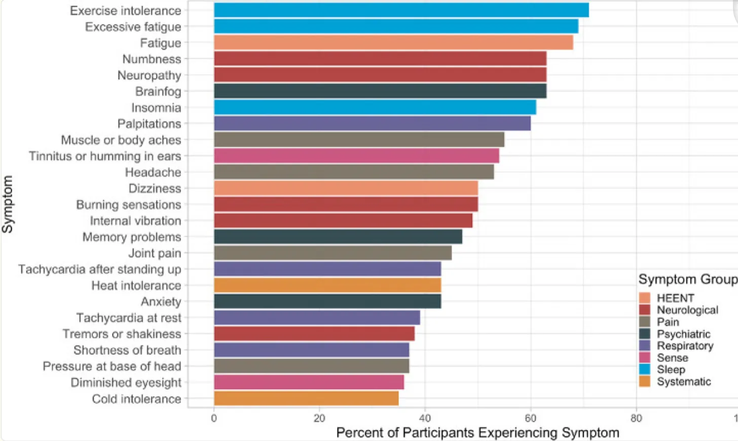
Symptoms

Symptoms vary. Different people can have different symptoms.
Some people have very mild symptoms, others can have life-threatening symptoms.
Diagnosis can be difficult because Sarcoidosis symptoms can mimic symptoms of diabetes, hypopituitarism, optic neuritis, meningitis, tumours, or neurologic disorders.
Some sarcoidosis patients have Lofgren’s syndrome with symptoms such as fever, enlarged lymph nodes, arthritic ankles, erythema nodosum.
Erythema nodosum is a rash on the ankles and/or shins, red or red-purple bumps, often warm and tender.
Sufferers can have any or all of the following symptoms, and some of these symptoms may be caused by other conditions:

  • Nodules (lumps, granulomas)
  • Constant fatigue
  • Glandular problems
  • Swollen or painful lymph nodes
  • Vision problems
  • Tiredness and lethargy
  • Facial swelling
  • Painful red lumps called erythema nodosum on the front of the legs.
  • Dry mouth, often very dry
  • Metallic taste in the mouth
  • Constant thirst
  • Increased Urinary Frequency
  • Dry Eye
  • A slight, moderate, or high fever
  • Occasional or frequent unexplained fevers
  • Sudden shortness of breath
  • Chronic or recent productive cough
  • Chronic or recent non-productive cough
  • Chest pain or discomfort
  • Arthritis, joint pain, swelling or stiffness
  • Skin rashes, sometime red or blue patches
  • Hyperprolactinemia (high prolactin)
  • Hyperparathyroidism (too much parathyroid hormone)
  • Monocytes Elevated
  • Headaches
  • Neuritis or Neuropathy (pins and needles or numbness)
  • Pulmonary Fibrosis
  • Interstitial Lung Disease
  • Cardiovascular problems
  • Enlarged spleen
  • Enlarged liver
  • Enlarged gall bladder
  • Blocked bile duct
  • Nervous system disorders

Complications and Prognosis

Lung and lymph node involvement is the most common problem, occurring in up to 90% of cases.

X-Rays, CT scans and lung biopsies are often used to confirm the diagnosis.
Sometimes only the lymph nodes are swollen with no other symptoms.
Involvement is not uncommon in the liver, blood, skin, musculoskeletal, eyes, kidneys and endocrine and reproductive organs.
Although most patients find the symptoms disappear by themselves, medical supervision is required. Left untreated, serious problems with any or all organs affected can cause complications.
Cardiovascular involvement may occur, where granulomas form in and around the heart.
ECG / EKG (Electrocardiogram) generally does not help the diagnosis, but MRI, Echocardiography, or even Endomyocardial biopsy may be required.
Although rare, this can be serious with symptoms leading to heart arrhythmia, palpitations, chest pain, or even sudden cardiac arrest.
Neurological involvement occurs in around 5% of cases, and can cause mild to serious problems in the CNS (Central Nervous System) and related areas if left untreated.
High calcium in the blood and urine can cause kidney stones, kidney damage, blocked blood vessels and osteoporosis.

Hypercalcemia

Sarcoidosis patients often suffer from Hypercalcemia (excess calcium in the blood).
However, other diseases can also cause hypercalcemia, including:

  • Tuberculosis
  • Berylliosis
  • Coccidioidomycosis
  • Histoplasmosis
  • Candidiasis
  • Crohn’s disease
  • Langerhans-cell histiocytosis (also Histiocytosis X, often with Eosinophilic Granuloma)
  • Silicone-induced granulomas
  • Cat-scratch disease
  • Wegener’s granulomatosis
  • Pneumocystis carinii pneumonia

Hypercalcemia, Hypercalciuria, Vitamin D2/D3, Parathyroid Hormone and Phosphates

Up to 50% of Sarcoidosis patients may have hypercalciuria (excess calcium in the urine).
Up to 20% may have hypercalcemia (excess calcium in the blood).
First, it is important to understand how the body makes and regulates Vitamin D3.
There are many stages and forms of vitamin D, and this is the simplest explanation I can put together, although by no means complete:
7-Dehydrocholesterol is made in the liver and migrates to the epidermis layer of the skin, where it is changed by UVB (Ultra-Violet in the B range) from sunlight into:
Cholecalciferol which then migrates back to the liver, where it is hydroxylated into: calcifediol, or 25-hydroxyvitamin D3 or 25-hydroxycholecalciferol or 25(OH).
The process of hydroxylation is adding the hydroxide (OH) molecule to the vitamin D molecule.
Calcifediol then migrates to the kidneys, where the enzyme 1-alpha-hydroxylase produces yet another hydroxylation process, (adding yet another hydroxide molecule, making it a dihydroxy form), converting into the active form calcitriol, or 1,25-dihydroxyvitamin D3, or 25(OH)2D
Vitamin D2 (and to a lesser extent D3) are also taken in from the diet, but in smaller quantities, and dietary D2 and D3 are poorly absorbed in the intestine.
Note that if prescription statin medication is taken, this prevents the liver from manufacturing as much 7-Dehydrocholesterol, preventing enough vitamin D from being made, regardless of the amount of sunlight exposure.
The pre-storage (un-hydroxylated) version of D3 can be called:

  • Calcifediol
  • Cholecalciferol
  • Calciferol (can refer to D2 and D3 collectively)
  • Toxiferol
  • Calciol

The storage form can be called several names, such as:

  • 25(OH)D
  • 25(OH)D3
  • 25(OH) vitamin D
  • 25-hydroxyvitamin D3
  • 25-hydroxycholecalciferol
  • Calcidiol

Vitamin D2 – mostly artificial supplements in “vitamin D fortified” foods and some natural foods.
This version is to be avoided because it displaces the real vitamin D3 which is superior for immunity.
Vitamin D2 is acquired from plant food by the body, and this is the version that is VERY BAD for Sarcoidosis.
Unfortunately, when doctors prescribe supplemental vitamin D, it is almost always the worst kind: Vitamin D2, which must be avoided, even for healthy people.
D2 does nothing for bones, and nothing for immunity, and makes Sarcoidosis worse.
Other names for D2:

  • Vitamin D2
  • Ergocalciferol

D2 is converted in the liver to become:

  • 25-hydroxyergocalciferol
  • 25-hydroxyvitamin D2
  • 25(OH)D2

The active form of vitamin D3 has many names:

  • 1,25-D
  • 1,25 2D
  • 1,25 2(OH) D
  • 1,25-dihydroxyvitamin D3
  • 1,25 dihydroxycholecalciferol
  • Calcitriol

In normal, healthy people, cytokines (macrophages, special white blood cells), also process Calcidiol into Calcitriol, increasing the active D3.
When the patient has Sarcoidosis, this process is magnified, especially in the granulomas where the macrophages accumulate.
The normal immune system has become compromised, leading to an over-abundance of the active D3, in turn raising calcium levels in blood and urine, “stealing” the calcium from the bones (and teeth).
Because Sarcoidosis granules increase production of 1,25-dihydroxyvitamin D (the active metabolite form of vitamin D), increased intestinal calcium absorption is induced.
Higher 1,25 dihydroxyvitamin D increases bone resorption, where the body’s osteoclast cells break down bone into it’s constituent minerals, releasing the minerals into the bloodstream.
This can be aggravated by sunlight exposure and/or supplements, which produce even more vitamin D.
In healthy people, 25(OH) vitamin D (calcidiol) is converted to 1,25 dihydroxyvitamin D (calcitriol) by the enzyme 1-alpha-hydroxylase in the kidneys.
The conversion process is controlled by:

  • PTH (Parathyroid Hormone)
  • Fibroblast growth factor 23 (FGF23)
  • Serum phosphate concentration

Normally, as calcium levels rise, the Parathyroid glands suppress the release of PTH (Parathyroid Hormone), which reduces 1,25 dihydroxyvitamin D production (calcitriol).
All hormones in the body are regulated. If too much or too little is produced, other hormones or enzymes come in to fix the problem, and for the most part, the system works well.
For Sarcoidosis patients, macrophage cells, typically in the granulomas in lungs and lymph nodes, convert 25(OH) or calcidiol into 1,25 dihydroxyvitamin D or calcitriol, without the normal control of PTH (Parathyroid Hormone).
This unregulated production of 1,25-dihydroxyvitamin D can cause hypercalciuria (high calcium in urine), or if the level of 1,25-dihydroxyvitamin D goes over 43.5 pg/ml (pica-grams per ml of blood), causes hypercalcemia (high blood calcium) because at this level, calcium is mobilised from bones into the blood.
If blood calcium levels are high, and parathyroid hormone levels are low, and 1,25-dihydroxyvitamin D is high, then Sarcoidosis or other granular disease may well be the diagnosis.
When 25(OD)D drops too low, the body increases 1,25D to increase calcium absorption, because the body needs calcium for the blood.
Calcium levels in blood are normally low in healthy people, but very important in the blood for electrical and clotting ability.
If we get more 25(OH)D from sunlight or supplements, this forces up 1,25(OH)2D.
The body is then fooled into believing it is low in calcium, and moves calcium from bones to blood, which increases fracture risk.
In this case, PTH (Parathyroid Hormone) will be low, telling the body it has more D and calcium than it needs.
Sarcoidosis patients typically have low PTH (Parathyroid Hormone), slowing production of 25(OH)D, because of the extra source of 1,25D from granulomas.
If the patient has high urine calcium levels, even if both vitamin D levels are normal and serum calcium is normal, high urine calcium is bad.
Blood calcium can be normal is because the kidneys are excreting excess calcium in the urine.
If the kidneys were not able to do this, the patient would have a high blood calcium and become sick.
Even if both vita,in D’s are low, high urine calcium means that no additional vitamin D or calcium supplements should be taken until urine calcium is resolved.
Nearly 60% of Sarcoidosis patients will have high urine calcium at some stage, which is not a concern if the rise in urine calcium is temporary.
If high urine calcium is persistently elevated, it must be treated, otherwise this can cause kidney stones, and eventually can lead to calcification of kidneys and loss of kidney function.
Parathyroid levels show if the body is suppressing vitamin D production as a response to increased blood calcium, which must be filtered out by the kidneys and excreted into the urine.
High blood calcium levels signal the parathyroid gland to decrease liver production of vitamin 25D, whose precursors come from food and from sunlight on skin.
This is why vitamin D levfels aree kept low.
Vitamin D increases calcium absorption from food, and if there is high blood calcium, the parathyroid reduces production of vitamin D to reduce blood calcium.
Excretion of calcium and reduced vitamin D is the body’s protective mechanism for high blood calcium.
However, without treatment, this cannot be kept up permanently, as kidney failure would eventually happen, perhaps 5 to 20 years down the track.
Many symptomless sarcoidosis patients who were never treated suddenly get symptoms of kidney failure, sometimes 20 years after original diagnosis.
During that time, silent hypercalciuria was occurring, was never tested, and then kidney damage becomes apparent.
Hypercalciuria is a silent and insidious symptom of sarcoidosis.
Generally, Sarcoidosis patients do not need to avoid calcium, as calcium is so abundant in so many foods.
But oxalates and phosphates must be avoided.
Phosphorus is also in almost every food we eat.
Phosphorus levels can rise when the kidneys are attempting to lower blood calcium levels.
When blood phosphorus levels are high, calcium and phosphorus bind together to form crystals, which can be deposited anywhere, but often in arteries, causing calcification of atherosclerotic or cholesterol plaques, also can increase stiffening of blood vessels.
Keep off the beach, water, snow and ice, where reflections magnify the suns rays, and limit exposure to the sun.
Carbonated soft drinks contain high phosphorus, especially colas, also chocolate, coffee, tea and orange juice are best avoided.
Doctors often forget the phosphate problems and only concentrate on calcium, but phosphorus is insidious in its actions and has fewer symptoms than calcium when it is high.

Diagnosis

Unfortunately there are no absolute tests available for a conclusive diagnosis, but there are tests which, combined with symptoms and/or visible nodules, can lead to diagnosis.
Often, the nodules are visible, but when they are internal, ultrasounds and blood tests become more important.
If nodules are easily accessible, they can be surgically removed and a biopsy performed.
One of the dangers of Sarcoidosis in Hypercalcemia (too much calcium in the blood) There are several essential blood tests:

  • 25(OH) vitamin D3
  • 1,25 (OH)2D (active vitamin D3)
    Note:
    We need BOTH 25(OH) and 1,25 (OH)2D tested because the kidneys and the nodules can make extra 1,25(OD)2D so this may be high, even when the regular test 25(OH) comes in low.
    Without this knowledge we do not know whether to supplement Vitamin D3 or to totally abstain from D3
  • ACE (Angiotensin Converting Enzyme) – often elevated with sarcoidosis, used to diagnose and monitor sarcoidosis, and monitor response to treatment.
  • Liver Panel or CMP (Comprehensive Metabolic Panel) – to evaluate liver and/or kidney function and to determine if those organs are affected.
  • CBC (Complete Blood Count) – to evaluate red and white blood cell changes
  • ESR (Erythrocyte Sedimentation Rate) – to detect inflammation
  • PTH (Parathyroid Hormone)
  • Calcium
  • Phosphates
  • Albumin
  • Calculation of estimated ionised calcium level
  • hs-CRP (high-sensitivity C-Reactive Protein) – monitors inflammation levels
    And 24-hour urine tests (IMPORTANT):
  • Calcium
  • Creatinine
    Note: Serum (blood) Calcium level tests are not enough, as this varies considerably even in healthy people. We need the 24-hour urine calcium test to determine the extent of any Hypercalcemia, which can cause kidney stones, kidney damage, blocked arteries, etc.
    Other tests:
  • CSF analysis – evaluates cerebrospinal fluid when brain or nervous system involvement is suspected
  • AFB cultures, sputum cultures, fungal tests – to distinguish between sarcoidosis and other lung or granuloma conditions
  • Chest X-ray – to detect granulomas in lungs, sometimes found in people who have an X-ray for some other reason.
  • Lung function test – to evaluate lung involvement, lung capacity, and condition severity.
  • CT (Computed Tomography), MRI (Magnetic Resonance Imaging), Gallium scan – to help diagnose and evaluate sarcoidosis
  • EKG / ECG (electrocardiogram) – when heart involvement is suspected
  • If Cardiac involvement is suspected, heterogeneous myocardial FDG uptake may be a useful diagnostic marker of disease activity
  • SAA (serum amyloid A protein – a measure if inflammation, normally corresponds with CRP
  • sIL2R(soluble interleukin-2 receptor) – perhaps a better measure of inflammation, mainly used to measure and monitor problems in lungs

Interpretation of test results

This is where we need a doctor experienced in treating Sarcoidosis, as most General Practitioners just do not know or may get it wrong.
If Parathyroid hormone is low and 25(OH) vitamin D3 is low, then no supplemental vitamin D3 should be taken.
PTH stimulates 1,25 D production in the kidneys.
If PTH (Parathyroid Hormone) is high (hyperparathyroidism) and blood calcium is high (hypercalcemia) then corticosteroids should be prescribed, but if the hypercalcemia fails to resolve, then further investigation is required.
If tests detect renal (kidney) impairment, hypercalcemia or significant hypercalcuria (over 400 mg per 24 hours), then a 24-hour creatinine clearance test should be carried out.
Also abdominal ultrasound investigations should be performed to exclude urolithiasis (stones in the kidney, bladder, or urethra) or nephrocalcinosis (calcium deposits in the kidneys).
Hypercalcemia in patients formally diagnosed with acute sarcoidosis require no further testing, and corticosteroid therapy with monitored response is the next step.
Hypercalcemia in patients without confirmation of sarcoidosis require more testing because lymphoma can cause lymphadenopathy (swollen or enlarged lymph nodes) and raise serum calcium levels.
If normal blood tests show no hypercalcemia (high blood calcium), this does not exclude hypercalcuria (high calcium excreted in the urine), so the 24-hour urine calcium test must always be carried out in every patient.
If liver tests show abnormal results, an ultrasound should be performed to exclude any damage to the liver.
If C-Reactive Protein levels are high or increasing, attention should be paid to reducing levels, through Sarcoidosis treatment, and using traditional CRP supplements:

Although Homocysteine is another marker of inflammation, there appears to be no correlation with Sarcoidosis.
High homoscsteine is generally used as a marker for future cardiovascular events.

Treatment

In most people, no treatment is required and the condition disappears, sometimes in a few weeks, months, or years.
Around one third of patients will have no more symptoms within 3 years, and another third will have no more symptoms within ten years.
Those with mild symptoms may not need treatment, but must be monitored to ensure the condition does not get worse.
The main goal is to decrease inflammation, relieve symptoms, and minimize organ damage.
Those with moderate to severe symptoms or at risk for organ damage are usually given corticosteroids such as Prednisone.
Corticosteriods are anti-inflammatory medications, given orally, topically, injected, or through an inhaler.
Corticosteroids mimic the hormones of the adrenal glands, which sit on top of the kidneys. This suppresses inflammation, reducing Sarcoidosis symptoms.
Also reduces symptoms of arthritis, asthma, lupus, allergies, etc and help excrete excess calcium.
The immune system is also suppressed, helping prevent diseases where the body’s immune system attacks it’s own healthy cells.
Other immune-suppressing medications may also be prescribed.
Long-term corticosteroid use will bring significant side-effects.
Side effects – Oral corticosteroids:

  • Glaucoma (elevated pressure in the eyes)
  • Fluid retention, causing swelling in lower legs
  • Increased blood pressure
  • Weight gain, fat deposits or swelling, mainly in abdomen, face (moon face) back of neck, fingers, hands, feet or lower legs
    And oral medications taken long-term:
  • Cataracts (clouding of the lens in one or both eyes)
  • Diabetes (high blood sugar)
  • Increased infection risk
  • Osteoporosis (thinning bones, fracture risk)
  • Suppressed adrenal gland hormone production
  • Thin skin, bruising, poor wound healing
  • Insomnia
  • Hunger leading to weight gain
  • Agitation, anger, depression, mood swings, increased risk of suicidal behaviour
  • Neuropsychiatric disorders
  • Potassium loss
  • Muscle weakness
  • Peptic ulcers, intestinal bleeding
  • Pounding in the ears
  • Shortness of breath, troubled breathing at rest
  • Trouble thinking, speaking, or walking
  • Sluggishness (or hyperactivity)
    Side effects – Inhaled corticosteroids:
    When using an inhaler, corticosteroids may deposit in the mouth and throat, on it’s way to the lungs, causing:
  • Oral Thrush (fungal infection in the mouth)
  • Hoarseness
    Avoid these problems by a gargle and rinse with water (do not swallow) after each puff of the inhaler
    Inhaled corticosteroid drugs for asthma slow growth rates in children, although they do not appear to affect their final adult height.
    Side effects – Topical corticosteroids:
  • Thin skin
  • Red skin lesions
  • Acne
    Side effects – Injected corticosteroids:
    Near the site of the injection, effects may include:
  • Pain
  • Infection
  • Shrinking of soft tissue
  • Loss of skin colour
    Corticosteroid injections are generally limited to 3 to 4 annually.
    To minimise side effects:
  • Try lower doses or intermittent dosing
  • Use low-dose, short-term medications
  • Use oral corticosteroids rather than injections as the dose can be varied daily
  • For lung problems, the inhaled version go directly to the lungs, limiting exposure to the rest of the body
  • Reduce calories and increase exercise to prevent weight gain
  • Exercise will reduce muscle weakness and osteoporosis risk
  • Eat an alkaline-forming diet to avoid acids leaching calcium from the bones
  • Get sunlight and/or Vitamin D supplements if there is no hyperglycemia
  • Discontinue slowly so that adrenal glands can recover

HP Acthar gel
Normally injected to help regulate adrenal gland function.
In the past, used to treat allergy-related conditions, breathing, blood, endocrine issues, arthritis, skin or eye problems, bowel inflammation, MS (multiple sclerosis), some cancers, and more recently for Sarcoidosis, either with or without corticosteroids.
HP Acthar gel is an adrenal hormone, and stimulates the body to produce more adrenocortical hormones including corticosteroids and glucocorticoids.

NSAIDs (nonsteroidal anti-inflammatory drugs) like ibuprofen are used for pain and inflammation (again with side-effects).
Methotrexate and other immune-suppressing drugs are used for lung, skin, or eye involvement.
Hydroxychloroquine (anti-malarial drug) used for skin and nervous system involvement, especially in those with high calcium levels.
Other prescription medications include Remicade (Infliximab) and Enbrel (Etanercept).
Most people can be successfully treated, but medications may be required for extended periods.
Very rarely, patients may need an organ transplant if lungs, liver or kidneys are severely damaged.
If Sarcoidosis is found in chest x-rays, corticosteroids are usually given, even if there are no other symptoms.
Arthritis and muscle weakness can be helped with physiotherapy or mobility aids.
ARB (Angiotensin Receptor Blockers) are normally used to control high blood pressure, it is involved in the inflammation of the granulomas.
ARBs may help reduce severity of symptoms.
Antibiotics – Low-dose, pulsed, bacteriostatic antibiotics can often help the immune system defeat Sarcoidosis.
ARB – Angiotensin Receptor Blockers are commonly used to control high blood pressure, but have shown benefit for Sarcoidosis patients.
Anti-Fungal Treatment – Beta-Glucan is a constituent of fungal cell walls, and has been found in BAL (broncho-alveolar lavage) fluid in sarcoidosis patients.
Anti-fungal medication may be prescribed for newly diagnosed, mild cases. More studies are required before higher anti-fungal doses are given to more severe cases.

Natural Treatments

There are many natural treatments, and most will reduce severity, speed recovery and reduce chances of a relapse, and can be used in conjunction with prescription medication.
Given that fungi may be a issue, steps taken to reduce fungi around the home or work environment may help:

  • Allow plenty of light and fresh air into the building to reduce mould.
  • Replace old carpets with tiles, floating timber or other clean floors
  • Use a central vacuum system to exhaust all air outside the building
  • Avoid having animals in the home, as they bring in fungi on their feet and fur.

Oregano Oil has excellent anti-fungal properties.
Brushing teeth with a mixture of a few drops of oil diluted with a quantity of grapeseed oil is enough to impact fungi, parasites and bad bacteria in the mouth, where the immune system starts.
Garlic an odourless capsule or a natural food contains Ajoene which has potent antifungal properties.
Garlic is also anti-bacterial, anti-viral and can help lower blood pressure and improve cardiovascular health.
Coconut Oil is another natural anti-fungal product, also anti-bacterial, anti-viral, and helps reduce excess body fat, as it contains MCT (Medium Chain Triglycerides) which go straight to the liver to be burnt as fuel, and cannot be stored as fat in the body.
Grapefruit Seed Extract has powerful anti-fungal properties, but works best in conjunction with others above rather than alone.

Other Natural Treatments

d-Mannose is a form of sugar, but is safe for diabetics, and reports by Sarcoidosis patients claim relief of symptoms.

UTI – Urinary Tract Infections

There can be a relationship between Urinary Tract Infections and Sarcoidosis.
UTI tests are available from the doctor.
Some Sarcoidosis patients have a genetic variant in MRC1 (Mannose Receptor gene) called carbohydrate-deficient glycoprotein syndrome, an inherited metabolic disorder, and supplementing with d-Mannose may help control UTI.
The active ingredient in Cranberries (and juice) contains natural d-Mannose, which is a type of sugar, but unlike sugar, it has almost zero effect on blood glucose levels, so is safe for diabetics, and shown to improve UTI (Urinary Tract Infections), and safer than cranberries which also contain fructose and other sugars.
The benefit for UTI appears to be it’s ability to affect the lining on the internal walls of the bladder, urethra and related areas to prevent harmful bacteria from “sticking” to those walls, preventing colonisation by allowing them to be flushed away in the urine.
Many reports by Sarcoidosis patients claim relief of symptoms.
Sarcoidosis patients, especially women over 60, and those on corticosteroids, are more prone to UTI.
UTI is mainly caused by the bacteria E.coli which attaches to the walls of the bladder and urinary passages, and in serious cases can work it’s way up to the kidneys, where it can cause more serious damage.
Mannan-Binding Lectin (MBL), also called Mannose-binding Lectin, Mannose-Binding Protein or Mannan-Binding Protein (MBP), is a lectin that is important for correct immune function.
The function of MBL appears to be pattern recognition in the immune system.
MBL recognizes carbohydrate patterns which are found on the surface of many pathogenic micro-organisms such as including bacteria, viruses, protozoa and fungi.
MBL then binds to carbohydrates found on the surfaces of these pathogens, preventing them from attaching to parts of the body.
In the case of urinary infections, E.coli is attracted to the lectins instead of the urinary linings, allowing the urine system to then flush away the bound E.coli and this is a more effective and safer treatment than antibiotics which is the normal treatment given by doctors.
Note that in the Marshall Protocol (see below), d-Mannose is prohibited, but I believe Marshall is wrong.
All of the science indicates d-Mannose> improves inflammation, regulation of the immune system, and removal of:

  • Bacteria such as Salmonella, Streptococci, E.coli
  • Yeasts such as Candida Albicans
  • Viruses such as HIV, Coronavirus and Influenza A
  • Parasites like Leishmania

More info: Mannan Binding Lectin

Stem Cell Therapy

Promising results with stem cell treatment so far for patients with lung damage, but it may be some time before this therapy is widely available.
Currently it is several thousand dollars, and most Health Insurance companies will not cover this treatment.
CytoSorb® – a Cytokine Filter CytoSorb® is used in immunotherapy to improve ability to prevent multiple organ failure, which is a leading cause of death in the ICU (intensive care unit).
In response to serious sepsis, infection, trauma, burns, lung injury, pancreatitis, and inflammation, the body can over-react by producing a massive “cytokine storm”.
Cytokines normally help an injured body, but cytokine storms drive major inflammation and severe pathophysiologic cell damage, organ failure and even death.
Cytokine storm reduction can help limit the cascading of events, helping patients to survive.
In the past, there was no significant method to control cytokine storms, but CytoSorb® is an extracorporeal cytokine adsorber, now approved in the European Union.
Use in any condition where cytokines are elevated, such as Sarcoidosis, and compatible with hemodialysis machines and blood pumps.
Blood is pumped from the body, through the CytoSorb® cartridge, and purified blood is retuned to the patient.
In six hours, the entire blood volume can be filtered 20 times.

Oxalates and Kidney Stones

Avoid foods high in oxalates. Kidney stones can cause excruciating pain, and Sarcoidosis patients with high calcium are more prone to kidney stones.
Also, a diet high in oxalates increases kidney stone risk for even normal, healthy people.
Many foods contain oxalates, but the following are the foods proven to increase kidney stone risk:

  • Beets (beetroot)
  • Spinach
  • Rhubarb
  • Strawberries
  • Nuts
  • Chocolate
  • Tea
  • Wheat bran
  • All beans (fresh, canned, or cooked), excluding lima and green beans

Alternative Protocols

Stop Sarcoidosis Physician’s Treatment Protocol.
Marshall protocol developed Trevor Marshall, an electrical engineer.
Marshall hypothesizes that bacteria lacking cell walls can live inside immune cells and thus evade detection and elimination by the immune system.
Not accepted by most doctors and supported only through anecdotal evidence, but worth reading.
Aden Protocol
Said by many to be a scam because to only way to obtain it is to buy the e-book at around US$37 but still contains some useful information.
Most reports I have read say it does not work, but I agree with the Serrapeptase supplementation and some of the other foods to include and avoid.
Serrapeptase has the ability to break down cysts, dead cells, granulomas and some tumours.

Risk factors

The best way of preventing Sarcoidosis is to reduce risk of the condition starting:
Ensure healthy levels of vitamin D3 (but reduce vitamin D3 if diagnosed)
Lose excess weight
Eat a healthy diet of fresh vegetables, especially leafy greens for vitamin K2, or supplement with K2 and B12.
Avoid processed foods, excess carbohydrates, all added sugar, mouldy food
Build a solid immune system by:

  • Incorporate fresh, natural food in the diet, avoid all processed food
  • Avoid sugar and high-carbohydrate foods
  • Work on oral hygiene as the mouth is the first line of defense against invaders
  • Improve intestinal health, the second and most important part of the immune system: Acidophilus Probiotic Blend


References

Erythema Nodosum – lumps on legs Confusion with Vitamin D The production of vitamin D (which is actually not a vitamin, but a steroid hormone) is a complex process, but the process is different in those with sarcoidosis.
I will attempt to explain the difference:
If the doctor orders a vitamin D blood test, the standard test is called 25(OH) which is not vitamin D, but a precursor to the ACTIVE form of vitamin D (1,25(OH)2D).
In normal, healthy people, this is not a problem, but the Vitamin D creation process is a long and complex, and many things can go wrong along the way.
In sarcoidosis patients who are untreated, the normal test of 25(OD)D comes back as low, and it is low because of the sarcoidosis disease process.
However, doctors must test the 1,25D because in untreated sarcoidosis, it is usually high.
Unfortunately, doctors inexperienced in treating sarcoidosis don’t do the correct test and don’t understand the difference even if they do.
The D3 hormone
Because vitamin D3 is essentially a steroid hormone and not a vitamin, it follows hormone rules in the body, meaning there is always a feedback system for all hormones.
If there is a low hormone level, a signal is sent to increase production of that hormone.
If there is a high hormone level, another signal is sent to slow or stop production, and this feedback system results in a state of equilibrium, or homeostasis.
Vitamin D3 receptors
In healthy people, almost all of the 60 trillion cells in the body have Vitamin D3 receptors.
This means that every time a D3 molecule goes past a cell, there is a special “key” in D3 which exactly fits the corresponding “lock” on the cell, and the two unite.
The cell now has much greater benefits for immunity, bone building and many other health benefits.
But this process allows a gene transcription of CYP24, an enzyme which can break down excess 1,25 D.
In sarcoidosis patients, the vitamin D3 receptor is blocked, often by 25D which attenuates inflammation and immunity, which is good thing in moderation.
However, the 25D is converted into 1,25 to excess, causing weight gain, fluid retention, headaches, nausea, and sometimes anorexia or neuropathies, all sarcoidosis symptoms.
With 25D blocking the VDR it cannot work properly to transcribe the genes which perform other body functions like making enzymes, antibacterial peptides or regulation of the hormone system generally.
This is those with sarcoidosis may have problems with menstrual periods or have thyroid issues.
Another reason why 25D is low in sarcoidosis patients is that where 1,25D normally binds to vitamin D receptors, it instead binds to PXR receptors.
The 1,25D binds where the 25D should bind and vice versa, and when PXR is activated by 1,25D it causes the CYP27A1 enzyme to be produced, stopping liver production of 25D from pre-D3.
Further, 25D is low because a another enzyme CYP27B1 produced by sarcoidosis granulomas, activated by PKA (Protein Kinase A), increases the conversion rate of 25D into 1,25D, so any available 25D is quickly converted into 1,25 D.
If the doctor prescribes vitamin D3 supplements, it blocks the VDR, stopping production of gene transcription for thousands of bodily functions, because PKA produced in the granulomas will cause enzymatic action to rapidly convert more 25D into 1,25 D.
Because the VDR is blocked, the enzyme CYP24 which breaks down excess 1,25 D cannot be transcribed, resulting in excess 1,25D, leading to development of calcium metabolism. problems.
When 1,25D reaches a high level in blood, osteoclasts in the bone are stimulated to release calcium, and the intestines are also stimulated to absorb more calcium, leading to high calcium levels in both urine and blood.
In sarcoidosis PTH has little effect, and often PTH is low when urine calcium is high, and less often, blood calcium is also high.
High calcium can lead to bone loss, osteoporosis and osteopenia, and calcium can be deposited in organs, especially kidneys, where calcium causes kidney stones or worse, blocked tubules causing kidney failure.
Calcium may also deposit in the brain, blood vessels, skin and other organs. Medication can help prevent excess calcium buildup by blocking the enzyme actions.
Prednisone, hydroxychloroquine and ketoconazole all help. Prednisone or hydroxychloroquine can bring down 1,25 D levels to normal.
Bisphosphonates are sometimes given to postmenopausal women but they are ineffective if given with supplemental vitamin D, and are not recommended for premenopausal women in any case.
Giving vitamin D supplements or cod liver oil to an untreated sarcoidosis patient can cause a hypercalcaemic state in a few weeks, and because few symptoms will appear until hypercalcemia is well advanced, urine and blood testing is essential.
LeanMachine does not recommend bisphosphonates in any case, as the method of action is to prevent osteoclasts from breaking down old bone. This will increase bone density, but makes bones more brittle, as it prevents osteoblasts from building new bone, as the bones become full of old bone.
Bones become “dead” allowing infection to become a problem, and many patients on bisphosphonates have lost their bottom jaw, which is not a pleasant thing.

Vitamin D Summary

If the patient has low vitamin D and a corresponding low PTH (parathyroid hormone), they should NOT take vitamin D.
In healthy people, if vitamin D is low, PTH increases, instructing kidneys to provide extra hydroxylation of 1,25D.
With sarcoidosis, PTH remains low because 1,25D is high even though 25D is high.
If we are truly deficient in 1.25D, PTH would never be low.
So if PTH is high, supplemental vitamin D3 is indicated, but if PTH is low, vitamin D3 must be avoided because it can make the sarcoidosis worse.
Extra D3 will add to the extra 1,25D produced by granulomatous inflammation, and will not broken down because 25D blocks the receptors that 1,25D should be binding to.

Outlook

Over 50% of patients recover within three years, and up to 70% recover in 10 years, with little or no problems.
Sarcoidosis may return a year or more after recovery, but generally in less than 5% of cases.
Lofgren’s syndrome patients usually all recover fully.
Sarcoidosis can damage organs in around 33% of cases, usually where the condition is untreated where damage takes many years and involves multiple organs.
Death is rare, usually as a result of complications with the lungs, heart, brain, or related to another condition.

Risk Factors for untreated Sarcoidosis


  • Chronic, long-term Sarcoidosis
  • Lung scarring, heart or brain complications
  • Lupus pernio, a skin condition caused by Sarcoidosis
  • Eye, brain, heart, liver damage


Eye problems

Annual eye exams are important for all Sarcoidosis patients.
For changes in vision or poor eyesight or poor colour definition, seek medical assistance immediately.
Any eye symptoms like burning, itching, tearing, pain, or light sensitivity also require medical assistance.
.

Risk factors for increased calcium and vitamin D in sarcoidosis

  • Disease is not in remission
  • Disease is spread to organs outside of hilar lymph nodes
  • Disease currently active and receiving no treatment
  • Disease is currently active and being treated with drugs other than prednisone and/or plaquenil
  • Male Gender
  • Previous history of kidney stones
  • Previous history of hypercalcemia
  • Working outdoors
  • Taking supplemental vitamin D or calcium
  • Has uncontrolled diabetes
  • Has uncontrolled kidney disease
  • Diet high in phosphorus and/or calcium and/or oxalate
  • Dehydration

The more things apply to the patient, the higher the risk for even worse Sarcoidosis.

Prescription Medication

Drugs that can interact negatively with sarcoidosis, including some common blood pressure drugs.
Search this list for any medication you are taking: www.ehealthme.com/symptom/sarcoidosis

Testing for Sarcoidosis

There is no definitive single test to diagnose Sarcoidosis.
Generally, a diagnosis is made after evaluating family history, symptoms, and various tests which may not mean much on their own, but point to Sarcoidosis when all of the facts and results are put together.
Sarcoidosis is different again when blood tests, urine tests and symptoms differ between individuals and differ also depending on the severity or mildness of the condition.

Ruling out what it is NOT

Part of the diagnosis is to rule out other things.
For example, Sarcoidosis is a CTD (Connective Tissue Disease), but there are a number of other diseases with often similar symptoms such as Wegener’s granulomatosis, RA (Rheumatoid Arthritis), SLE (Systemic Lupus Erythematosus), Polymyositis, CREST (Calcinosis, Raynaud disease, Esophageal motility disorder, Sclerodactyly, and Telangiectasia), CNS (Central Nervous System), Sjögren syndrome, Scleroderma, or different combinations of everything.
See the diagnostic road map (where Sarcoidosis is not even mentioned): Mayo Medical Laboratories)
Wegener’s granulomatosis is a connective tissue disease but has different symptoms: Blood flow is restricted when the vessels swell.
Common symptoms are nosebleeds, sinus pain, inflammation, constantly runny nose, pus-filled nasal discharge.